Department of Molecular Neuropathology, Centro de Biología Molecular "Severo Ochoa" (CBMSO), Spanish Research Council (CSIC), Universidad Autónoma de Madrid (UAM) (Campus de Cantoblanco), c/Nicolás Cabrera 1, 28049, Madrid, Spain.
Center for Networked Biomedical Research On Neurodegenerative Diseases (CIBERNED), Madrid, Spain.
Acta Neuropathol Commun. 2022 Aug 29;10(1):125. doi: 10.1186/s40478-022-01431-7.
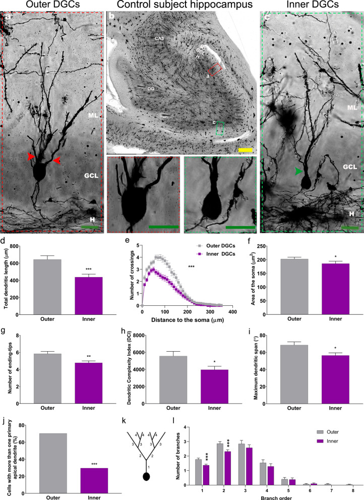
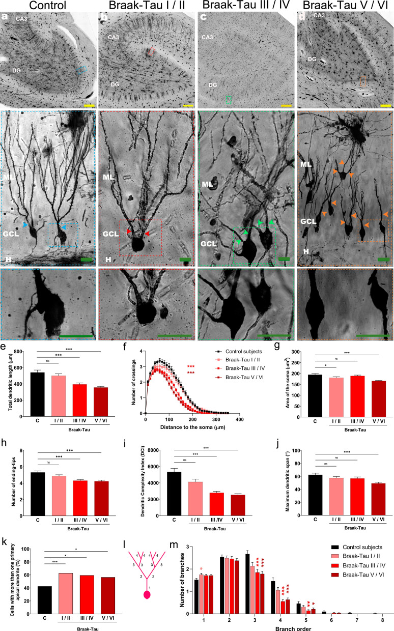
Alzheimer´s disease (AD), the most common form of dementia in industrialized countries, severely targets the hippocampal formation in humans and mouse models of this condition. The adult hippocampus hosts the continuous addition of new dentate granule cells (DGCs) in numerous mammalian species, including humans. Although the morphology and positioning of DGCs within the granule cell layer (GCL) match their developmental origin in rodents, a similar correlation has not been reported in humans to date. Our data reveal that DGCs located in inner portions of the human GCL show shorter and less complex dendrites than those found in outer portions of this layer, which are presumably generated developmentally. Moreover, in AD patients, DGCs show early morphological alterations that are further aggravated as the disease progresses. An aberrantly increased number of DGCs with several primary apical dendrites is the first morphological change detected in patients at Braak-Tau I/II stages. This alteration persists throughout AD progression and leads to generalized dendritic atrophy at late stages of the disease. Our data reveal the distinct vulnerability of several morphological characteristics of DGCs located in the inner and outer portions of the GCL to AD and support the notion that the malfunction of the hippocampus is related to cognitive impairments in patients with AD.
阿尔茨海默病(AD)是工业化国家最常见的痴呆症形式,严重影响人类和该疾病小鼠模型的海马结构。成年海马体在包括人类在内的许多哺乳动物物种中持续产生新的颗粒细胞(DGC)。尽管 DGC 在颗粒细胞层(GCL)中的形态和位置与其在啮齿动物中的发育起源相匹配,但迄今为止尚未在人类中报道类似的相关性。我们的数据显示,位于人类 GCL 内部的 DGC 的树突比位于该层外部的 DGC 短且复杂程度较低,这些 DGC 可能是发育产生的。此外,在 AD 患者中,DGC 表现出早期的形态改变,随着疾病的进展进一步加重。在 Braak-Tau I/II 阶段的患者中,首先检测到 DGC 数量异常增加,具有多个主树突的异常改变。这种改变在 AD 进展过程中持续存在,并导致疾病晚期的广泛树突萎缩。我们的数据揭示了 GCL 内、外部 DGC 的几个形态特征对 AD 的明显易损性,并支持海马体功能障碍与 AD 患者认知障碍有关的观点。