Plateforme Imagerie In Vitro, CNRS UAR 3156, Neuropôle, University of Strasbourg, Strasbourg, France.
Department of Physiological Genomics, BioMedical Center BMC, Ludwig-Maximilian University, Planegg-Martinsried, Germany.
J Extracell Vesicles. 2022 Sep;11(9):e12254. doi: 10.1002/jev2.12254.
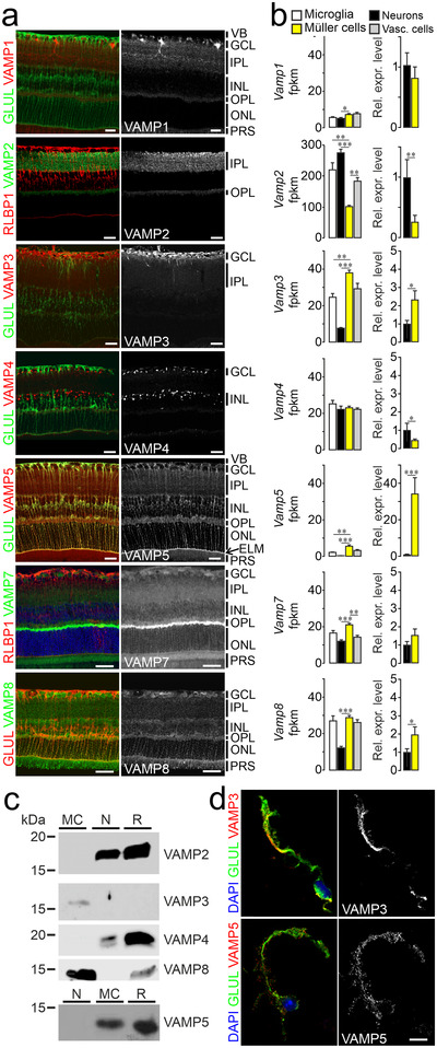
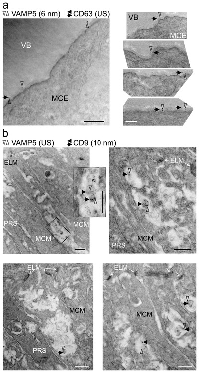
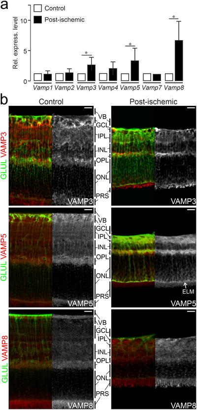
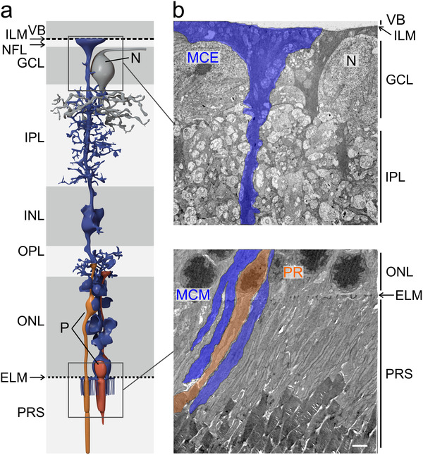
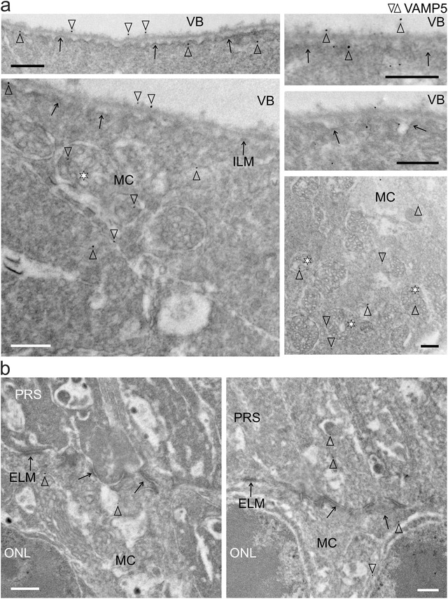
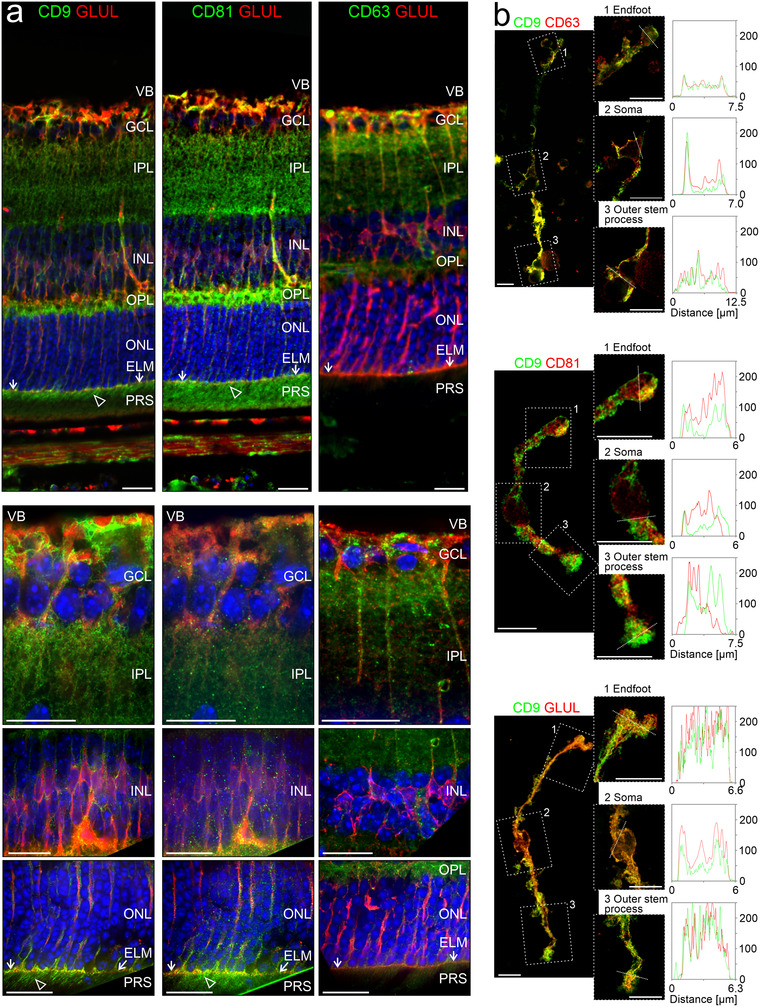
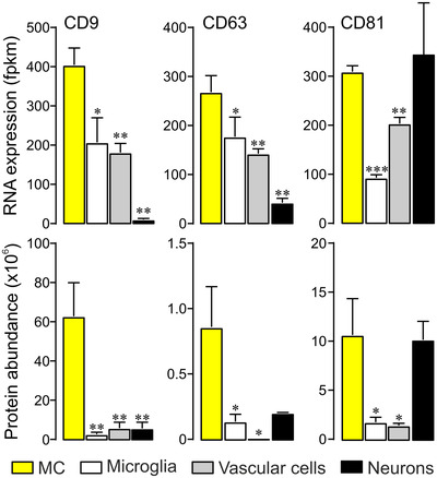
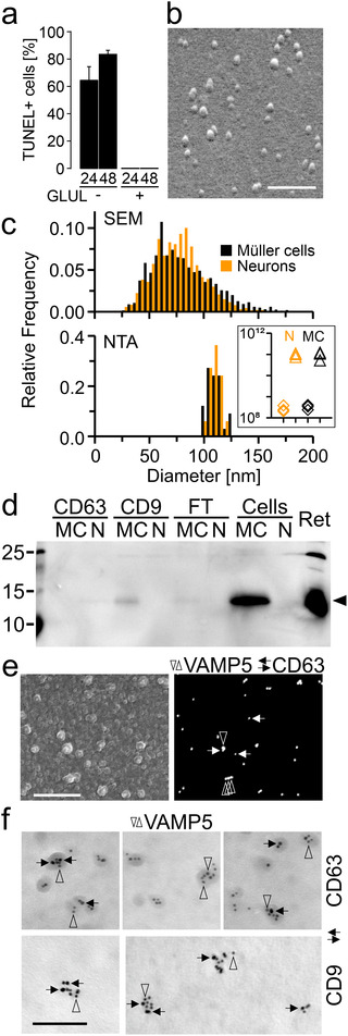
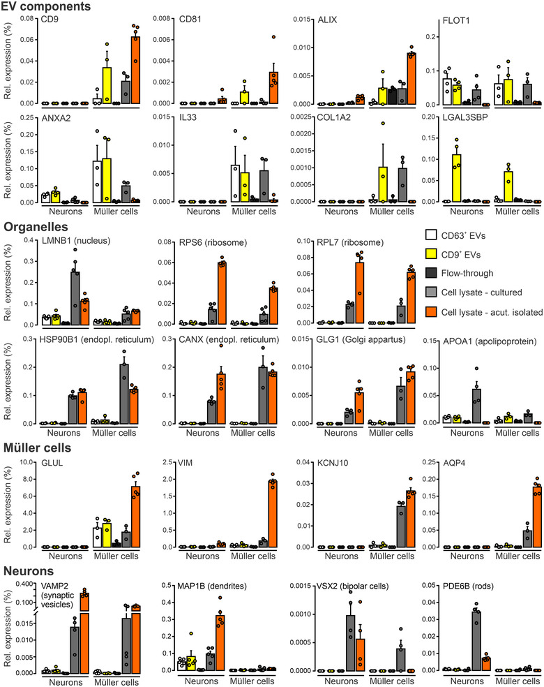
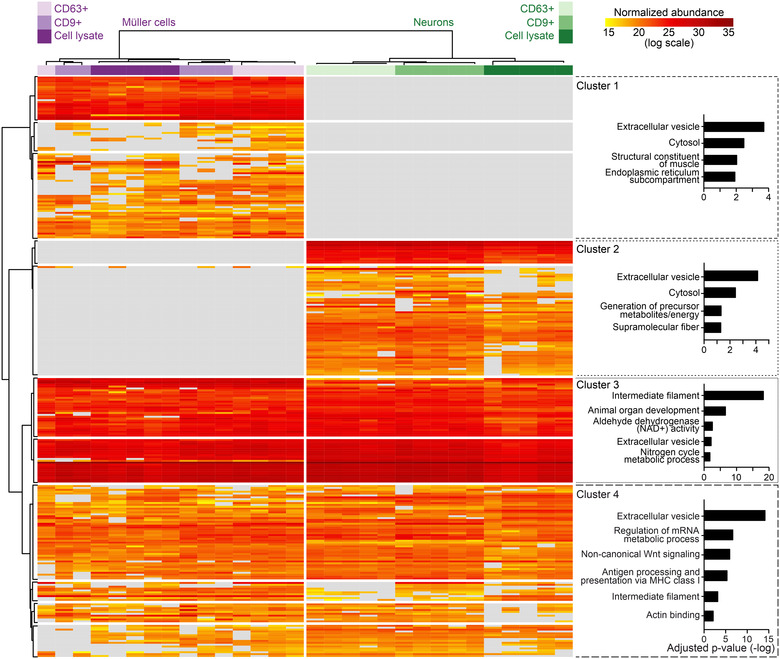
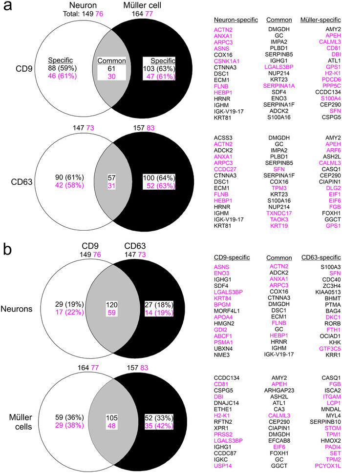
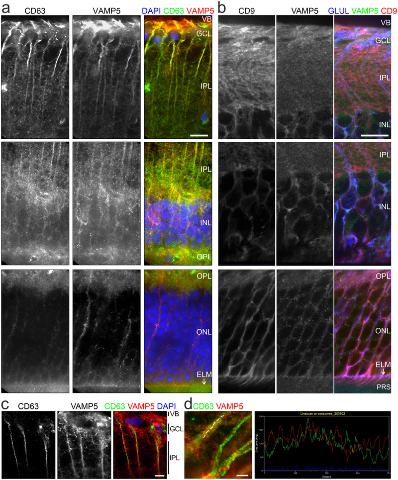
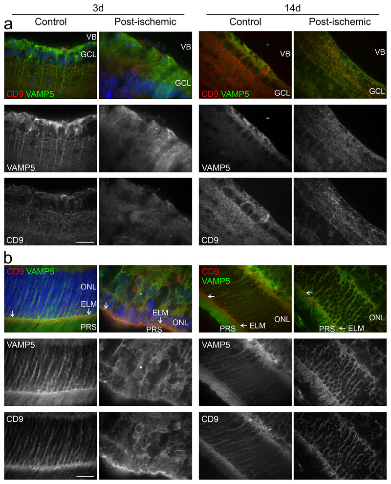
Cell-cell interactions in the central nervous system are based on the release of molecules mediating signal exchange and providing structural and trophic support through vesicular exocytosis and the formation of extracellular vesicles. The specific mechanisms employed by each cell type in the brain are incompletely understood. Here, we explored the means of communication used by Müller cells, a type of radial glial cells in the retina, which forms part of the central nervous system. Using immunohistochemical, electron microscopic, and molecular analyses, we provide evidence for the release of distinct extracellular vesicles from endfeet and microvilli of retinal Müller cells in adult mice in vivo. We identify VAMP5 as a Müller cell-specific SNARE component that is part of extracellular vesicles and responsive to ischemia, and we reveal differences between the secretomes of immunoaffinity-purified Müller cells and neurons in vitro. Our findings suggest extracellular vesicle-based communication as an important mediator of cellular interactions in the retina.
中枢神经系统中的细胞间相互作用基于释放介导信号交换的分子,通过囊泡胞吐和形成细胞外囊泡为结构和营养支持提供保障。大脑中每种细胞类型所采用的特定机制尚未完全清楚。在这里,我们探索了视网膜中的一种放射状胶质细胞—— Müller 细胞所使用的通讯手段,Müller 细胞是中枢神经系统的一部分。通过免疫组织化学、电子显微镜和分子分析,我们为成年小鼠活体中源自视网膜 Müller 细胞的足突和微绒毛的特定细胞外囊泡的释放提供了证据。我们鉴定出 VAMP5 作为一种 Müller 细胞特异性 SNARE 组成部分,它是细胞外囊泡的一部分,并对缺血有反应,并且我们揭示了体外免疫亲和纯化的 Müller 细胞和神经元的分泌组之间的差异。我们的研究结果表明,基于细胞外囊泡的通讯是视网膜中细胞相互作用的重要介质。