Department of Biomedical Engineering, School of Medicine, Kyungpook National University, Daegu, South Korea.
Department of Neurology, School of Medicine, Kyungpook National University, 680 Gukchaebosang-ro, Jung-gu, Daegu, 41944, South Korea.
Fluids Barriers CNS. 2022 Sep 1;19(1):66. doi: 10.1186/s12987-022-00362-8.
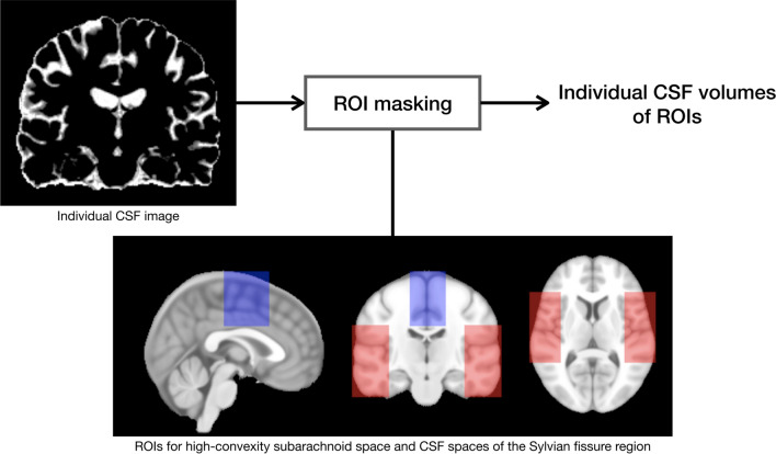
The aims of the study were to measure the cerebrospinal fluid (CSF) volumes in the lateral ventricle, high-convexity subarachnoid space, and Sylvian fissure region in patients with idiopathic normal-pressure hydrocephalus (INPH) and Alzheimer's disease (AD), and to evaluate differences in these volumes between INPH and AD groups and healthy controls.
Forty-nine INPH patients, 59 AD patients, and 26 healthy controls were imaged with automated three-dimensional volumetric MRI.
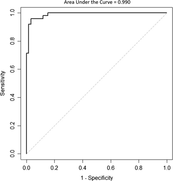
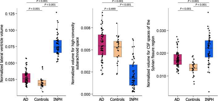
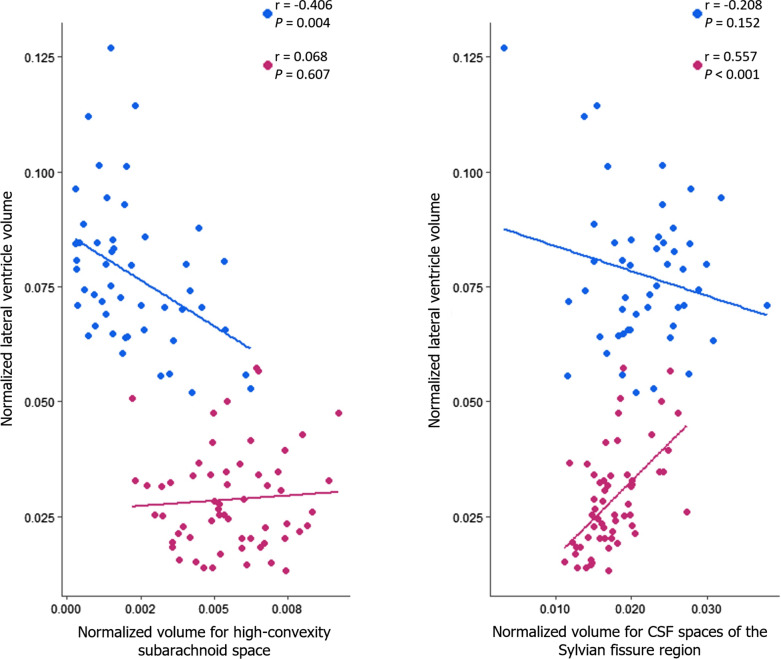
INPH patients had larger lateral ventricles and CSF spaces of the Sylvian fissure region and smaller high-convexity subarachnoid spaces than other groups, and AD patients had larger lateral ventricles and CSF spaces of the Sylvian fissure region than the control group. The INPH group showed a negative correlation between lateral ventricle and high-convexity subarachnoid space volumes, while the AD group showed a positive correlation between lateral ventricle volume and volume for CSF spaces of the Sylvian fissure region. The ratio of lateral ventricle to high-convexity subarachnoid space volumes yielded an area under the curve of 0.990, differentiating INPH from AD.
Associations between CSF volumes suggest that there might be different mechanisms between INPH and AD to explain their respective lateral ventricular dilations. The ratio of lateral ventricle to high-convexity subarachnoid space volumes distinguishes INPH from AD with good diagnostic sensitivity and specificity. We propose to refer to this ratio as the VOSS (ventricle over subarachnoid space) index.
本研究旨在测量特发性正常压力脑积水(INPH)和阿尔茨海默病(AD)患者的侧脑室、高凸部蛛网膜下腔和大脑外侧裂区脑脊液(CSF)体积,并评估这些体积在 INPH 和 AD 组与健康对照组之间的差异。
对 49 例 INPH 患者、59 例 AD 患者和 26 名健康对照者进行了自动三维容积 MRI 成像。
与其他组相比,INPH 患者的侧脑室和大脑外侧裂区 CSF 空间较大,高凸部蛛网膜下腔较小,AD 患者的侧脑室和大脑外侧裂区 CSF 空间大于对照组。INPH 组侧脑室与高凸部蛛网膜下腔体积之间呈负相关,而 AD 组侧脑室体积与大脑外侧裂区 CSF 空间体积之间呈正相关。侧脑室与高凸部蛛网膜下腔体积之比的曲线下面积为 0.990,可将 INPH 与 AD 区分开来。
CSF 体积之间的关联表明,INPH 和 AD 之间可能存在不同的机制来解释其各自的侧脑室扩张。侧脑室与高凸部蛛网膜下腔体积之比可区分 INPH 和 AD,具有良好的诊断敏感性和特异性。我们建议将该比值称为 VOSS(脑室与蛛网膜下腔)指数。