Wallenberg Centre for Molecular and Translational Medicine, University of Gothenburg, Gothenburg, Sweden.
Department of Psychiatry and Neurochemistry, Institute of Physiology and Neuroscience, University of Gothenburg, Gothenburg, Sweden.
Alzheimers Dement. 2024 Jan;20(1):629-640. doi: 10.1002/alz.13445. Epub 2023 Sep 28.
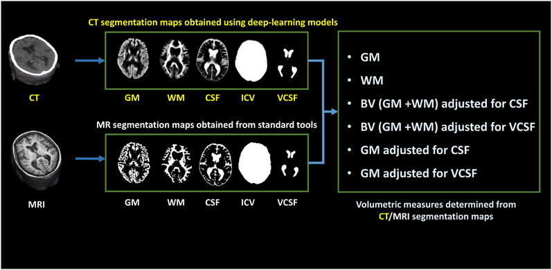
Cranial computed tomography (CT) is an affordable and widely available imaging modality that is used to assess structural abnormalities, but not to quantify neurodegeneration. Previously we developed a deep-learning-based model that produced accurate and robust cranial CT tissue classification.
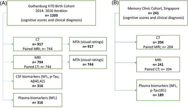
We analyzed 917 CT and 744 magnetic resonance (MR) scans from the Gothenburg H70 Birth Cohort, and 204 CT and 241 MR scans from participants of the Memory Clinic Cohort, Singapore. We tested associations between six CT-based volumetric measures (CTVMs) and existing clinical diagnoses, fluid and imaging biomarkers, and measures of cognition.
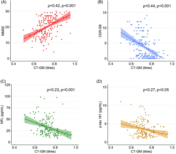
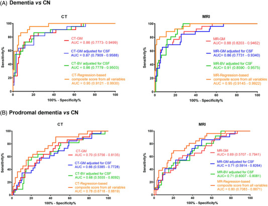
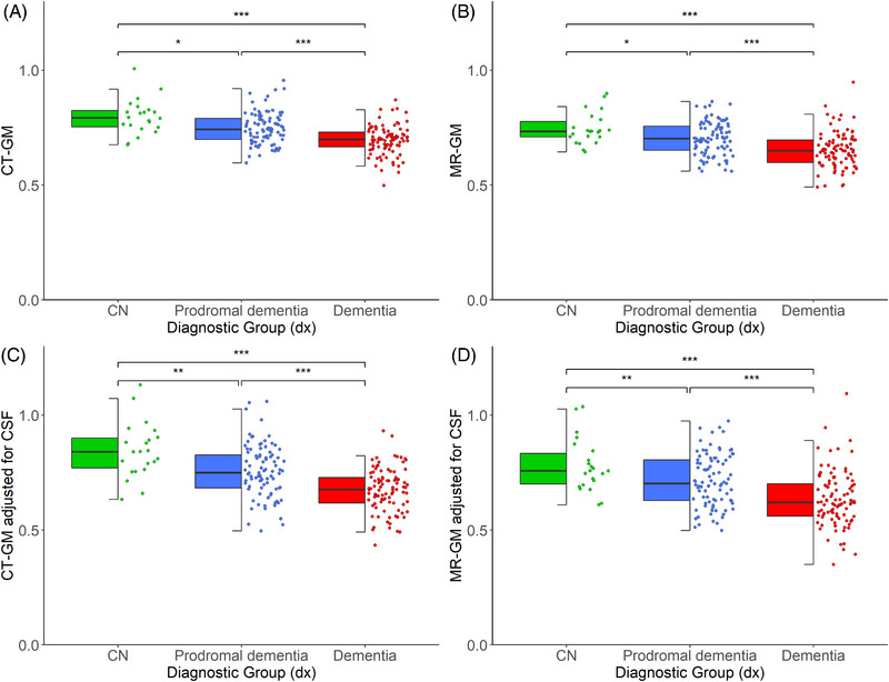
CTVMs differentiated cognitively healthy individuals from dementia and prodromal dementia patients with high accuracy levels comparable to MR-based measures. CTVMs were significantly associated with measures of cognition and biochemical markers of neurodegeneration.
These findings suggest the potential future use of CT-based volumetric measures as an informative first-line examination tool for neurodegenerative disease diagnostics after further validation.
Computed tomography (CT)-based volumetric measures can distinguish between patients with neurodegenerative disease and healthy controls, as well as between patients with prodromal dementia and controls. CT-based volumetric measures associate well with relevant cognitive, biochemical, and neuroimaging markers of neurodegenerative diseases. Model performance, in terms of brain tissue classification, was consistent across two cohorts of diverse nature. Intermodality agreement between our automated CT-based and established magnetic resonance (MR)-based image segmentations was stronger than the agreement between visual CT and MR imaging assessment.
头颅计算机断层扫描(CT)是一种经济实惠且广泛应用的成像方式,用于评估结构异常,但无法定量评估神经退行性变。我们之前开发了一种基于深度学习的模型,该模型可以准确、稳健地对头颅 CT 组织进行分类。
我们分析了来自哥德堡 H70 出生队列的 917 例 CT 和 744 例磁共振(MR)扫描,以及来自新加坡记忆诊所队列的 204 例 CT 和 241 例 MR 扫描。我们测试了 6 种基于 CT 的容积测量值(CTVM)与现有临床诊断、液体和成像生物标志物以及认知测量值之间的关联。
CTVM 以与基于 MR 的测量值相当的高精度水平,将认知健康个体与痴呆和前驱痴呆患者区分开来。CTVM 与认知测量值和神经退行性变的生化标志物显著相关。
这些发现表明,在进一步验证后,基于 CT 的容积测量值可能作为神经退行性疾病诊断的有价值的一线检查工具。
基于 CT 的容积测量值可以区分神经退行性疾病患者和健康对照者,以及前驱痴呆患者和对照者。基于 CT 的容积测量值与神经退行性疾病相关的认知、生化和神经影像学标志物密切相关。在两个性质不同的队列中,我们的自动基于 CT 的模型在脑组织分类方面的性能表现一致。我们的自动基于 CT 的和已建立的基于 MR 的图像分割之间的模态间一致性强于视觉 CT 和 MR 成像评估之间的一致性。