Department of Psychiatry, Schulich School of Medicine and Dentistry, Western University, London, Canada.
Department of Medical Biophysics, Western University, London, Canada.
Transl Psychiatry. 2022 Sep 1;12(1):358. doi: 10.1038/s41398-022-02136-0.
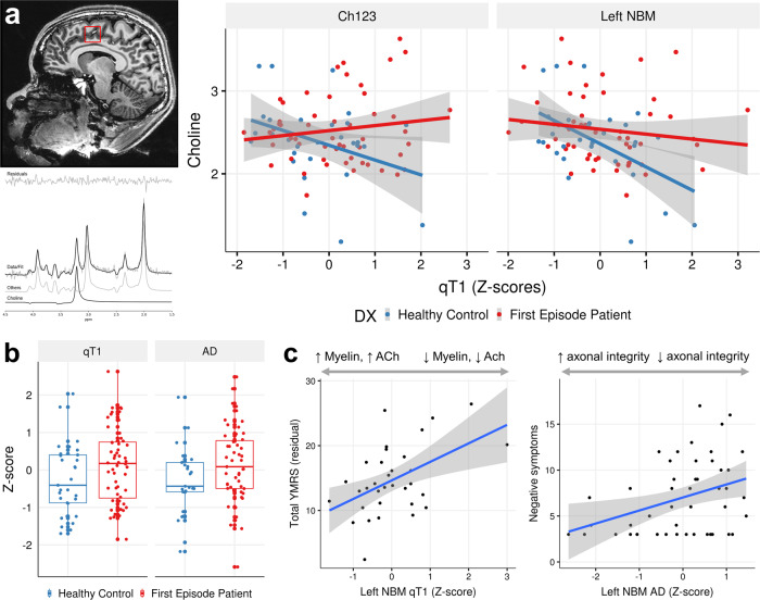
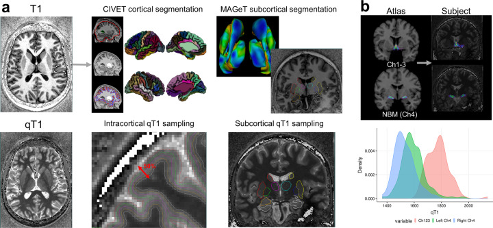
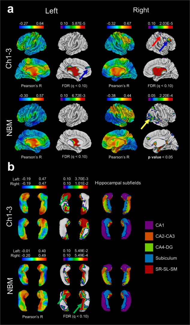
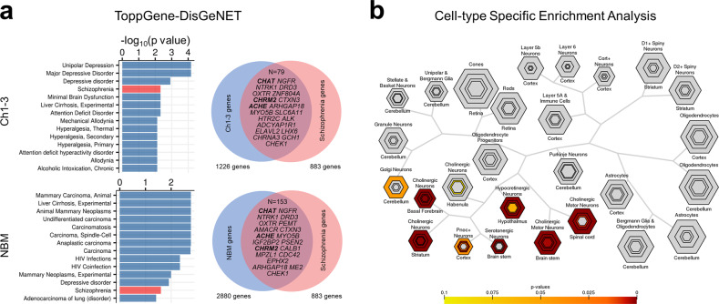
Cholinergic dysfunction has been implicated in the pathophysiology of psychosis and psychiatric disorders such as schizophrenia, depression, and bipolar disorder. The basal forebrain (BF) cholinergic nuclei, defined as cholinergic cell groups Ch1-3 and Ch4 (Nucleus Basalis of Meynert; NBM), provide extensive cholinergic projections to the rest of the brain. Here, we examined microstructural neuroimaging measures of the cholinergic nuclei in patients with untreated psychosis (~31 weeks of psychosis, <2 defined daily dose of antipsychotics) and used magnetic resonance spectroscopy (MRS) and transcriptomic data to support our findings. We used a cytoarchitectonic atlas of the BF to map the nuclei and obtained measures of myelin (quantitative T1, or qT1 as myelin surrogate) and microstructure (axial diffusion; AxD). In a clinical sample (n = 85; 29 healthy controls, 56 first-episode psychosis), we found significant correlations between qT1 of Ch1-3, left NBM and MRS-based dorsal anterior cingulate choline in healthy controls while this relationship was disrupted in FEP (p > 0.05). Case-control differences in qT1 and AxD were observed in the Ch1-3, with increased qT1 (reflecting reduced myelin content) and AxD (reflecting reduced axonal integrity). We found clinical correlates between left NBM qT1 with manic symptom severity, and AxD with negative symptom burden in FEP. Intracortical and subcortical myelin maps were derived and correlated with BF myelin. BF-cortical and BF-subcortical myelin correlations demonstrate known projection patterns from the BF. Using data from the Allen Human Brain Atlas, cholinergic nuclei showed significant enrichment for schizophrenia and depression-related genes. Cell-type specific enrichment indicated enrichment for cholinergic neuron markers as expected. Further relating the neuroimaging correlations to transcriptomics demonstrated links with cholinergic receptor genes and cell type markers of oligodendrocytes and cholinergic neurons, providing biological validity to the measures. These results provide genetic, neuroimaging, and clinical evidence for cholinergic dysfunction in schizophrenia.
胆碱能功能障碍与精神分裂症、抑郁症和双相情感障碍等精神疾病的病理生理学有关。基底前脑(BF)胆碱能核,定义为胆碱能细胞群 Ch1-3 和 Ch4(Meynert 基底核;NBM),为大脑的其余部分提供广泛的胆碱能投射。在这里,我们检查了未经治疗的精神病患者(~31 周的精神病,<2 种定义的每日抗精神病药物剂量)的胆碱能核的微观结构神经影像学测量值,并使用磁共振波谱(MRS)和转录组数据来支持我们的发现。我们使用 BF 的细胞构筑图集来绘制核,并获得了髓鞘的测量值(定量 T1,或作为髓鞘替代物的 qT1)和微观结构(轴向扩散;AxD)。在一个临床样本中(n=85;29 名健康对照,56 名首发精神病),我们发现健康对照组中 Ch1-3、左 NBM 和基于 MRS 的背侧前扣带回胆碱的 qT1 之间存在显著相关性,而这种相关性在 FEP 中被破坏(p>0.05)。Ch1-3 中观察到 qT1 和 AxD 的病例对照差异,qT1 增加(反映髓鞘含量减少)和 AxD 减少(反映轴突完整性降低)。我们发现左 NBM qT1 与 FEP 中的躁狂症状严重程度以及 AxD 与阴性症状负担之间存在临床相关性。推导了皮质内和皮质下髓鞘图,并与 BF 髓鞘相关联。BF-皮质和 BF-皮质下髓鞘相关性显示了来自 BF 的已知投射模式。使用 Allen 人类大脑图谱的数据,胆碱能核显示出与精神分裂症和抑郁症相关基因的显著富集。细胞类型特异性富集表明,如预期的那样,胆碱能神经元标志物的富集。进一步将神经影像学相关性与转录组学相关联,证明了与胆碱能受体基因以及少突胶质细胞和胆碱能神经元的细胞类型标志物的联系,为这些测量值提供了生物学有效性。这些结果为精神分裂症中的胆碱能功能障碍提供了遗传、神经影像学和临床证据。