State Key Laboratory of Ultrasound in Medicine and Engineering, College of Biomedical Engineering, Chongqing Medical University, Chongqing, China.
Chongqing Key Laboratory of Biomedical Engineering, Chongqing Medical University, Chongqing, China.
Ultrasound Obstet Gynecol. 2022 Nov;60(5):681-692. doi: 10.1002/uog.26053.
To develop and evaluate magnetic resonance imaging (MRI)-based radiomics models for predicting residual myoma regrowth within 1 year after high-intensity focused ultrasound (HIFU) ablation of uterine myomas.
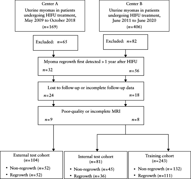
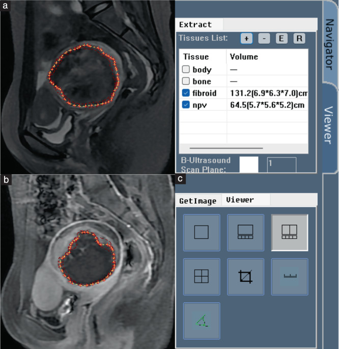
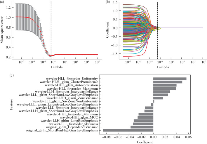
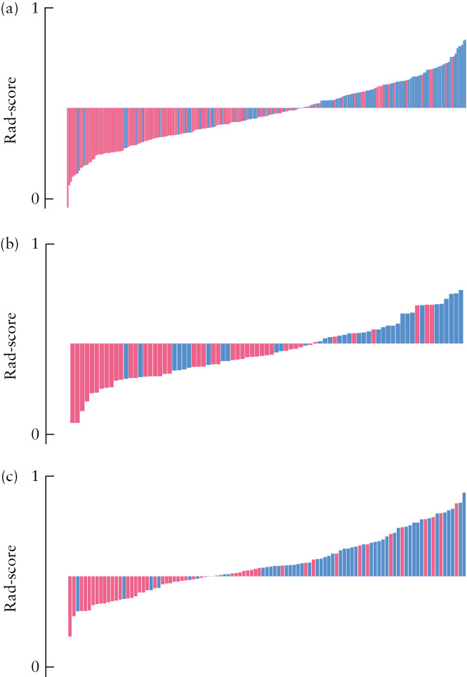
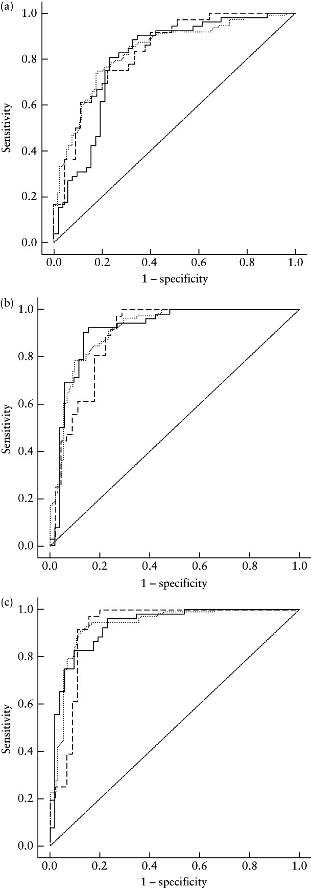
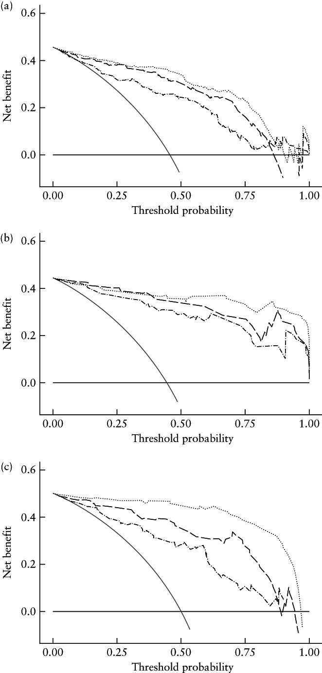
A retrospective analysis of residual myoma regrowth within 1 year was performed on 428 myomas in 339 patients who were diagnosed with uterine myoma and treated with HIFU ablation in two hospital centers. In total, 851 radiomics features were extracted from T2-weighted images (T2WI) obtained 1 day after HIFU ablation, and the least absolute shrinkage and selection operator in the training cohort (n = 243) was employed to select radiomics features. Support vector machines were adopted to develop radiomics, clinicoradiological and combined radiomics-clinical models to predict residual myoma regrowth, defined as an increase in residual myoma volume of > 10% between that at day 1 post HIFU and that at follow-up MRI within 1 year. These models were validated in both internal (n = 81) and external (n = 104) test cohorts. The predictive performance and clinical application of these models were assessed using receiver-operating-characteristics-curve analysis, the area under the curve (AUC) and decision-curve analysis.
The AUCs of the T2WI-based radiomics prediction model in the internal and external test cohorts were 0.834 (95% CI, 0.747-0.920) and 0.801 (95% CI, 0.712-0.889), respectively, and those of the clinicoradiological model were 0.888 (95% CI, 0.816-0.960) and 0.912 (95% CI, 0.851-0.973), respectively. The combined model had better predictive performance than either the radiomics or the clinicoradiological model, with AUC values of 0.922 (95% CI, 0.857-0.987) and 0.930 (95% CI, 0.880-0.980) in the internal and external test cohorts, respectively. Decision-curve analysis also indicated that application of the combined model has clinical value, this model achieving more net benefits than the other two models.
T2WI-based radiomics features can predict effectively the occurrence of residual myoma regrowth within 1 year after HIFU ablation of uterine myomas, which serves as an accurate and convenient reference for clinical decision-making. © 2022 The Authors. Ultrasound in Obstetrics & Gynecology published by John Wiley & Sons Ltd on behalf of International Society of Ultrasound in Obstetrics and Gynecology.
开发并评估基于磁共振成像(MRI)的放射组学模型,以预测高强度聚焦超声(HIFU)消融子宫肌瘤后 1 年内肌瘤残留的再生长。
对 2 家医院中心接受 HIFU 消融治疗的 339 例子宫肌瘤患者的 428 个肌瘤进行了 1 年内残留肌瘤再生长的回顾性分析。共从 HIFU 消融后 1 天获得的 T2 加权图像(T2WI)中提取了 851 个放射组学特征,在训练队列(n=243)中采用最小绝对收缩和选择算子选择放射组学特征。采用支持向量机建立放射组学、临床放射学和联合放射组学-临床模型,以预测残留肌瘤再生长,定义为 HIFU 后第 1 天和 1 年内的随访 MRI 之间残留肌瘤体积增加>10%。这些模型在内部(n=81)和外部(n=104)测试队列中进行了验证。使用受试者工作特征曲线分析、曲线下面积(AUC)和决策曲线分析评估这些模型的预测性能和临床应用。
内部和外部测试队列中基于 T2WI 的放射组学预测模型的 AUC 分别为 0.834(95%CI,0.747-0.920)和 0.801(95%CI,0.712-0.889),临床放射学模型的 AUC 分别为 0.888(95%CI,0.816-0.960)和 0.912(95%CI,0.851-0.973)。联合模型的预测性能优于放射组学或临床放射学模型,内部和外部测试队列的 AUC 值分别为 0.922(95%CI,0.857-0.987)和 0.930(95%CI,0.880-0.980)。决策曲线分析还表明,联合模型的应用具有临床价值,该模型比其他两个模型获得更多的净效益。
基于 T2WI 的放射组学特征可有效预测 HIFU 消融子宫肌瘤后 1 年内肌瘤残留的再生长,为临床决策提供了准确、便捷的参考。[© 2022 作者。超声在妇产科由 John Wiley & Sons Ltd 代表国际妇产科超声学会出版。]