• 文献检索
  • 文档翻译
  • 深度研究
  • 学术资讯
  • Suppr Zotero 插件Zotero 插件
  • 邀请有礼
  • 套餐&价格
  • 历史记录
应用&插件
Suppr Zotero 插件Zotero 插件浏览器插件Mac 客户端Windows 客户端微信小程序
定价
高级版会员购买积分包购买API积分包
服务
文献检索文档翻译深度研究API 文档MCP 服务
关于我们
关于 Suppr公司介绍联系我们用户协议隐私条款
关注我们

Suppr 超能文献

核心技术专利:CN118964589B侵权必究
粤ICP备2023148730 号-1Suppr @ 2026

文献检索

告别复杂PubMed语法,用中文像聊天一样搜索,搜遍4000万医学文献。AI智能推荐,让科研检索更轻松。

立即免费搜索

文件翻译

保留排版,准确专业,支持PDF/Word/PPT等文件格式,支持 12+语言互译。

免费翻译文档

深度研究

AI帮你快速写综述,25分钟生成高质量综述,智能提取关键信息,辅助科研写作。

立即免费体验

一种半自动容积方法(5D CNS+™)在异常胎儿中枢神经系统解剖结构详细产前重建中的适用性。

Applicability of a semiautomated volumetric approach (5D CNS+™) for detailed antenatal reconstruction of abnormal fetal CNS anatomy.

机构信息

Division of Prenatal Medicine, Department of Obstetrics and Gynecology, University Hospital of Schleswig-Holstein, Campus Luebeck, Ratzeburger Allee 160, 23538, Luebeck, Germany.

出版信息

BMC Med Imaging. 2022 Sep 2;22(1):154. doi: 10.1186/s12880-022-00888-1.

DOI:10.1186/s12880-022-00888-1
PMID:36056307
原文链接:https://pmc.ncbi.nlm.nih.gov/articles/PMC9438215/
Abstract

BACKGROUND

The aim of this study was to evaluate the accuracy and reliability of a semiautomated volumetric approach (5D CNS+™) when examining fetuses with an apparent abnormal anatomy of the central nervous system (CNS).

METHODS

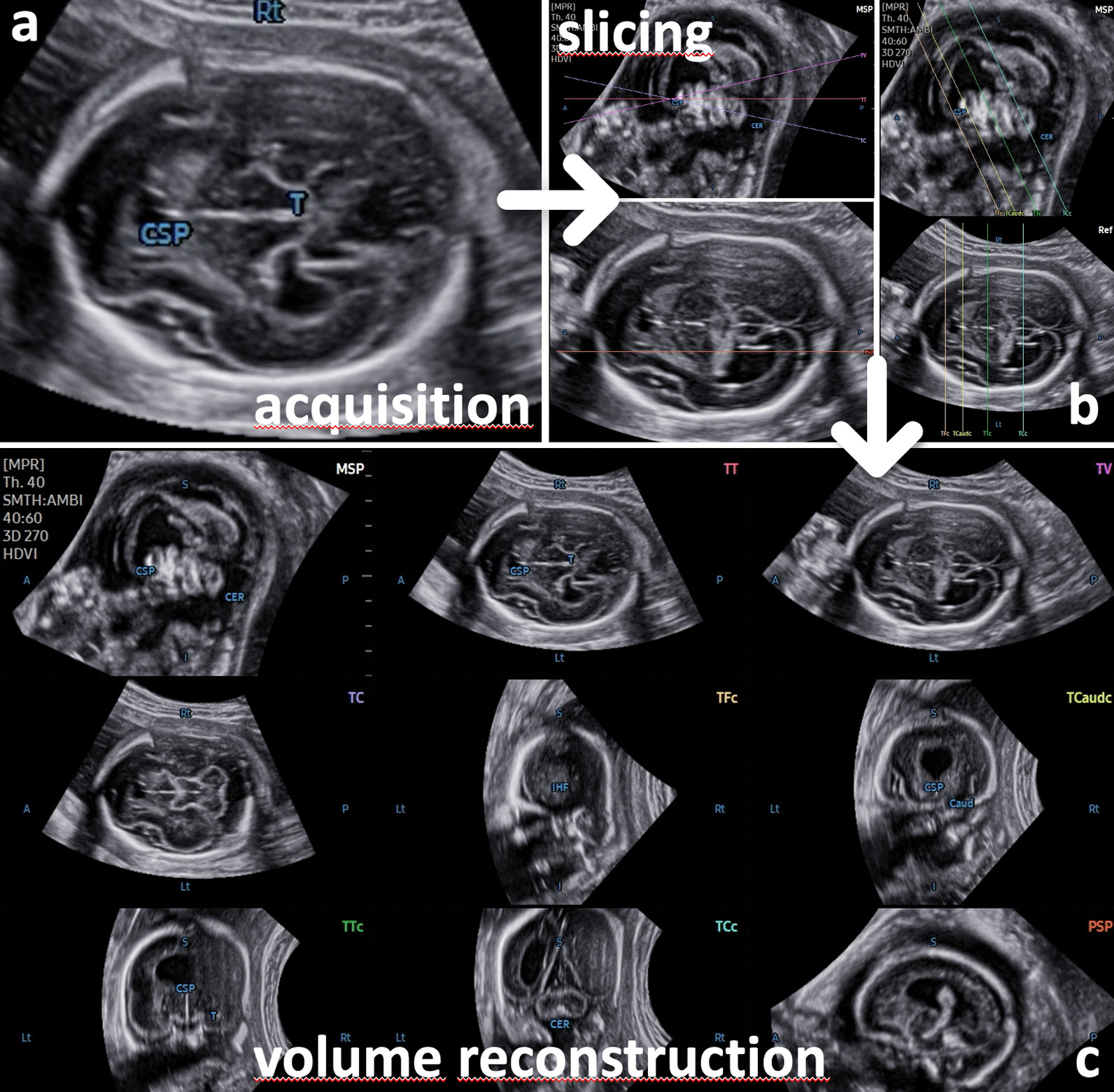
Stored 3D volumes extracted from a cohort of > 1.400 consecutive 2nd and 3rd trimester pregnancies (range 15-36 gestational weeks) were analyzed using the semiautomatic software tool 5D CNS+™, enabling detailed reconstruction of nine diagnostic planes of the fetal brain. All 3D data sets were examined and judged for plane accuracy, the need for manual adjustment, and fetal CNS anomalies affecting successful plane reconstruction.

RESULTS

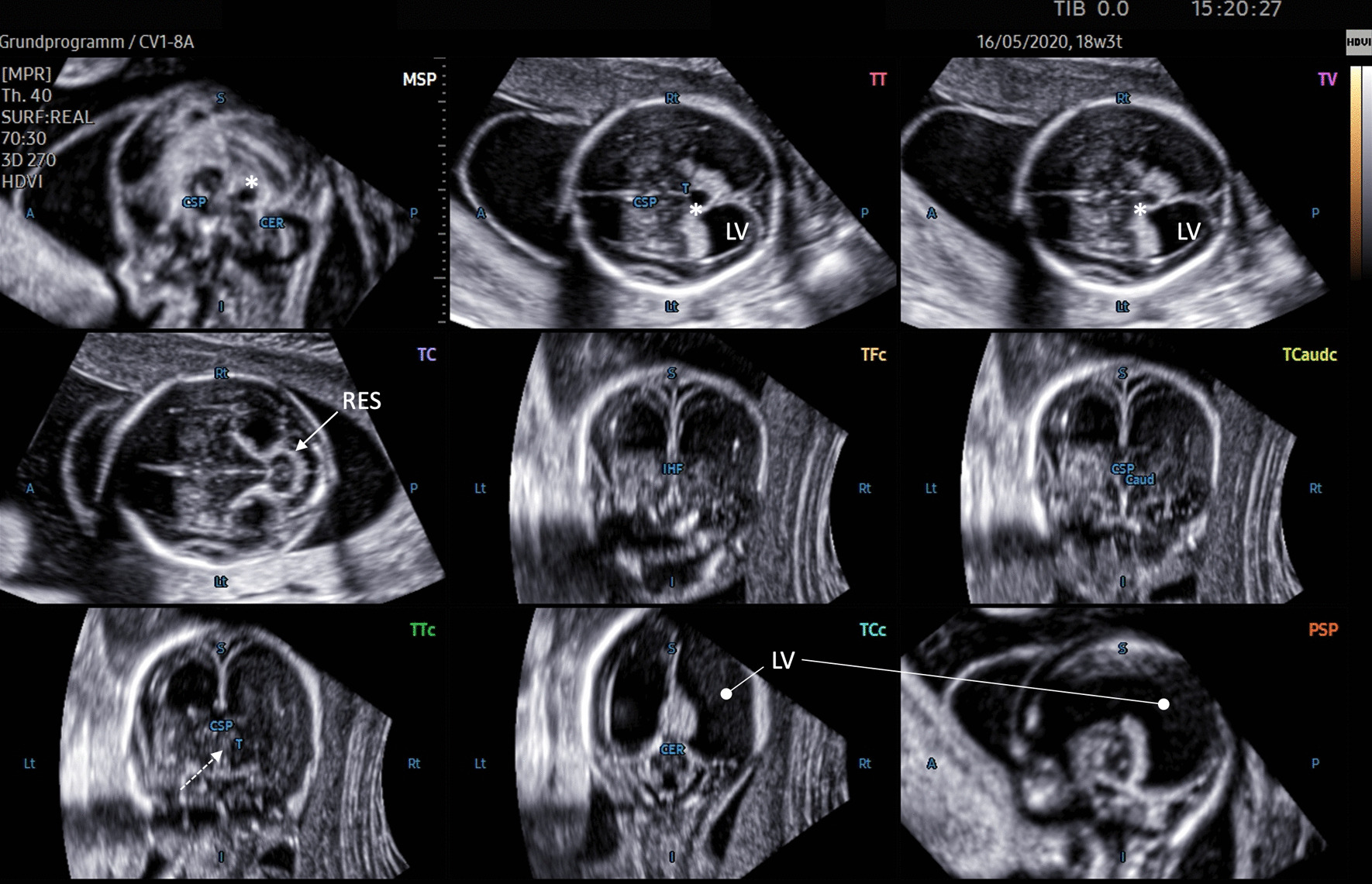
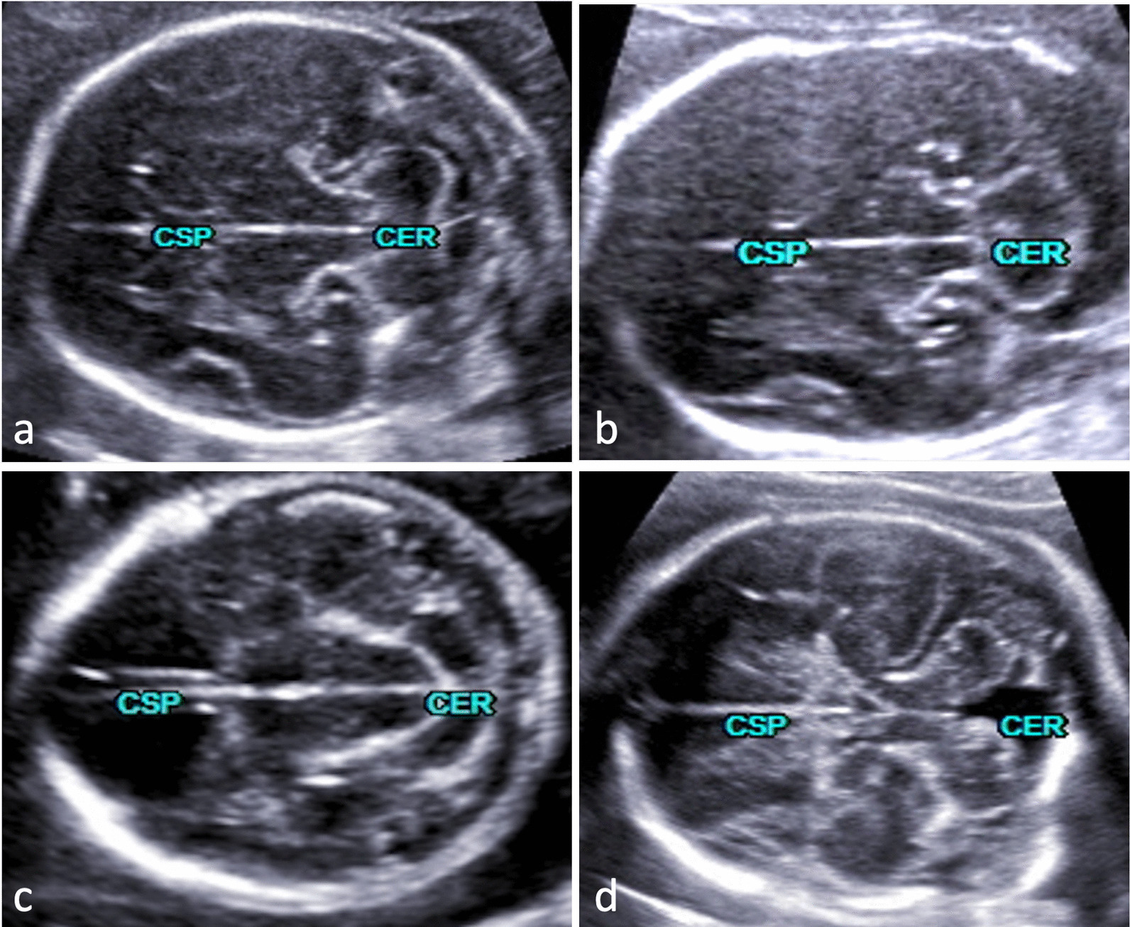
Based on our data of 91 fetuses with structural cerebral anomalies, we were able to reveal details of a wide range of CNS anomalies with application of the 5D CNS+™ technique. The corresponding anatomical features and consecutive changes of neighboring structures could be clearly demonstrated. Thus, a profound assessment of the entire altered CNS anatomy could be achieved in nearly all cases. The comparison with matched controls showed a significant difference in volume acquisition (p < 0.001) and in need for manual adjustment (p < 0.001) but not in the drop-out rates (p = 0.677) of both groups.

CONCLUSION

5D CNS+™ is applicable in the majority of cases with brain lesions and constitutes a reliable tool even if the integrity of the fetal CNS is compromised by structural anomalies. Using volume data that were acquired in identical cutting sections needed for conventional biometry allows for detailed anatomic surveys grossly independent of the examiner's experience.

摘要

背景

本研究旨在评估一种半自动容积分析方法(5D CNS+™)在检查中枢神经系统(CNS)解剖结构异常的胎儿时的准确性和可靠性。

方法

从超过 1400 例连续的 2 至 3 期妊娠(15-36 孕周)的存储三维体积中提取数据,使用半自动软件工具 5D CNS+™ 对其进行分析,从而实现胎儿大脑的九个诊断平面的详细重建。对所有的三维数据集进行检查,并评估平面准确性、手动调整的必要性以及影响成功平面重建的胎儿 CNS 异常。

结果

基于我们 91 例有结构脑异常的胎儿数据,我们能够应用 5D CNS+™ 技术揭示广泛的 CNS 异常的细节。可以清楚地显示相应的解剖特征和相邻结构的连续变化。因此,几乎所有病例都能对整个改变的 CNS 解剖结构进行深入评估。与匹配的对照组相比,两组在体积采集(p<0.001)和手动调整的需求(p<0.001)方面存在显著差异,但在两组的脱落率(p=0.677)方面无差异。

结论

5D CNS+™ 适用于大多数有脑病变的病例,即使胎儿 CNS 的完整性受到结构异常的影响,它也是一种可靠的工具。使用在获取传统生物计量所需的相同切割平面获取的容积数据,可以大大减少对检查者经验的依赖,实现详细的解剖学检查。

https://cdn.ncbi.nlm.nih.gov/pmc/blobs/d9c6/9438215/6889d1d36d24/12880_2022_888_Fig5_HTML.jpg
https://cdn.ncbi.nlm.nih.gov/pmc/blobs/d9c6/9438215/93bf28f7f6db/12880_2022_888_Fig1_HTML.jpg
https://cdn.ncbi.nlm.nih.gov/pmc/blobs/d9c6/9438215/363124531fae/12880_2022_888_Fig2_HTML.jpg
https://cdn.ncbi.nlm.nih.gov/pmc/blobs/d9c6/9438215/3cad8789b024/12880_2022_888_Fig3_HTML.jpg
https://cdn.ncbi.nlm.nih.gov/pmc/blobs/d9c6/9438215/e9cebdbc98e8/12880_2022_888_Fig4_HTML.jpg
https://cdn.ncbi.nlm.nih.gov/pmc/blobs/d9c6/9438215/6889d1d36d24/12880_2022_888_Fig5_HTML.jpg

相似文献

1
Applicability of a semiautomated volumetric approach (5D CNS+™) for detailed antenatal reconstruction of abnormal fetal CNS anatomy.一种半自动容积方法(5D CNS+™)在异常胎儿中枢神经系统解剖结构详细产前重建中的适用性。
BMC Med Imaging. 2022 Sep 2;22(1):154. doi: 10.1186/s12880-022-00888-1.
2
Validation of a semiautomated volumetric approach for fetal neurosonography using 5DCNS+ in clinical data from > 1100 consecutive pregnancies.使用5DCNS+对超过1100例连续妊娠的临床数据进行胎儿神经超声检查的半自动容积测量方法的验证。
Childs Nerv Syst. 2020 Dec;36(12):2989-2995. doi: 10.1007/s00381-020-04607-5. Epub 2020 Apr 30.
3
5D CNS+ Software for Automatically Imaging Axial, Sagittal, and Coronal Planes of Normal and Abnormal Second-Trimester Fetal Brains.用于自动成像孕中期正常和异常胎儿脑部轴位、矢状位和冠状位平面的5D中枢神经系统+软件。
J Ultrasound Med. 2016 Oct;35(10):2263-72. doi: 10.7863/ultra.15.11013. Epub 2016 Aug 31.
4
A Clinical Approach to Semiautomated Three-Dimensional Fetal Brain Biometry-Comparing the Strengths and Weaknesses of Two Diagnostic Tools: 5DCNS+ and SonoCNS.半自动三维胎儿脑生物测量的临床方法——比较两种诊断工具5DCNS+和SonoCNS的优缺点
J Clin Med. 2023 Aug 16;12(16):5334. doi: 10.3390/jcm12165334.
5
The feasibility of using 5D CNS software in obtaining standard fetal head measurements from volumes acquired by three-dimensional ultrasonography: comparison with two-dimensional ultrasound.使用5D中枢神经系统软件从三维超声采集的容积中获取标准胎儿头部测量值的可行性:与二维超声的比较
J Matern Fetal Neonatal Med. 2016;29(14):2217-22. doi: 10.3109/14767058.2015.1081891. Epub 2015 Sep 11.
6
How to obtain diagnostic planes of the fetal central nervous system using three-dimensional ultrasound and a context-preserving rendering technology.如何利用三维超声和保形渲染技术获取胎儿中枢神经系统的诊断切面。
Am J Obstet Gynecol. 2019 Mar;220(3):215-229. doi: 10.1016/j.ajog.2018.11.1088. Epub 2018 Nov 14.
7
[First trimester prenatal diagnosis of structural fetal anomalies with three dimensional ultrasound--possibilities and limitations].[孕早期应用三维超声对胎儿结构异常进行产前诊断——可能性与局限性]
Akush Ginekol (Sofiia). 2010;49(7):4-10.
8
Role of magnetic resonance imaging in fetuses with mild or moderate ventriculomegaly in the era of fetal neurosonography: systematic review and meta-analysis.磁共振成像在胎儿神经超声时代轻度或中度脑室扩张胎儿中的作用:系统评价和荟萃分析。
Ultrasound Obstet Gynecol. 2019 Aug;54(2):164-171. doi: 10.1002/uog.20197. Epub 2019 Jul 11.
9
Impact of 3D ultrasound on fetal CNS examination.三维超声对胎儿中枢神经系统检查的影响。
Ceska Gynekol. 2019 Spring;84(3):222-228.
10
Diagnostic accuracy of ultrasonography and magnetic resonance imaging for the detection of fetal anomalies: a blinded case-control study.超声检查和磁共振成像检测胎儿异常的诊断准确性:一项盲法病例对照研究。
Ultrasound Obstet Gynecol. 2016 Aug;48(2):185-92. doi: 10.1002/uog.15774. Epub 2016 Jul 10.

引用本文的文献

1
Accuracy of ultrasonographic transcerebellar diameter for dating in third trimester of pregnancy in Nigerian women: a cross-sectional study.尼日利亚女性妊娠晚期经小脑超声测量胎儿双顶径用于预产期推算的准确性:一项横断面研究
BMC Med Imaging. 2025 Mar 25;25(1):100. doi: 10.1186/s12880-025-01634-z.
2
Advancements in Artificial Intelligence for Fetal Neurosonography: A Comprehensive Review.胎儿神经超声检查中人工智能的进展:综述
J Clin Med. 2024 Sep 22;13(18):5626. doi: 10.3390/jcm13185626.
3
A Clinical Approach to Semiautomated Three-Dimensional Fetal Brain Biometry-Comparing the Strengths and Weaknesses of Two Diagnostic Tools: 5DCNS+ and SonoCNS.

本文引用的文献

1
Early second-trimester three-dimensional transvaginal neurosonography of fetal midbrain and hindbrain: normative data and technical aspects.早孕期经阴道三维超声检查胎儿中脑和后脑:正常值及技术要点。
Ultrasound Obstet Gynecol. 2022 Mar;59(3):317-324. doi: 10.1002/uog.23691.
2
Prenatal diagnosis of rhombencephalosynapsis: neuroimaging features and severity of vermian anomaly.菱形脑融合的产前诊断:神经影像学特征及蚓部异常的严重程度
Ultrasound Obstet Gynecol. 2021 Dec;58(6):864-874. doi: 10.1002/uog.23660.
3
ISUOG Practice Guidelines (updated): sonographic examination of the fetal central nervous system. Part 2: performance of targeted neurosonography.
半自动三维胎儿脑生物测量的临床方法——比较两种诊断工具5DCNS+和SonoCNS的优缺点
J Clin Med. 2023 Aug 16;12(16):5334. doi: 10.3390/jcm12165334.
国际妇产科超声学会实践指南(更新版):胎儿中枢神经系统超声检查。第2部分:针对性神经超声检查的操作
Ultrasound Obstet Gynecol. 2021 Apr;57(4):661-671. doi: 10.1002/uog.23616. Epub 2021 Mar 18.
4
Interpeduncular angle: A new parameter for assessing intracranial hypotension in fetuses with spinal dysraphism.脚间窝角:评估脊柱裂胎儿颅内低压的新参数。
Prenat Diagn. 2021 Jul;41(8):942-948. doi: 10.1002/pd.5905. Epub 2021 Feb 5.
5
Fetal neurosonography detects differences in cortical development and corpus callosum in late-onset small fetuses.胎儿神经超声检查可检测出晚发型小胎儿皮质发育和胼胝体的差异。
Ultrasound Obstet Gynecol. 2021 Jul;58(1):42-47. doi: 10.1002/uog.23592.
6
Prenatal sonographic assessment of Sylvian fissure operculization (SFO): importance of distinguishing between screening and diagnostic tools and selecting precise anatomical landmarks.产前超声评估大脑外侧裂盖化(SFO):区分筛查和诊断工具以及选择精确解剖标志的重要性。
Ultrasound Obstet Gynecol. 2020 Nov;56(5):786-788. doi: 10.1002/uog.23113.
7
Three-dimensional ultrasonography for advanced neurosonography (neurosofe-3D): Validation of a brain volume acquisition guideline.三维超声在高级神经超声中的应用(neurosofe-3D):脑容量采集指南的验证。
Acta Obstet Gynecol Scand. 2021 Jan;100(1):84-90. doi: 10.1111/aogs.13996. Epub 2020 Oct 8.
8
ISUOG Practice Guidelines (updated): sonographic examination of the fetal central nervous system. Part 1: performance of screening examination and indications for targeted neurosonography.国际妇产科超声学会(ISUOG)实践指南(更新版):胎儿中枢神经系统的超声检查。第1部分:筛查检查的实施及针对性神经超声检查的指征
Ultrasound Obstet Gynecol. 2020 Sep;56(3):476-484. doi: 10.1002/uog.22145.
9
Accuracy of automated three-dimensional ultrasound imaging technique for fetal head biometry.自动化三维超声成像技术在胎儿头部生物测量中的准确性。
Ultrasound Obstet Gynecol. 2021 May;57(5):798-803. doi: 10.1002/uog.22171.
10
Fetal Open and Closed Spina Bifida on a Routine Scan at 11 Weeks to 13 Weeks 6 Days.孕11周 至 13周6天常规超声检查发现的胎儿开放性和闭合性脊柱裂
J Ultrasound Med. 2021 Feb;40(2):237-247. doi: 10.1002/jum.15392. Epub 2020 Jul 21.