Amouei Mehrnam, Momtazmanesh Sara, Kavosi Hoda, Davarpanah Amir H, Shirkhoda Ali, Radmard Amir Reza
Department of Radiology, Shariati Hospital, Tehran University of Medical Sciences, North Kargar St., Tehran, 14117, Iran.
Department of Rheumatology, Shariati Hospital, Tehran University of Medical Sciences, Tehran, Iran.
Insights Imaging. 2022 Sep 4;13(1):143. doi: 10.1186/s13244-022-01284-7.
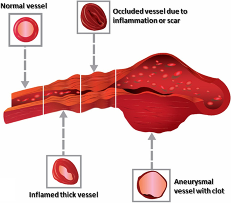
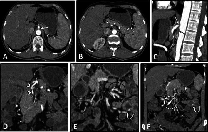
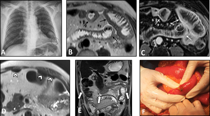
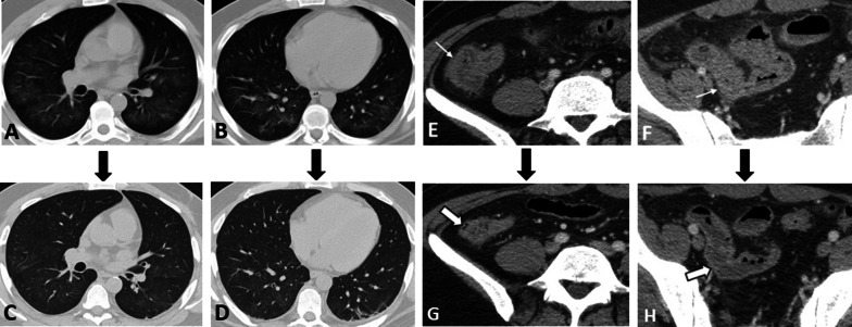
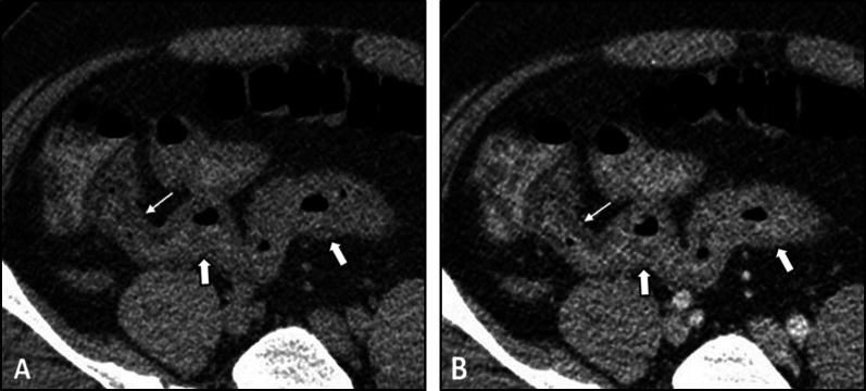
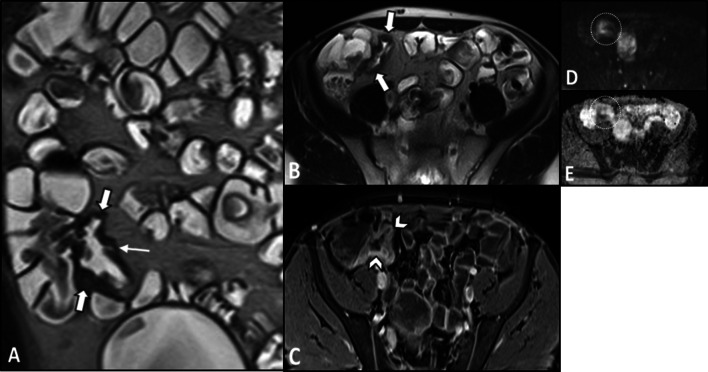
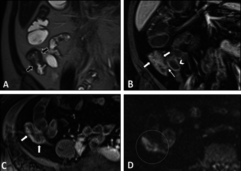
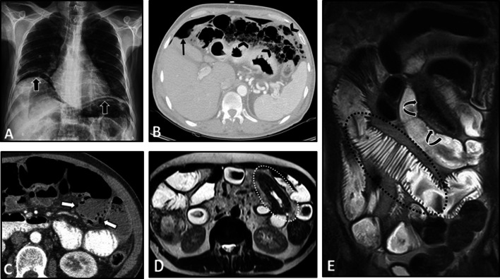
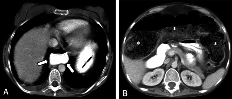
Diagnosis of intestinal vasculitis is often challenging due to the non-specific clinical and imaging findings. Vasculitides with gastrointestinal (GI) manifestations are rare, but their diagnosis holds immense significance as late or missed recognition can result in high mortality rates. Given the resemblance of radiologic findings with some other entities, GI vasculitis is often overlooked on small bowel studies done using computed tomography/magnetic resonance enterography (CTE/MRE). Hereon, we reviewed radiologic findings of vasculitis with gastrointestinal involvement on CTE and MRE. The variety of findings on MRE/CTE depend upon the size of the involved vessels. Signs of intestinal ischemia, e.g., mural thickening, submucosal edema, mural hyperenhancement, and restricted diffusion on diffusion-weighted imaging, are common in intestinal vasculitis. Involvement of the abdominal aorta and the major visceral arteries is presented as concentric mural thickening, transmural calcification, luminal stenosis, occlusion, aneurysmal changes, and collateral vessels. Such findings can be observed particularly in large- and medium-vessel vasculitis. The presence of extra-intestinal findings, including within the liver, kidneys, or spleen in the form of focal areas of infarction or heterogeneous enhancement due to microvascular involvement, can be another radiologic clue in diagnosis of vasculitis. The link between the clinical/laboratory findings and MRE/CTE abnormalities needs to be corresponded when it comes to the diagnosis of intestinal vasculitis.
由于临床和影像学表现不具有特异性,肠道血管炎的诊断往往具有挑战性。伴有胃肠道(GI)表现的血管炎较为罕见,但其诊断具有极其重要的意义,因为诊断延迟或漏诊可能导致高死亡率。鉴于放射学表现与其他一些疾病相似,在使用计算机断层扫描/磁共振小肠造影(CTE/MRE)进行的小肠检查中,胃肠道血管炎常常被忽视。在此,我们回顾了CTE和MRE上伴有胃肠道受累的血管炎的放射学表现。MRE/CTE上的各种表现取决于受累血管的大小。肠道缺血的征象,如肠壁增厚、黏膜下水肿、肠壁强化增强以及扩散加权成像上的扩散受限,在肠道血管炎中很常见。腹主动脉和主要内脏动脉受累表现为同心性肠壁增厚、透壁钙化、管腔狭窄、闭塞、动脉瘤样改变以及侧支血管。这些表现尤其在大、中血管血管炎中可见。肝、肾或脾等肠外表现,如因微血管受累而出现的梗死灶或不均匀强化,可能是血管炎诊断的另一个放射学线索。在诊断肠道血管炎时,临床/实验室检查结果与MRE/CTE异常之间的联系需要相互对应。