Devan Sean P, Luo Guozhen, Jiang Xiaoyu, Xie Jingping, Dean Daniel, Johnson Levi S, Morales-Paliza Manuel, Harmsen Hannah, Xu Junzhong, Kirschner Austin N
Chemical and Physical Biology Program, Vanderbilt University, Nashville, Tennessee.
Vanderbilt University Institute of Imaging Science.
Adv Radiat Oncol. 2022 Jul 19;7(6):101014. doi: 10.1016/j.adro.2022.101014. eCollection 2022 Nov-Dec.
Our purpose was to develop a rodent model of brain radionecrosis using clinical linear accelerator based stereotactic radiosurgery.
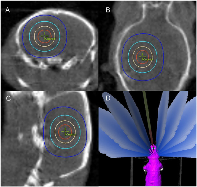
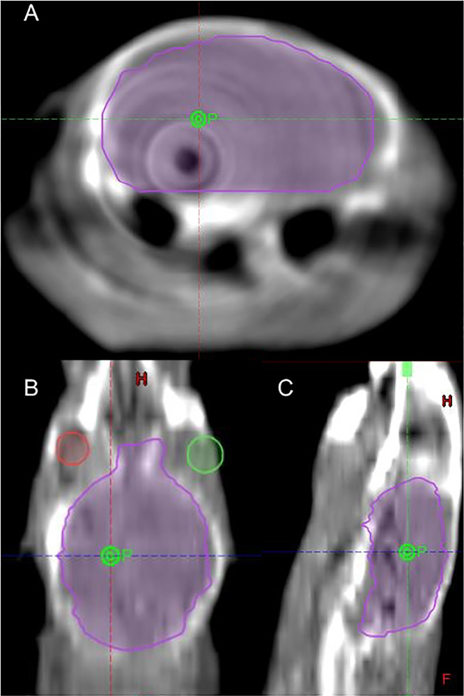
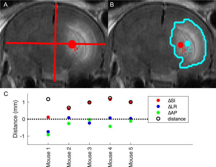
Single fraction maximum prescription points in the mouse's left hemisphere were irradiated using linear accelerator-based stereotactic radiosurgery with multiple arcs at 60 (n = 5), 100 (n = 5), and 140 (n = 5) Gy. Rats (n = 6) were similarly treated with 140 Gy. Gadolinium (Gd)-enhanced magnetic resonance imaging (MRI) was used to track radiation injury in mice over weeks (100 and 140 Gy) or months (60 Gy). Target accuracy was measured by the distance from the prescription point to the center of the earliest Gd-MRI enhancement. Confirmation of necrosis via histology was performed at the subject endpoints.
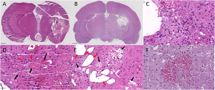
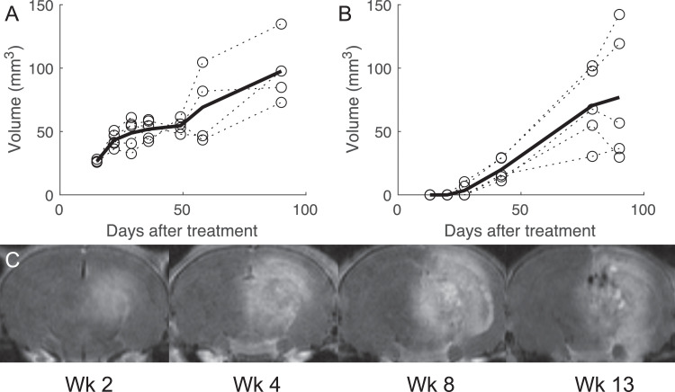
Radiation injury as indicated by Gd-MRI was first identified at 2 weeks (140 Gy), 4 to 6 weeks (100 Gy), and 8 months (60 Gy). A volumetric time course showed rapid growth in the volume of Gd-MRI signal enhancement after the appearance of apparent necrosis. Histopathologic features were consistent with radionecrosis.
The presented method uses a commonly available clinical linear accelerator to induce radiation necrosis in both mice and rats. The treatment is modeled after patient therapy for a more direct model of human tissue under a range of doses used in clinical neuro-ablation techniques. The short time to onset of apparent necrosis, accurate targeting of the prescription point, high incidence of necrosis, and similar pathologic features make this a suitable animal model for further research in radionecrosis.
我们的目的是利用基于临床直线加速器的立体定向放射外科技术建立脑放射性坏死的啮齿动物模型。
使用基于直线加速器的立体定向放射外科技术,以60(n = 5)、100(n = 5)和140(n = 5)Gy的剂量对小鼠左半球的单分割最大处方点进行多弧照射。对大鼠(n = 6)同样给予140 Gy的照射。使用钆(Gd)增强磁共振成像(MRI)来追踪小鼠在数周(100和140 Gy)或数月(60 Gy)内的放射性损伤。通过从处方点到最早Gd-MRI增强中心的距离来测量靶点准确性。在实验终点通过组织学确认坏死情况。
Gd-MRI显示的放射性损伤在2周(140 Gy)、4至6周(100 Gy)和8个月(60 Gy)时首次被发现。体积随时间变化的过程显示,在明显坏死出现后,Gd-MRI信号增强的体积迅速增大。组织病理学特征与放射性坏死一致。
所提出的方法使用常见的临床直线加速器在小鼠和大鼠中诱导放射性坏死。该治疗方法以患者治疗为模型,在临床神经消融技术使用的一系列剂量下,为人类组织提供了更直接的模型。明显坏死出现的时间短、处方点靶向准确、坏死发生率高以及相似的病理特征,使其成为放射性坏死进一步研究的合适动物模型。