Department of Radiology and Biomedical Imaging, Yale University, New Haven, Connecticut, USA.
High-Field MR Centre, Max Planck Institute for Biological Cybernetics, Tübingen, Germany.
Magn Reson Med. 2023 Jan;89(1):29-39. doi: 10.1002/mrm.29439. Epub 2022 Sep 5.
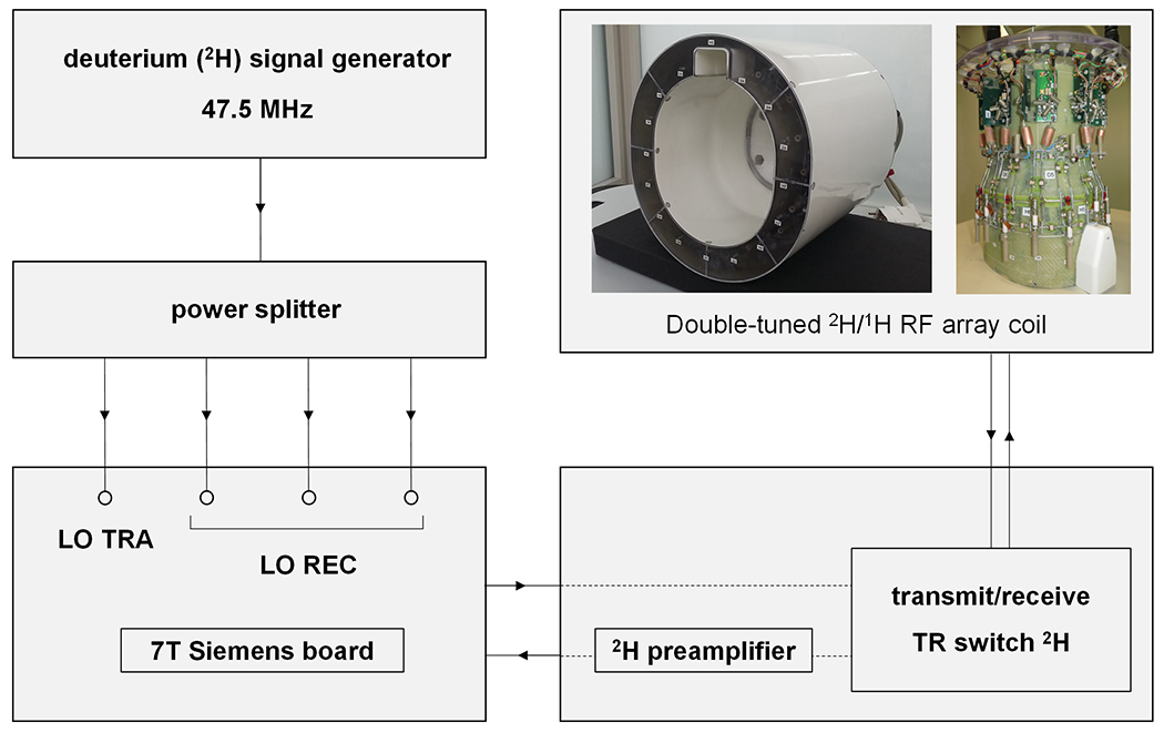
To explore the potential of deuterium metabolic imaging (DMI) in the human brain in vivo at 7 T, using a multi-element deuterium ( H) RF coil for 3D volume coverage.
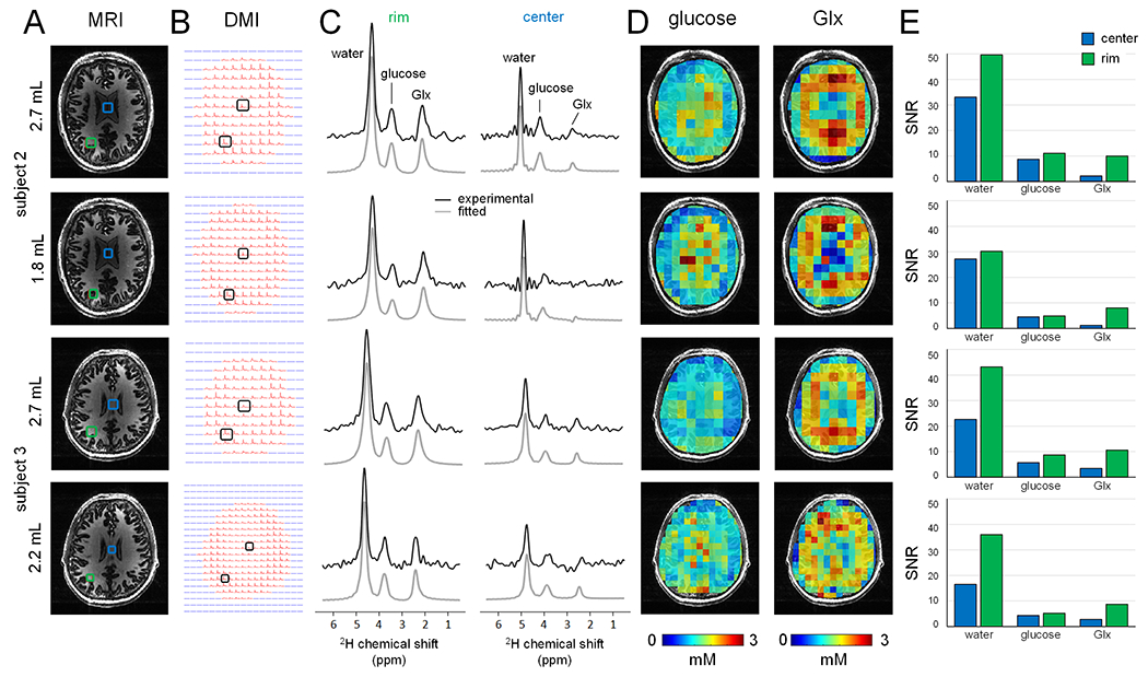
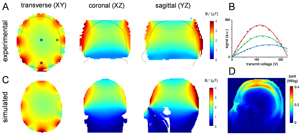
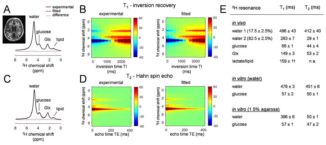
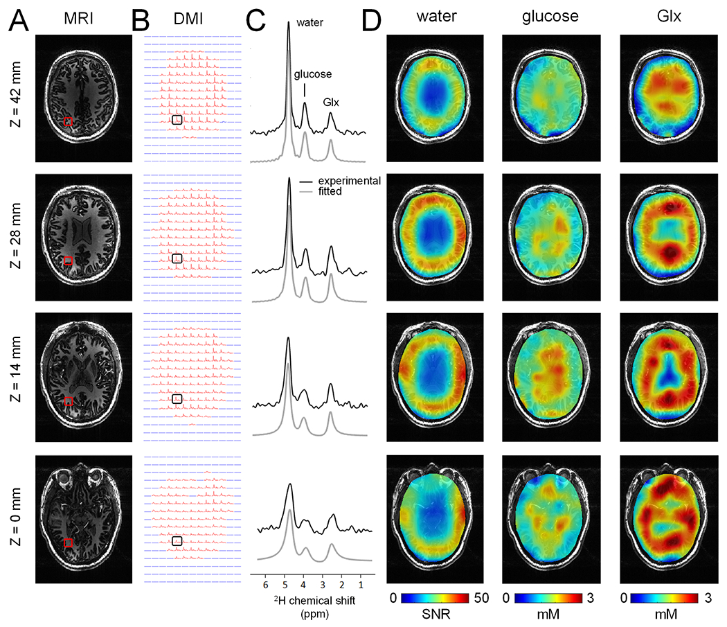
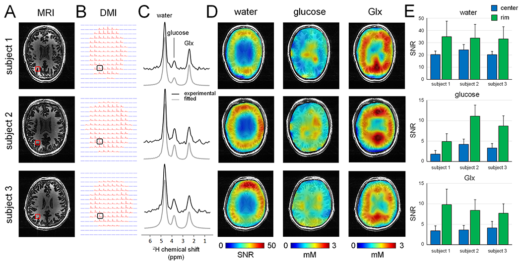
H-MR images and localized H MR spectra were acquired in vivo in the human brain of 3 healthy subjects to generate DMI maps of H-labeled water, glucose, and glutamate/glutamine (Glx). In addition, non-localized H-MR spectra were acquired both in vivo and in vitro to determine T and T relaxation times of deuterated metabolites at 7 T. The performance of the H coil was assessed through numeric simulations and experimentally acquired B maps.
3D DMI maps covering the entire human brain in vivo were obtained from well-resolved deuterated ( H) metabolite resonances of water, glucose, and Glx. The T and T relaxation times were consistent with those reported at adjacent field strengths. Experimental B maps were in good agreement with simulations, indicating efficient and homogeneous B transmission and low RF power deposition for H, consistent with a similar array coil design reported at 9.4 T.
Here, we have demonstrated the successful implementation of 3D DMI in the human brain in vivo at 7 T. The spatial and temporal nominal resolutions achieved at 7 T (i.e., 2.7 mL in 28 min, respectively) were close to those achieved at 9.4 T and greatly outperformed DMI at lower magnetic fields. DMI at 7 T and beyond has clear potential in applications dealing with small brain lesions.
探索在 7T 下使用多元素氘( H)射频线圈进行 3D 容积覆盖的人体大脑中氘代谢成像(DMI)的潜力。
在 3 名健康受试者的人体大脑中采集 H-MR 图像和局部 H MR 谱,以生成 H 标记水、葡萄糖和谷氨酸/谷氨酰胺(Glx)的 DMI 图。此外,在体内和体外采集非局部 H-MR 谱,以确定 7T 下氘代谢物的 T 和 T 弛豫时间。通过数值模拟和实验获得的 B 图评估 H 线圈的性能。
从水、葡萄糖和 Glx 的氘( H)代谢物共振中获得了分辨率良好的整个人体大脑的 3D DMI 图。T 和 T 弛豫时间与相邻场强下的报道一致。实验 B 图与模拟结果吻合良好,表明 H 的 B 传输高效且均匀,RF 功率沉积低,与在 9.4T 下报道的类似阵列线圈设计一致。
本文成功地在 7T 下实现了人体大脑的 3D DMI。在 7T 下实现的空间和时间分辨率(即 28 分钟内 2.7ml)接近在 9.4T 下的分辨率,大大优于较低磁场下的 DMI。7T 及更高磁场下的 DMI 在处理小的脑部病变的应用中有很大的潜力。