• 文献检索
  • 文档翻译
  • 深度研究
  • 学术资讯
  • Suppr Zotero 插件Zotero 插件
  • 邀请有礼
  • 套餐&价格
  • 历史记录
应用&插件
Suppr Zotero 插件Zotero 插件浏览器插件Mac 客户端Windows 客户端微信小程序
定价
高级版会员购买积分包购买API积分包
服务
文献检索文档翻译深度研究API 文档MCP 服务
关于我们
关于 Suppr公司介绍联系我们用户协议隐私条款
关注我们

Suppr 超能文献

核心技术专利:CN118964589B侵权必究
粤ICP备2023148730 号-1Suppr @ 2026

文献检索

告别复杂PubMed语法,用中文像聊天一样搜索,搜遍4000万医学文献。AI智能推荐,让科研检索更轻松。

立即免费搜索

文件翻译

保留排版,准确专业,支持PDF/Word/PPT等文件格式,支持 12+语言互译。

免费翻译文档

深度研究

AI帮你快速写综述,25分钟生成高质量综述,智能提取关键信息,辅助科研写作。

立即免费体验

酵母细胞在激光生物打印过程中所受冲击的理论与实验评估。

Theoretical and Experimental Assay of Shock Experienced by Yeast Cells during Laser Bioprinting.

机构信息

Bach Institute of Biochemistry, Federal Research Center of Biotechnology of the RAS, 119071 Moscow, Russia.

Moscow Institute of Physics and Technology (National Research University), 141700 Dolgoprudny, Russia.

出版信息

Int J Mol Sci. 2022 Aug 29;23(17):9823. doi: 10.3390/ijms23179823.

DOI:10.3390/ijms23179823
PMID:36077218
原文链接:https://pmc.ncbi.nlm.nih.gov/articles/PMC9456252/
Abstract

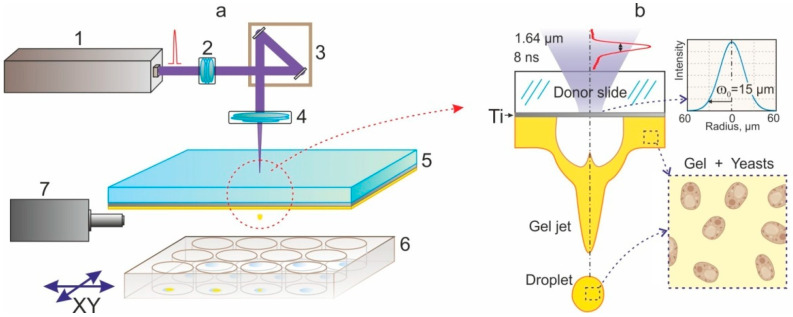
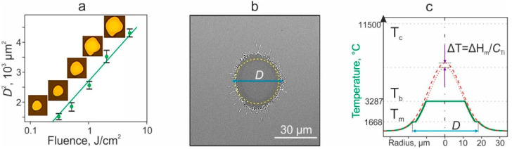
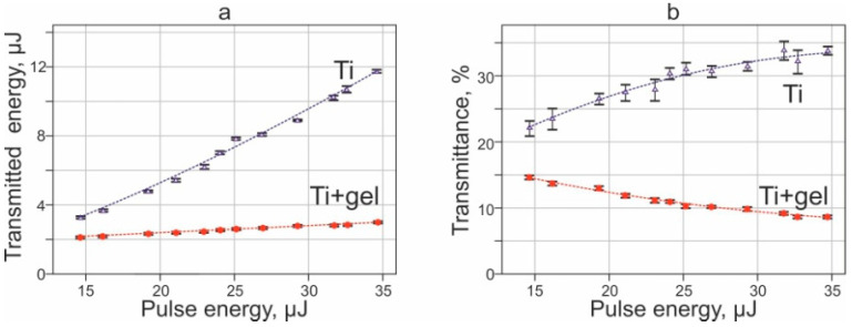
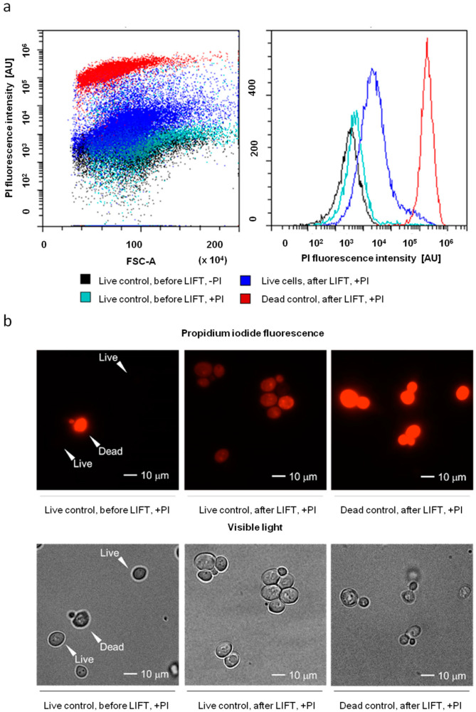
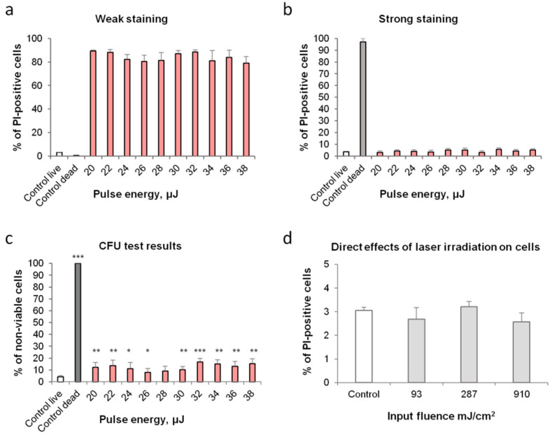
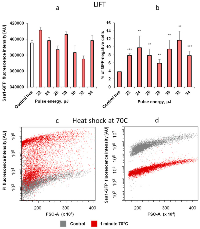
Laser-induced forward transfer (LIFT) is a useful technique for bioprinting using gel-embedded cells. However, little is known about the stresses experienced by cells during LIFT. This paper theoretically and experimentally explores the levels of laser pulse irradiation and pulsed heating experienced by yeast cells during LIFT. It has been found that only 5% of the cells in the gel layer adjacent to the absorbing Ti film should be significantly heated for fractions of microseconds, which was confirmed by the fact that a corresponding population of cells died during LIFT. This was accompanied by the near-complete dimming of intracellular green fluorescent protein, also observed in response to heat shock. It is shown that microorganisms in the gel layer experience laser irradiation with an energy density of ~0.1-6 J/cm. This level of irradiation had no effect on yeast on its own. We conclude that in a wide range of laser fluences, bioprinting kills only a minority of the cell population. Importantly, we detected a previously unobserved change in membrane permeability in viable cells. Our data provide a wider perspective on the effects of LIFT-based bioprinting on living organisms and might provide new uses for the procedure based on its effects on cell permeability.

摘要

激光诱导正向传输(LIFT)是一种使用凝胶嵌入细胞进行生物打印的有用技术。然而,人们对细胞在 LIFT 过程中所经历的应力知之甚少。本文从理论和实验两方面探讨了酵母细胞在 LIFT 过程中经历的激光脉冲辐照和脉冲加热水平。研究发现,只有靠近吸收钛膜的凝胶层中的 5%的细胞应该在几微秒的时间内被显著加热,这一事实得到了证实,即有相应数量的细胞在 LIFT 过程中死亡。这伴随着细胞内绿色荧光蛋白的几乎完全变暗,这也可以观察到对热休克的反应。结果表明,凝胶层中的微生物经历了约 0.1-6 J/cm 的激光辐照能量密度。这种辐照水平本身对酵母没有影响。我们得出的结论是,在广泛的激光强度范围内,生物打印只会杀死少数细胞群体。重要的是,我们在存活细胞中检测到了以前未观察到的膜通透性变化。我们的数据为基于 LIFT 的生物打印对生物体的影响提供了更广泛的视角,并可能基于其对细胞通透性的影响为该方法提供新的用途。

https://cdn.ncbi.nlm.nih.gov/pmc/blobs/f197/9456252/330e9ad529f9/ijms-23-09823-g010.jpg
https://cdn.ncbi.nlm.nih.gov/pmc/blobs/f197/9456252/fb5cba8ae00e/ijms-23-09823-g001.jpg
https://cdn.ncbi.nlm.nih.gov/pmc/blobs/f197/9456252/edb7d5a5df99/ijms-23-09823-g002.jpg
https://cdn.ncbi.nlm.nih.gov/pmc/blobs/f197/9456252/a53b8c5d3504/ijms-23-09823-g003.jpg
https://cdn.ncbi.nlm.nih.gov/pmc/blobs/f197/9456252/939ed29d8f28/ijms-23-09823-g004.jpg
https://cdn.ncbi.nlm.nih.gov/pmc/blobs/f197/9456252/4167a2138080/ijms-23-09823-g005.jpg
https://cdn.ncbi.nlm.nih.gov/pmc/blobs/f197/9456252/c56c06b5176e/ijms-23-09823-g006.jpg
https://cdn.ncbi.nlm.nih.gov/pmc/blobs/f197/9456252/ede1335572cd/ijms-23-09823-g007.jpg
https://cdn.ncbi.nlm.nih.gov/pmc/blobs/f197/9456252/218c3691d305/ijms-23-09823-g008.jpg
https://cdn.ncbi.nlm.nih.gov/pmc/blobs/f197/9456252/d31422d70586/ijms-23-09823-g009.jpg
https://cdn.ncbi.nlm.nih.gov/pmc/blobs/f197/9456252/330e9ad529f9/ijms-23-09823-g010.jpg

相似文献

1
Theoretical and Experimental Assay of Shock Experienced by Yeast Cells during Laser Bioprinting.酵母细胞在激光生物打印过程中所受冲击的理论与实验评估。
Int J Mol Sci. 2022 Aug 29;23(17):9823. doi: 10.3390/ijms23179823.
2
Fluorescence enhanced BA-LIFT for single cell detection and isolation.荧光增强 BA-LIFT 用于单细胞检测和分离。
Biofabrication. 2020 Feb 26;12(2):025019. doi: 10.1088/1758-5090/ab6138.
3
Laser-assisted bioprinting of microorganisms with hydrogel microdroplets: peculiarities of Ascomycota and Basidiomycota yeast transfer.利用水凝胶微滴的激光辅助微生物生物打印:子囊菌和担子菌酵母转移的特点。
World J Microbiol Biotechnol. 2022 Nov 28;39(1):29. doi: 10.1007/s11274-022-03478-z.
4
Study of gelatin as an effective energy absorbing layer for laser bioprinting.明胶作为激光生物打印有效能量吸收层的研究。
Biofabrication. 2017 Jun 9;9(2):024103. doi: 10.1088/1758-5090/aa74f2.
5
Extending Single Cell Bioprinting from Femtosecond to Picosecond Laser Pulse Durations.将单细胞生物打印从飞秒激光脉冲持续时间扩展到皮秒激光脉冲持续时间。
Micromachines (Basel). 2021 Sep 29;12(10):1172. doi: 10.3390/mi12101172.
6
Laser printing of microbial systems: effect of absorbing metal film.微生物系统的激光打印:吸收性金属膜的影响
Lett Appl Microbiol. 2018 Dec;67(6):544-549. doi: 10.1111/lam.13074. Epub 2018 Oct 18.
7
Laser-assisted bioprinting at different wavelengths and pulse durations with a metal dynamic release layer: A parametric study.使用金属动态释放层在不同波长和脉冲持续时间下的激光辅助生物打印:一项参数研究。
Int J Bioprint. 2017 Jan 25;3(1):001. doi: 10.18063/IJB.2017.01.001. eCollection 2017.
8
Metal Nanoparticles in Laser Bioprinting.激光生物打印中的金属纳米颗粒
Nanomaterials (Basel). 2021 Sep 30;11(10):2584. doi: 10.3390/nano11102584.
9
Laser-assisted bioprinting of targeted cartilaginous spheroids for high density bottom-up tissue engineering.激光辅助靶向软骨球的生物打印在高密度自下而上的组织工程中的应用。
Biofabrication. 2024 Aug 22;16(4). doi: 10.1088/1758-5090/ad6e1a.
10
Femtosecond versus picosecond laser pulses for film-free laser bioprinting.用于无膜激光生物打印的飞秒与皮秒激光脉冲
Appl Opt. 2017 Nov 1;56(31):8648-8655. doi: 10.1364/AO.56.008648.

引用本文的文献

1
High-Affinity Plasma Membrane Ca Channel Cch1 Modulates Adaptation to Sodium Dodecyl Sulfate-Triggered Rise in Cytosolic Ca Concentration in .高亲和力血浆膜 Ca 通道 Cch1 调节对十二烷基硫酸钠引发的胞质 Ca 浓度升高的适应。
Int J Mol Sci. 2024 Oct 25;25(21):11450. doi: 10.3390/ijms252111450.
2
Development and Prospective Applications of 3D Membranes as a Sensor for Monitoring and Inducing Tissue Regeneration.作为监测和诱导组织再生的传感器的3D膜的开发及潜在应用
Membranes (Basel). 2023 Sep 18;13(9):802. doi: 10.3390/membranes13090802.
3
Laser Bioprinting with Cell Spheroids: Accurate and Gentle.

本文引用的文献

1
Extracellular pH and high concentration of potassium regulate the primary necrosis in the yeast Saccharomyces cerevisiae.细胞外pH值和高浓度钾调节酿酒酵母中的原发性坏死。
Arch Microbiol. 2021 Dec 20;204(1):35. doi: 10.1007/s00203-021-02708-6.
2
A Systematic Survey of Characteristic Features of Yeast Cell Death Triggered by External Factors.外部因素引发酵母细胞死亡特征的系统调查
J Fungi (Basel). 2021 Oct 20;7(11):886. doi: 10.3390/jof7110886.
3
Metal Nanoparticles in Laser Bioprinting.激光生物打印中的金属纳米颗粒
使用细胞球体的激光生物打印:精准且温和。
Micromachines (Basel). 2023 May 30;14(6):1152. doi: 10.3390/mi14061152.
Nanomaterials (Basel). 2021 Sep 30;11(10):2584. doi: 10.3390/nano11102584.
4
Laser-induced Forward Transfer Hydrogel Printing: A Defined Route for Highly Controlled Process.激光诱导正向转移水凝胶打印:一种高度可控过程的精确途径。
Int J Bioprint. 2020 Apr 23;6(3):271. doi: 10.18063/ijb.v6i3.271. eCollection 2020.
5
New microorganism isolation techniques with emphasis on laser printing.以激光打印为重点的新型微生物分离技术。
Int J Bioprint. 2018 Dec 14;5(1):165. doi: 10.18063/ijb.v5i1.165. eCollection 2019.
6
Analysis of novel hyperosmotic shock response suggests 'beads in liquid' cytosol structure.新型高渗休克反应分析表明存在“液体中的珠子”样胞质溶胶结构。
Biol Open. 2019 Jul 10;8(7):bio044529. doi: 10.1242/bio.044529.
7
Increasing throughput of manual microscopy of cell suspensions using solid medium pads.使用固体培养基垫提高细胞悬液手动显微镜检查的通量。
MethodsX. 2019 Feb 15;6:329-332. doi: 10.1016/j.mex.2019.02.010. eCollection 2019.
8
Glucose causes primary necrosis in exponentially grown yeast Saccharomyces cerevisiae.葡萄糖导致指数生长的酵母酿酒酵母发生原发性坏死。
FEMS Yeast Res. 2019 May 1;19(3). doi: 10.1093/femsyr/foz019.
9
Direct Writing of Tunable Living Inks for Bioprocess Intensification.直接书写可调活细胞墨水用于生物工艺强化。
Nano Lett. 2019 Sep 11;19(9):5829-5835. doi: 10.1021/acs.nanolett.9b00066. Epub 2019 Feb 20.
10
A novel cell transfection platform based on laser optoporation mediated by Au nanostar layers.基于金纳米星层介导的激光电穿孔的新型细胞转染平台。
J Biophotonics. 2019 Jan;12(1):e201800166. doi: 10.1002/jbio.201800166. Epub 2018 Oct 5.