• 文献检索
  • 文档翻译
  • 深度研究
  • 学术资讯
  • Suppr Zotero 插件Zotero 插件
  • 邀请有礼
  • 套餐&价格
  • 历史记录
应用&插件
Suppr Zotero 插件Zotero 插件浏览器插件Mac 客户端Windows 客户端微信小程序
定价
高级版会员购买积分包购买API积分包
服务
文献检索文档翻译深度研究API 文档MCP 服务
关于我们
关于 Suppr公司介绍联系我们用户协议隐私条款
关注我们

Suppr 超能文献

核心技术专利:CN118964589B侵权必究
粤ICP备2023148730 号-1Suppr @ 2026

文献检索

告别复杂PubMed语法,用中文像聊天一样搜索,搜遍4000万医学文献。AI智能推荐,让科研检索更轻松。

立即免费搜索

文件翻译

保留排版,准确专业,支持PDF/Word/PPT等文件格式,支持 12+语言互译。

免费翻译文档

深度研究

AI帮你快速写综述,25分钟生成高质量综述,智能提取关键信息,辅助科研写作。

立即免费体验

脑龄与记忆门诊患者向痴呆的进展相关。

Brain-age is associated with progression to dementia in memory clinic patients.

机构信息

Department of Neuroimaging, Institute of Psychiatry, Psychology and Neuroscience, King's College London, SE5 8AF, UK; South London and Maudsley NHS Foundation Trust, UK; Centre for Medical Image Computing, Department of Computer Science, University College London, WC1V 6LJ, UK.

South London and Maudsley NHS Foundation Trust, UK.

出版信息

Neuroimage Clin. 2022;36:103175. doi: 10.1016/j.nicl.2022.103175. Epub 2022 Aug 30.

DOI:10.1016/j.nicl.2022.103175
PMID:36087560
原文链接:https://pmc.ncbi.nlm.nih.gov/articles/PMC9467894/
Abstract

BACKGROUND

Biomarkers for the early detection of dementia risk hold promise for better disease monitoring and targeted interventions. However, most biomarker studies, particularly in neuroimaging, have analysed artificially 'clean' research groups, free from comorbidities, erroneous referrals, contraindications and from a narrow sociodemographic pool. Such biases mean that neuroimaging samples are often unrepresentative of the target population for dementia risk (e.g., people referred to a memory clinic), limiting the generalisation of these studies to real-world clinical settings. To facilitate better translation from research to the clinic, datasets that are more representative of dementia patient groups are warranted.

METHODS

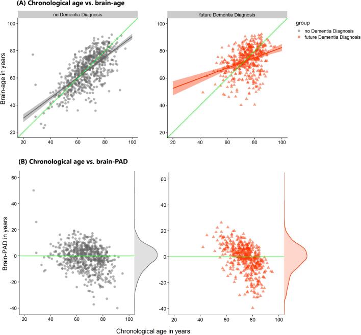
We analysed T1-weighted MRI scans from a real-world setting of patients referred to UK memory clinic services (n = 1140; 60.2 % female and mean [SD] age of 70.0[10.8] years) to derive 'brain-age'. Brain-age is an index of age-related brain health based on quantitative analysis of structural neuroimaging, largely reflecting brain atrophy. Brain-predicted age difference (brain-PAD) was calculated as brain-age minus chronological age. We determined which patients went on to develop dementia between three months and 7.8 years after neuroimaging assessment (n = 476) using linkage to electronic health records.

RESULTS

Survival analysis, using Cox regression, indicated a 3 % increased risk of dementia per brain-PAD year (hazard ratio [95 % CI] = 1.03 [1.02,1.04], p < 0.0001), adjusted for baseline age, age, sex, Mini Mental State Examination (MMSE) score and normalised brain volume. In sensitivity analyses, brain-PAD remained significant when time-to-dementia was at least 3 years (hazard ratio [95 % CI] = 1.06 [1.02, 1.09], p = 0.0006), or when baseline MMSE score ≥ 27 (hazard ratio [95 % CI] = 1.03 [1.01, 1.05], p = 0.0006).

CONCLUSIONS

Memory clinic patients with older-appearing brains are more likely to receive a subsequent dementia diagnosis. Potentially, brain-age could aid decision-making during initial memory clinic assessment to improve early detection of dementia. Even when neuroimaging assessment was more than 3 years prior to diagnosis and when cognitive functioning was not clearly impaired, brain-age still proved informative. These real-world results support the use of quantitative neuroimaging biomarkers like brain-age in memory clinics.

摘要

背景

用于痴呆风险早期检测的生物标志物有望更好地进行疾病监测和针对性干预。然而,大多数生物标志物研究,特别是神经影像学研究,分析的都是人为“干净”的研究组,没有共病、错误转诊、禁忌症和来自狭窄的社会人口群体。这些偏倚意味着神经影像学样本通常不能代表痴呆风险的目标人群(例如,被转介到记忆诊所的人群),从而限制了这些研究在真实临床环境中的推广。为了促进研究向临床更好地转化,需要使用更能代表痴呆患者群体的数据集。

方法

我们分析了来自英国记忆诊所服务的实际患者队列的 T1 加权 MRI 扫描(n=1140;60.2%为女性,平均[标准差]年龄为 70.0[10.8]岁),以得出“大脑年龄”。大脑年龄是基于结构神经影像学的定量分析得出的与年龄相关的大脑健康指数,主要反映脑萎缩。脑预测年龄差(brain-PAD)的计算方法是大脑年龄减去实际年龄。我们通过与电子健康记录链接,确定了在神经影像学评估后 3 个月至 7.8 年内发展为痴呆的患者(n=476)。

结果

使用 Cox 回归的生存分析表明,每增加一个 brain-PAD 年,痴呆风险增加 3%(风险比[95%置信区间]为 1.03[1.02,1.04],p<0.0001),调整了基线年龄、年龄、性别、简易精神状态检查(MMSE)评分和标准化脑容量。在敏感性分析中,当痴呆发生时间至少为 3 年(风险比[95%置信区间]为 1.06[1.02,1.09],p=0.0006)或基线 MMSE 评分≥27 时(风险比[95%置信区间]为 1.03[1.01,1.05],p=0.0006),brain-PAD 仍然具有显著性。

结论

大脑年龄较大的记忆诊所患者更有可能被诊断为痴呆。脑年龄可能有助于改善痴呆的早期检测,在初始记忆诊所评估期间做出决策。即使神经影像学评估是在诊断前 3 年以上进行的,并且认知功能没有明显受损,脑年龄仍然具有信息性。这些真实世界的结果支持在记忆诊所中使用脑年龄等定量神经影像学生物标志物。

https://cdn.ncbi.nlm.nih.gov/pmc/blobs/15c2/9467894/78ba2509af10/gr5.jpg
https://cdn.ncbi.nlm.nih.gov/pmc/blobs/15c2/9467894/5b5675e27d7b/gr1.jpg
https://cdn.ncbi.nlm.nih.gov/pmc/blobs/15c2/9467894/410cb3d43262/gr2.jpg
https://cdn.ncbi.nlm.nih.gov/pmc/blobs/15c2/9467894/c4a7e63f408d/gr3.jpg
https://cdn.ncbi.nlm.nih.gov/pmc/blobs/15c2/9467894/3afb90d3d8cd/gr4.jpg
https://cdn.ncbi.nlm.nih.gov/pmc/blobs/15c2/9467894/78ba2509af10/gr5.jpg

相似文献

1
Brain-age is associated with progression to dementia in memory clinic patients.脑龄与记忆门诊患者向痴呆的进展相关。
Neuroimage Clin. 2022;36:103175. doi: 10.1016/j.nicl.2022.103175. Epub 2022 Aug 30.
2
Structural magnetic resonance imaging for the early diagnosis of dementia due to Alzheimer's disease in people with mild cognitive impairment.用于轻度认知障碍患者阿尔茨海默病所致痴呆早期诊断的结构磁共振成像
Cochrane Database Syst Rev. 2020 Mar 2;3(3):CD009628. doi: 10.1002/14651858.CD009628.pub2.
3
Interpreting Biomarker Results in Individual Patients With Mild Cognitive Impairment in the Alzheimer's Biomarkers in Daily Practice (ABIDE) Project.阿尔茨海默病生物标志物日常实践(ABIDE)项目中个体轻度认知障碍患者生物标志物结果的解读
JAMA Neurol. 2017 Dec 1;74(12):1481-1491. doi: 10.1001/jamaneurol.2017.2712.
4
Cognitive and neuroimaging features and brain β-amyloidosis in individuals at risk of Alzheimer's disease (INSIGHT-preAD): a longitudinal observational study.认知和神经影像学特征以及阿尔茨海默病风险个体的脑β-淀粉样蛋白(INSIGHT-preAD):一项纵向观察性研究。
Lancet Neurol. 2018 Apr;17(4):335-346. doi: 10.1016/S1474-4422(18)30029-2. Epub 2018 Feb 27.
5
Neuropsychiatric symptoms and neuroimaging-based brain age in mild cognitive impairment and early dementia: A multicenter study.轻度认知障碍和早期痴呆中的神经精神症状与基于神经影像学的脑龄:一项多中心研究。
Psychiatry Clin Neurosci. 2025 Apr;79(4):158-164. doi: 10.1111/pcn.13777. Epub 2025 Jan 17.
6
Combined Biomarker Prognosis of Mild Cognitive Impairment: An 11-Year Follow-Up Study in the Alzheimer's Disease Neuroimaging Initiative.联合生物标志物预测轻度认知障碍:阿尔茨海默病神经影像学倡议的 11 年随访研究。
J Alzheimers Dis. 2019;68(4):1549-1559. doi: 10.3233/JAD-181243.
7
A generalizable data-driven model of atrophy heterogeneity and progression in memory clinic settings.记忆门诊环境中萎缩异质性和进展的可推广数据驱动模型。
Brain. 2024 Jul 5;147(7):2400-2413. doi: 10.1093/brain/awae118.
8
Whole-brain atrophy rate and cognitive decline: longitudinal MR study of memory clinic patients.全脑萎缩率与认知衰退:记忆门诊患者的纵向磁共振成像研究
Radiology. 2008 Aug;248(2):590-8. doi: 10.1148/radiol.2482070938. Epub 2008 Jun 23.
9
Cross-sectional and longitudinal characterization of SCD patients recruited from the community versus from a memory clinic: subjective cognitive decline, psychoaffective factors, cognitive performances, and atrophy progression over time.从社区和记忆诊所招募的 SCD 患者的横断面和纵向特征:主观认知下降、心理情感因素、认知表现以及随时间的进展的萎缩。
Alzheimers Res Ther. 2019 Jul 8;11(1):61. doi: 10.1186/s13195-019-0514-z.
10
Dynamics of brain structure and cognitive function in the Alzheimer's disease neuroimaging initiative.阿尔茨海默病神经影像学倡议中的大脑结构和认知功能的动态变化。
J Neurol Neurosurg Psychiatry. 2013 Jan;84(1):71-8. doi: 10.1136/jnnp-2012-303579. Epub 2012 Nov 2.

引用本文的文献

1
Buffering brain aging: education moderates language impairment in Parkinson's disease.缓冲大脑衰老:教育可减轻帕金森病患者的语言障碍。
Front Cell Neurosci. 2025 Aug 20;19:1606451. doi: 10.3389/fncel.2025.1606451. eCollection 2025.
2
The effect of lesion filling on brain age estimation in multiple sclerosis.病灶填充对多发性硬化症脑年龄估计的影响。
BMC Med Imaging. 2025 Aug 27;25(1):356. doi: 10.1186/s12880-025-01897-6.
3
BrainAGE as a measure of maturation during early adolescence.脑龄作为青春期早期成熟度的一种衡量指标。
Imaging Neurosci (Camb). 2023 Nov 30;1. doi: 10.1162/imag_a_00037. eCollection 2023.
4
Real-world brain imaging in a population-based cohort enables accurate markers for dementia.在基于人群的队列中进行的真实世界脑成像能够为痴呆症提供准确的标志物。
Alzheimers Dement. 2025 Jul;21(7):e70227. doi: 10.1002/alz.70227.
5
Brain Age Prediction: Deep Models Need a Hand to Generalize.脑龄预测:深度模型需要助力以实现泛化。
Hum Brain Mapp. 2025 Aug 1;46(11):e70254. doi: 10.1002/hbm.70254.
6
DunedinPACNI estimates the longitudinal Pace of Aging from a single brain image to track health and disease.达尼丁太平洋神经影像衰老时钟(DunedinPACNI)通过单一脑图像估计衰老的纵向进程,以追踪健康与疾病状况。
Nat Aging. 2025 Jul 1. doi: 10.1038/s43587-025-00897-z.
7
Repeatability and reproducibility of brain age estimates in multiple sclerosis for three publicly available models.三种公开可用模型在多发性硬化症中脑年龄估计的可重复性和再现性。
Neuroimage Rep. 2025 Mar 21;5(2):100252. doi: 10.1016/j.ynirp.2025.100252. eCollection 2025 Jun.
8
Associations of history of traumatic brain injury with markers of brain health among older men in the Vietnam Era Twin Study of Aging.越战时期男性双生子衰老研究中,创伤性脑损伤史与老年男性脑健康标志物的关联。
Neuroimage Clin. 2025 Jun 21;47:103831. doi: 10.1016/j.nicl.2025.103831.
9
Relationships between measures of neurovascular integrity and fluid transport in aging: a multi-modal neuroimaging study.衰老过程中神经血管完整性指标与液体运输之间的关系:一项多模态神经影像学研究。
Fluids Barriers CNS. 2025 Jun 16;22(1):59. doi: 10.1186/s12987-025-00664-7.
10
Your Brain Doesn't Look a Day Past 70! Cross-Sectional Associations with Brain-Predicted Age in the Cognitively-Intact Oldest-Old.你的大脑看起来一点都不像超过70岁的!认知功能正常的高龄老人大脑预测年龄的横断面关联。
bioRxiv. 2025 May 30:2025.05.26.655855. doi: 10.1101/2025.05.26.655855.