Hacettepe University, Faculty of Dentistry, Department of Endodontics, Ankara, Turkey.
TOBB University of Economics and Technology, Faculty of Medicine, Department of Histology and Embryology, Ankara, Turkey.
J Appl Oral Sci. 2022 Sep 9;30:e20220086. doi: 10.1590/1678-7757-2022-0086. eCollection 2022.
Bioactive molecules present the potential to be used along with biomaterials in vital pulp therapy and regenerative endodontic treatment.
The aim of this study was to assess the effects of the combined use of bone morphogenetic protein-7 (BMP-7) and mineral trioxide aggregate (MTA) on the proliferation, migration, and differentiation of human dental pulp stem cells (DPSCs).
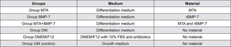
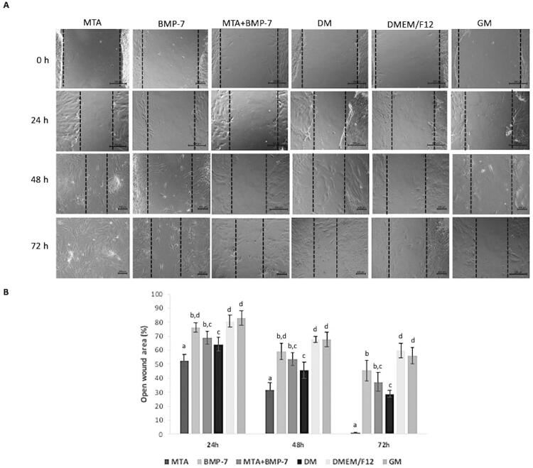
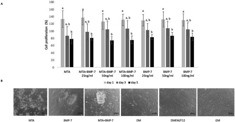
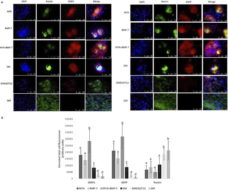
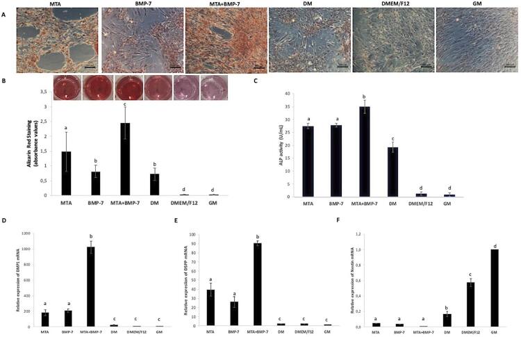
For the proliferation analysis, DPSCs were incubated with a growth medium and treated with MTA and/or BMP-7 at different concentrations. For the following analyses, DPSCs were incubated with a differentiation medium and treated with MTA and/or BMP-7. Moreover, there were groups in which DPSCs were incubated with the growth medium (control), the differentiation medium, or DMEM/F12 containing fetal bovine serum, and not treated with MTA or BMP-7. Cell proliferation was analyzed using the WST-1 assay. The odontogenic/osteogenic differentiation was evaluated by immunocytochemistry, alkaline phosphatase (ALP) activity assay, alizarin red staining, and reverse transcription-quantitative polymerase chain reaction (RT-qPCR). Cell migration was evaluated using a wound-healing assay. Data were analyzed using analysis of variance and Tukey test (p=0.05).
The use of BMP-7 with MTA presented no significant effect on cell proliferation in comparison with the treatment with MTA alone (p>0.05), but showed higher ALP activity, increased mineralization, and higher expression of DMP1 and DSPP when compared with other groups (p<0.05). Nestin expression was higher in the control group than in groups treated with MTA and/or BMP-7 (p<0.05). The cell migration rate increased after treatment with MTA when compared with other groups in all periods of time (p<0.05). At 72 hours, the wound area was smaller in groups treated with MTA and/or BMP-7 than in the control group (p<0.05).
The use of BMP-7 with MTA increased odontogenic/osteogenic differentiation without adversely affecting proliferation and migration of DPSCs. The use of BMP-7 with MTA may improve treatment outcomes by increasing repair and regeneration capacity of DPSCs.
生物活性分子具有与生物材料结合应用于活髓治疗和再生性牙髓治疗的潜力。
本研究旨在评估骨形成蛋白 7(BMP-7)和矿化三氧化物凝聚体(MTA)联合使用对人牙髓干细胞(DPSCs)增殖、迁移和分化的影响。
为了进行增殖分析,将 DPSCs 与生长培养基孵育,并以不同浓度用 MTA 和/或 BMP-7 处理。对于以下分析,将 DPSCs 与分化培养基孵育,并以 MTA 和/或 BMP-7 处理。此外,还有一些组将 DPSCs 与生长培养基(对照)、分化培养基或含胎牛血清的 DMEM/F12 孵育,且不使用 MTA 或 BMP-7 处理。使用 WST-1 测定法分析细胞增殖。通过免疫细胞化学、碱性磷酸酶(ALP)活性测定、茜素红染色和逆转录-定量聚合酶链反应(RT-qPCR)评估成牙/成骨分化。使用划痕愈合试验评估细胞迁移。使用方差分析和 Tukey 检验(p=0.05)进行数据分析。
与单独使用 MTA 相比,BMP-7 与 MTA 联合使用对细胞增殖没有显著影响(p>0.05),但与其他组相比,其具有更高的 ALP 活性、增加的矿化以及更高的 DMP1 和 DSPP 表达(p<0.05)。与用 MTA 和/或 BMP-7 处理的组相比,对照组中的巢蛋白表达更高(p<0.05)。与其他组相比,在所有时间段,用 MTA 处理后细胞迁移率均增加(p<0.05)。在 72 小时时,与对照组相比,用 MTA 和/或 BMP-7 处理的组的伤口面积更小(p<0.05)。
BMP-7 与 MTA 联合使用增加了牙源性/成骨性分化,而不会对 DPSCs 的增殖和迁移产生不利影响。BMP-7 与 MTA 的联合使用可能通过增加 DPSCs 的修复和再生能力来改善治疗效果。