• 文献检索
  • 文档翻译
  • 深度研究
  • 学术资讯
  • Suppr Zotero 插件Zotero 插件
  • 邀请有礼
  • 套餐&价格
  • 历史记录
应用&插件
Suppr Zotero 插件Zotero 插件浏览器插件Mac 客户端Windows 客户端微信小程序
定价
高级版会员购买积分包购买API积分包
服务
文献检索文档翻译深度研究API 文档MCP 服务
关于我们
关于 Suppr公司介绍联系我们用户协议隐私条款
关注我们

Suppr 超能文献

核心技术专利:CN118964589B侵权必究
粤ICP备2023148730 号-1Suppr @ 2026

文献检索

告别复杂PubMed语法,用中文像聊天一样搜索,搜遍4000万医学文献。AI智能推荐,让科研检索更轻松。

立即免费搜索

文件翻译

保留排版,准确专业,支持PDF/Word/PPT等文件格式,支持 12+语言互译。

免费翻译文档

深度研究

AI帮你快速写综述,25分钟生成高质量综述,智能提取关键信息,辅助科研写作。

立即免费体验

非小细胞肺癌纵隔淋巴结转移的机器学习分类:一项针对西欧患者群体的多中心研究

Machine learning classification of mediastinal lymph node metastasis in NSCLC: a multicentre study in a Western European patient population.

作者信息

Laros Sara S A, Dieckens Dennis, Blazis Stephan P, van der Heide Johannes A

机构信息

Department of Medical Physics and Engineering, Albert Schweitzer Hospital, Afdeling Klinische Fysica - Medische Techniek, Albert Schweitzerplaats 25, 3318 AT, Dordrecht, The Netherlands.

Department of Nuclear Medicine, Albert Schweitzer Hospital, Dordrecht, The Netherlands.

出版信息

EJNMMI Phys. 2022 Sep 24;9(1):66. doi: 10.1186/s40658-022-00494-8.

DOI:10.1186/s40658-022-00494-8
PMID:36153446
原文链接:https://pmc.ncbi.nlm.nih.gov/articles/PMC9509500/
Abstract

BACKGROUND

[F] FDG PET-CT has an important role in the initial staging of lung cancer; however, accurate differentiation between activity in malignant and benign intrathoracic lymph nodes on PET-CT scans can be challenging. The purpose of the current study was to investigate the effect of incorporating primary tumour data and clinical features to differentiate between [F] FDG-avid malignant and benign intrathoracic lymph nodes.

METHODS

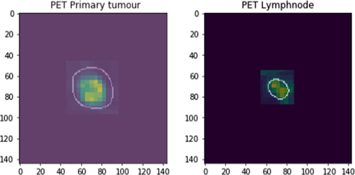
We retrospectively selected lung cancer patients who underwent PET-CT for initial staging in two centres in the Netherlands. The primary tumour and suspected lymph node metastases were annotated and cross-referenced with pathology results. Lymph nodes were classified as malignant or benign. From the image data, we extracted radiomic features and trained the classifier model using the extreme gradient boost (XGB) algorithm. Various scenarios were defined by selecting different combinations of data input and clinical features. Data from centre 1 were used for training and validation of the models using the XGB algorithm. To determine the performance of the model in a different hospital, the XGB model was tested using data from centre 2.

RESULTS

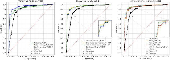
Adding primary tumour data resulted in a significant gain in the performance of the trained classifier model. Adding the clinical information about distant metastases did not lead to significant improvement. The performance of the model in the test set (centre 2) was slightly but statistically significantly lower than in the validation set (centre 1).

CONCLUSIONS

Using the XGB algorithm potentially leads to an improved model for the classification of intrathoracic lymph nodes. The inclusion of primary tumour data improved the performance of the model, while additional knowledge of distant metastases did not. In patients in whom metastases are limited to lymph nodes in the thorax, this may reduce costly and invasive procedures such as endobronchial ultrasound or mediastinoscopy procedures.

摘要

背景

[F] FDG PET-CT在肺癌的初始分期中具有重要作用;然而,在PET-CT扫描中准确区分恶性和良性胸内淋巴结的活性可能具有挑战性。本研究的目的是探讨纳入原发肿瘤数据和临床特征对区分[F] FDG摄取的恶性和良性胸内淋巴结的影响。

方法

我们回顾性选择了在荷兰两个中心接受PET-CT进行初始分期的肺癌患者。对原发肿瘤和疑似淋巴结转移灶进行标注,并与病理结果进行交叉对照。淋巴结被分类为恶性或良性。从图像数据中,我们提取了影像组学特征,并使用极端梯度提升(XGB)算法训练分类器模型。通过选择不同的数据输入和临床特征组合定义了各种场景。中心1的数据用于使用XGB算法训练和验证模型。为了确定模型在另一家医院的性能,使用中心2的数据对XGB模型进行测试。

结果

添加原发肿瘤数据使训练后的分类器模型性能显著提高。添加远处转移的临床信息并未带来显著改善。模型在测试集(中心2)中的性能略低于验证集(中心1),但具有统计学意义。

结论

使用XGB算法可能会改进胸内淋巴结分类模型。纳入原发肿瘤数据提高了模型的性能,而远处转移的额外信息则没有。对于转移仅限于胸部淋巴结的患者,这可能会减少诸如支气管内超声或纵隔镜检查等昂贵且有创的检查。

https://cdn.ncbi.nlm.nih.gov/pmc/blobs/6c14/9509500/114de7d6cf34/40658_2022_494_Fig3_HTML.jpg
https://cdn.ncbi.nlm.nih.gov/pmc/blobs/6c14/9509500/cbcef473ad01/40658_2022_494_Fig1_HTML.jpg
https://cdn.ncbi.nlm.nih.gov/pmc/blobs/6c14/9509500/a3769359e9d5/40658_2022_494_Fig2_HTML.jpg
https://cdn.ncbi.nlm.nih.gov/pmc/blobs/6c14/9509500/114de7d6cf34/40658_2022_494_Fig3_HTML.jpg
https://cdn.ncbi.nlm.nih.gov/pmc/blobs/6c14/9509500/cbcef473ad01/40658_2022_494_Fig1_HTML.jpg
https://cdn.ncbi.nlm.nih.gov/pmc/blobs/6c14/9509500/a3769359e9d5/40658_2022_494_Fig2_HTML.jpg
https://cdn.ncbi.nlm.nih.gov/pmc/blobs/6c14/9509500/114de7d6cf34/40658_2022_494_Fig3_HTML.jpg

相似文献

1
Machine learning classification of mediastinal lymph node metastasis in NSCLC: a multicentre study in a Western European patient population.非小细胞肺癌纵隔淋巴结转移的机器学习分类:一项针对西欧患者群体的多中心研究
EJNMMI Phys. 2022 Sep 24;9(1):66. doi: 10.1186/s40658-022-00494-8.
2
FDG PET/CT radiomics as a tool to differentiate between reactive axillary lymphadenopathy following COVID-19 vaccination and metastatic breast cancer axillary lymphadenopathy: a pilot study.COVID-19 疫苗接种后反应性腋窝淋巴结病与转移性乳腺癌腋窝淋巴结病的 FDG PET/CT 影像组学鉴别:一项初步研究。
Eur Radiol. 2022 Sep;32(9):5921-5929. doi: 10.1007/s00330-022-08725-3. Epub 2022 Apr 6.
3
Radiomic Analysis using Density Threshold for FDG-PET/CT-Based N-Staging in Lung Cancer Patients.基于密度阈值的FDG-PET/CT对肺癌患者进行N分期的影像组学分析
Mol Imaging Biol. 2017 Apr;19(2):315-322. doi: 10.1007/s11307-016-0996-z.
4
Combined endobronchial and esophageal endosonography for the diagnosis and staging of lung cancer: European Society of Gastrointestinal Endoscopy (ESGE) Guideline, in cooperation with the European Respiratory Society (ERS) and the European Society of Thoracic Surgeons (ESTS).支气管内与食管内超声联合用于肺癌诊断及分期:欧洲胃肠内镜学会(ESGE)指南,与欧洲呼吸学会(ERS)及欧洲胸外科医师学会(ESTS)合作制定
Endoscopy. 2015 Jun;47(6):545-59. doi: 10.1055/s-0034-1392040. Epub 2015 Jun 1.
5
Integrated FDG-PET/CT does not make invasive staging of the intrathoracic lymph nodes in non-small cell lung cancer redundant: a prospective study.18F-氟脱氧葡萄糖正电子发射断层显像/计算机断层扫描(FDG-PET/CT)一体化检查对于非小细胞肺癌胸内淋巴结的侵袭性分期并非多余:一项前瞻性研究。
Thorax. 2007 Aug;62(8):696-701. doi: 10.1136/thx.2006.072959.
6
Is mediastinoscopy still the gold standard to evaluate mediastinal lymph nodes in patients with non-small cell lung carcinoma?对于非小细胞肺癌患者,纵隔镜检查仍是评估纵隔淋巴结的金标准吗?
Thorac Cardiovasc Surg. 2012 Mar;60(2):116-21. doi: 10.1055/s-0030-1271148. Epub 2011 Jun 20.
7
MEDIASTinal staging of non-small cell lung cancer by endobronchial and endoscopic ultrasonography with or without additional surgical mediastinoscopy (MEDIASTrial): study protocol of a multicenter randomised controlled trial.通过支气管内和内镜超声检查联合或不联合额外的手术纵隔镜检查进行非小细胞肺癌的纵隔分期(MEDIAST试验):一项多中心随机对照试验的研究方案
BMC Surg. 2018 May 18;18(1):27. doi: 10.1186/s12893-018-0359-6.
8
Preoperative intrathoracic lymph node staging in patients with non-small-cell lung cancer: accuracy of integrated positron emission tomography and computed tomography.术前胸内淋巴结分期在非小细胞肺癌患者中的应用:正电子发射断层扫描和计算机断层扫描的准确性。
Eur J Cardiothorac Surg. 2009 Sep;36(3):440-5. doi: 10.1016/j.ejcts.2009.04.003. Epub 2009 May 22.
9
Development and Validation of a F-FDG PET-Based Radiomic Model for Evaluating Hypermetabolic Mediastinal-Hilar Lymph Nodes in Non-Small-Cell Lung Cancer.基于F-FDG PET的放射组学模型在评估非小细胞肺癌中高代谢纵隔-肺门淋巴结的开发与验证
Front Oncol. 2021 Sep 8;11:710909. doi: 10.3389/fonc.2021.710909. eCollection 2021.
10
Staging of lymph nodes with FDG dual-headed PET in patients with non-small-cell lung cancer.非小细胞肺癌患者使用氟代脱氧葡萄糖双头正电子发射断层显像术对淋巴结进行分期
Nucl Med Commun. 1999 Nov;20(11):1001-7. doi: 10.1097/00006231-199911000-00004.

引用本文的文献

1
The value of F-fluorodeoxyglucose positron emission tomography-based radiomics in non-small cell lung cancer.基于F-氟脱氧葡萄糖正电子发射断层扫描的影像组学在非小细胞肺癌中的价值
Tzu Chi Med J. 2024 Sep 3;37(1):17-27. doi: 10.4103/tcmj.tcmj_124_24. eCollection 2025 Jan-Mar.
2
Evaluating peritumoral and intratumoral radiomics signatures for predicting lymph node metastasis in surgically resectable non-small cell lung cancer.评估瘤周和瘤内影像组学特征以预测可手术切除的非小细胞肺癌中的淋巴结转移
Front Oncol. 2024 Oct 11;14:1427743. doi: 10.3389/fonc.2024.1427743. eCollection 2024.
3
Clinico-biological-radiomics (CBR) based machine learning for improving the diagnostic accuracy of FDG-PET false-positive lymph nodes in lung cancer.

本文引用的文献

1
The Role of Radiomics in Lung Cancer: From Screening to Treatment and Follow-Up.放射组学在肺癌中的作用:从筛查到治疗及随访
Front Oncol. 2021 May 5;11:603595. doi: 10.3389/fonc.2021.603595. eCollection 2021.
2
Impact of feature selection methods and subgroup factors on prognostic analysis with CT-based radiomics in non-small cell lung cancer patients.基于 CT 影像组学的特征选择方法和亚组因素对非小细胞肺癌患者预后分析的影响。
Radiat Oncol. 2021 Apr 30;16(1):80. doi: 10.1186/s13014-021-01810-9.
3
Machine learning-based diagnostic method of pre-therapeutic F-FDG PET/CT for evaluating mediastinal lymph nodes in non-small cell lung cancer.
基于临床-生物学-放射组学(CBR)的机器学习提高肺癌 FDG-PET 假阳性淋巴结的诊断准确性。
Eur J Med Res. 2023 Dec 2;28(1):554. doi: 10.1186/s40001-023-01497-6.
4
Clinical application of F-fluorodeoxyglucose positron emission tomography/computed tomography radiomics-based machine learning analyses in the field of oncology.基于 F-氟代脱氧葡萄糖正电子发射断层扫描/计算机断层扫描放射组学的机器学习分析在肿瘤学领域的临床应用。
Jpn J Radiol. 2024 Jan;42(1):28-55. doi: 10.1007/s11604-023-01476-1. Epub 2023 Aug 1.
基于机器学习的非小细胞肺癌治疗前 F-FDG PET/CT 纵隔淋巴结诊断方法。
Eur Radiol. 2021 Jun;31(6):4184-4194. doi: 10.1007/s00330-020-07523-z. Epub 2020 Nov 25.
4
Convolutional Neural Networks in Predicting Nodal and Distant Metastatic Potential of Newly Diagnosed Non-Small Cell Lung Cancer on FDG PET Images.卷积神经网络在预测 FDG PET 图像中新诊断的非小细胞肺癌的淋巴结和远处转移潜能中的应用。
AJR Am J Roentgenol. 2020 Jul;215(1):192-197. doi: 10.2214/AJR.19.22346. Epub 2020 Apr 29.
5
[Artificial intelligence in lung imaging].[肺部成像中的人工智能]
Radiologe. 2020 Jan;60(1):42-47. doi: 10.1007/s00117-019-00611-2.
6
Advances in Imaging and Automated Quantification of Malignant Pulmonary Diseases: A State-of-the-Art Review.恶性肺部疾病的影像学进展与自动化定量:最新综述。
Lung. 2018 Dec;196(6):633-642. doi: 10.1007/s00408-018-0156-0. Epub 2018 Oct 9.
7
Preoperative Staging by EBUS in cN0/N1 Lung Cancer: Systematic Review and Meta-Analysis.经支气管超声引导针吸活检术对cN0/N1期肺癌的术前分期:系统评价与Meta分析
J Bronchology Interv Pulmonol. 2019 Jul;26(3):155-165. doi: 10.1097/LBR.0000000000000545.
8
Computational Radiomics System to Decode the Radiographic Phenotype.用于解码影像学表型的计算放射组学系统
Cancer Res. 2017 Nov 1;77(21):e104-e107. doi: 10.1158/0008-5472.CAN-17-0339.
9
Comparison of machine learning methods for classifying mediastinal lymph node metastasis of non-small cell lung cancer from F-FDG PET/CT images.基于F-FDG PET/CT图像的非小细胞肺癌纵隔淋巴结转移机器学习分类方法比较
EJNMMI Res. 2017 Dec;7(1):11. doi: 10.1186/s13550-017-0260-9. Epub 2017 Jan 28.
10
Minimally Invasive Methods for Staging in Lung Cancer: Systematic Review and Meta-Analysis.肺癌分期的微创方法:系统评价与荟萃分析
Pulm Med. 2016;2016:1024709. doi: 10.1155/2016/1024709. Epub 2016 Oct 13.