Walker Cierra J, Batan Dilara, Bishop Carrie T, Ramirez Daniel, Aguado Brian A, Schroeder Megan E, Crocini Claudia, Schwisow Jessica, Moulton Karen, Macdougall Laura, Weiss Robert M, Allen Mary A, Dowell Robin, Leinwand Leslie A, Anseth Kristi S
Materials Science and Engineering Program University of Colorado Boulder Boulder Colorado USA.
BioFrontiers Institute, University of Colorado Boulder Boulder Colorado USA.
Bioeng Transl Med. 2022 Aug 22;7(3):e10394. doi: 10.1002/btm2.10394. eCollection 2022 Sep.
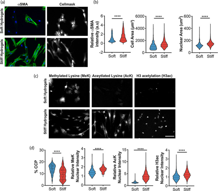
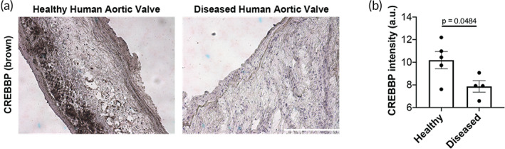
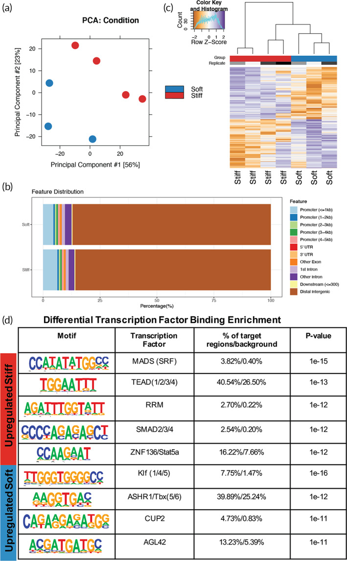
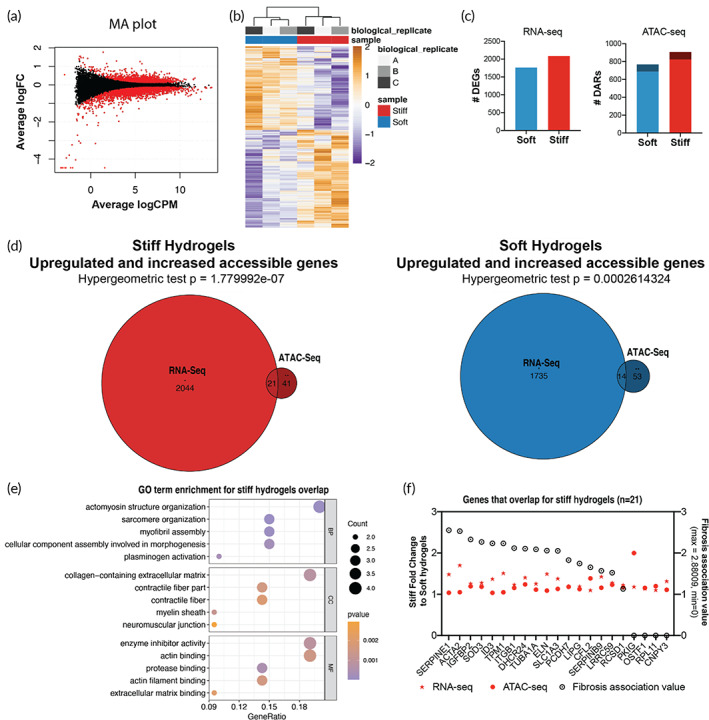
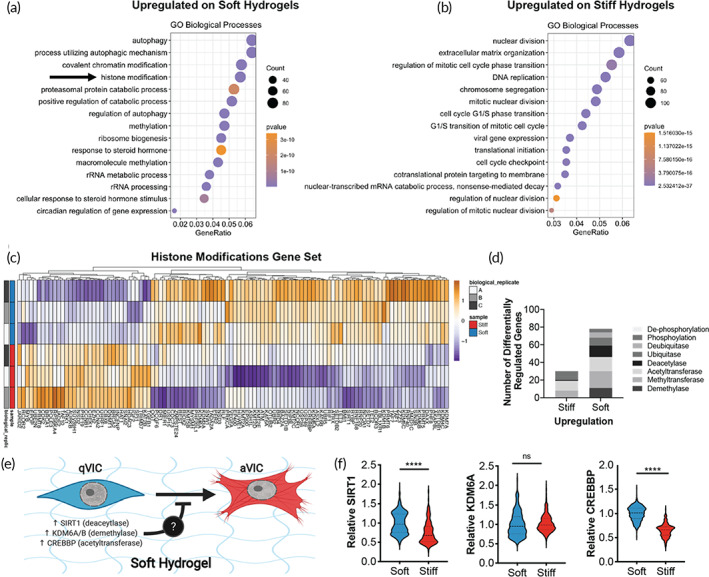
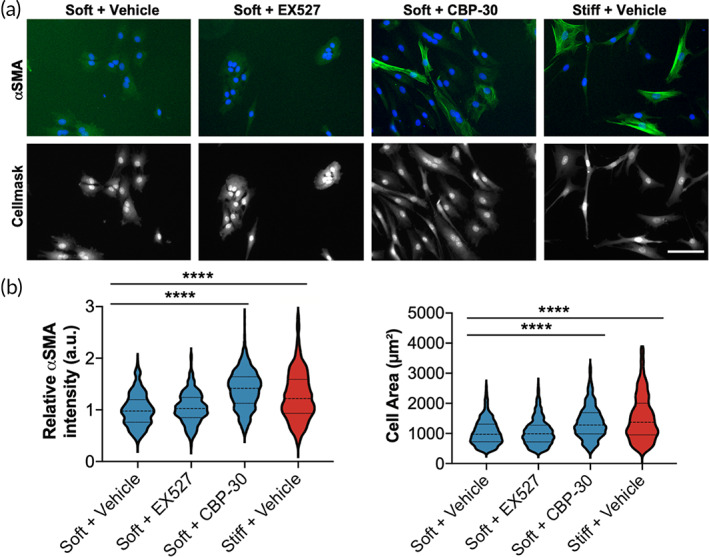
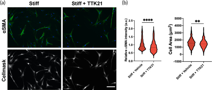
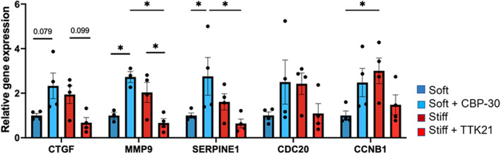
Aortic valve stenosis (AVS) is a progressive fibrotic disease that is caused by thickening and stiffening of valve leaflets. At the cellular level, quiescent valve interstitial cells (qVICs) activate to myofibroblasts (aVICs) that persist within the valve tissue. Given the persistence of myofibroblasts in AVS, epigenetic mechanisms have been implicated. Here, we studied changes that occur in VICs during myofibroblast activation by using a hydrogel matrix to recapitulate different stiffnesses in the valve leaflet during fibrosis. We first compared the chromatin landscape of qVICs cultured on soft hydrogels and aVICs cultured on stiff hydrogels, representing the native and diseased phenotypes respectively. Using assay for transposase-accessible chromatin sequencing (ATAC-Seq), we found that open chromatin regions in aVICs were enriched for transcription factor binding motifs associated with mechanosensing pathways compared to qVICs. Next, we used RNA-Seq to show that the open chromatin regions in aVICs correlated with pro-fibrotic gene expression, as aVICs expressed higher levels of contractile fiber genes, including myofibroblast markers such as alpha smooth muscle actin (αSMA), compared to qVICs. In contrast, chromatin remodeling genes were downregulated in aVICs compared to qVICs, indicating qVICs may be protected from myofibroblast activation through epigenetic mechanisms. Small molecule inhibition of one of these remodelers, CREB Binding Protein (CREBBP), prevented qVICs from activating to aVICs. Notably, CREBBP is more abundant in valves from healthy patients compared to fibrotic valves. Our findings reveal the role of mechanical regulation in chromatin remodeling during VIC activation and quiescence and highlight one potential therapeutic target for treating AVS.
主动脉瓣狭窄(AVS)是一种进行性纤维化疾病,由瓣膜小叶增厚和硬化引起。在细胞水平上,静止的瓣膜间质细胞(qVICs)激活成为肌成纤维细胞(aVICs),并持续存在于瓣膜组织中。鉴于肌成纤维细胞在AVS中的持续性,表观遗传机制被认为与之相关。在这里,我们通过使用水凝胶基质来模拟纤维化过程中瓣膜小叶的不同硬度,研究了VICs在肌成纤维细胞激活过程中发生的变化。我们首先比较了在柔软水凝胶上培养的qVICs和在坚硬水凝胶上培养的aVICs的染色质景观,分别代表天然和患病表型。使用转座酶可及染色质测序分析(ATAC-Seq),我们发现与qVICs相比,aVICs中的开放染色质区域富含与机械传感途径相关的转录因子结合基序。接下来,我们使用RNA-Seq表明,aVICs中的开放染色质区域与促纤维化基因表达相关,因为与qVICs相比,aVICs表达更高水平的收缩纤维基因,包括肌成纤维细胞标志物,如α平滑肌肌动蛋白(αSMA)。相比之下,与qVICs相比,aVICs中的染色质重塑基因下调,表明qVICs可能通过表观遗传机制免受肌成纤维细胞激活。对这些重塑因子之一的小分子抑制,即CREB结合蛋白(CREBBP),可防止qVICs激活为aVICs。值得注意的是,与纤维化瓣膜相比,CREBBP在健康患者的瓣膜中更为丰富。我们的研究结果揭示了机械调节在VIC激活和静止过程中染色质重塑中的作用,并突出了治疗AVS的一个潜在治疗靶点。