Mehan Anoushika, Anthony Michael Leonard, Paul Pranoy, Syed Anjum, Chowdhury Nilotpal, Rao Shalinee, Hussain Nuzhat, Ravi Bina
Department of Pathology and Laboratory Medicine, AIIMS, Rishikesh, Uttarakhand, India.
Department of Radiodiagnosis and Integrated Breast Care Centre (IBCC), AIIMS, Rishikesh, Uttarakhand, India.
J Lab Physicians. 2021 Nov 10;14(1):27-31. doi: 10.1055/s-0041-1736522. eCollection 2022 Mar.
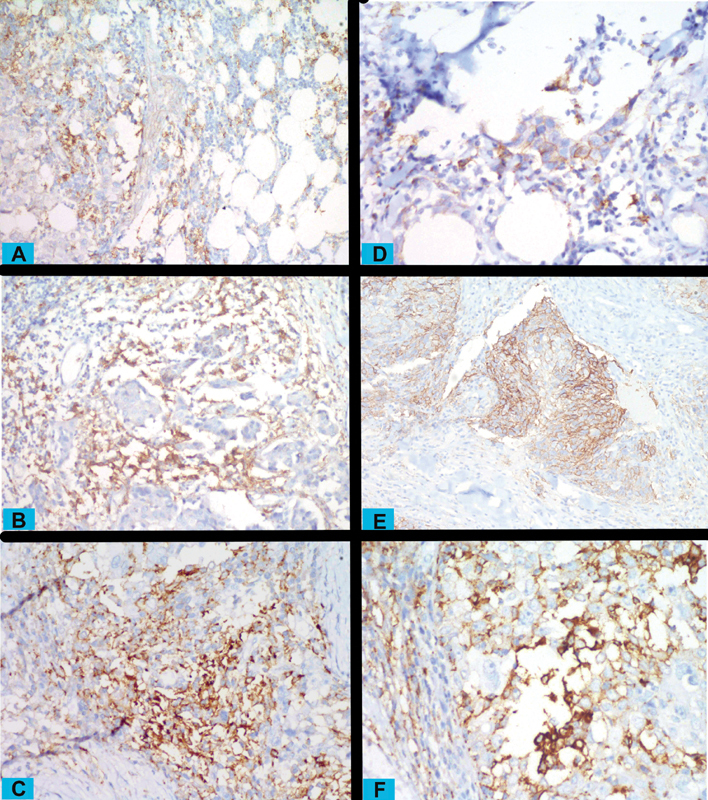
Cancer immunotherapy targeting the programmed cell death ligand 1 (PD-L1) and programmed cell death-1 (PD-1) axis has revolutionized cancer therapy. PD-L1 also serves as a predictive marker for such therapy. To assess the potential of such therapy in any cancer, the positivity of PD-1 and PD-L1 in such cancers needs to be assessed. However, such studies for breast cancer are lacking in South Asia. We aimed to estimate the positivity of PD-L1 and PD-1 receptors in breast cancer and its various clinicopathological groups in our patient population. We studied the immunoexpression of PD-1 and PD-L1 in 103 histologically proven invasive carcinoma breast cases from October 2018 to April 2019. The percent positivity of PD-1 and PD-L1 with 95% confidence intervals (CI) was estimated for all the cases as well as groups defined by stage, grade, molecular subtype, hormone receptor status, K -67, and age. PD-1 positivity was seen in 72 (69.9%) cases (95% CI: 60.1-78.6). PD-L1 immunoexpression was seen in 61 (59.2%) cases (95% CI: 49.1-68.8) in immune cells and in 39 (37.9%) cases (95% CI: 28.5-50.0) in tumor cells. No significant association was found between PD-1, PD-L1 and age, overall clinical stage, grade, size, estrogen receptor, progesterone receptor, human epidermal growth factor receptor 2, and K -67. Moderate-to-high PD-1 and PD-L1 immunopositivity was seen in all subtypes of breast cancer. PD-1 and PD-L1 is expressed in all subgroups of breast carcinoma. Patients in all such groups are amenable to immunotherapy, provided they are found suitable otherwise.
针对程序性细胞死亡配体1(PD-L1)和程序性细胞死亡蛋白1(PD-1)轴的癌症免疫疗法彻底改变了癌症治疗方式。PD-L1也是此类疗法的预测标志物。为评估这种疗法在任何癌症中的潜力,需要评估此类癌症中PD-1和PD-L1的阳性情况。然而,南亚地区缺乏针对乳腺癌的此类研究。我们旨在评估我们患者群体中乳腺癌及其各种临床病理分组中PD-L1和PD-1受体的阳性情况。
我们研究了2018年10月至2019年4月期间103例经组织学证实的浸润性乳腺癌病例中PD-1和PD-L1的免疫表达情况。对所有病例以及按分期、分级、分子亚型、激素受体状态、Ki-67和年龄定义的分组,估计了PD-1和PD-L1的阳性百分比及95%置信区间(CI)。
72例(69.9%)病例出现PD-1阳性(95%CI:60.1-78.6)。免疫细胞中61例(59.2%)病例出现PD-L1免疫表达(95%CI:49.1-68.8),肿瘤细胞中39例(37.9%)病例出现PD-L1免疫表达(95%CI:28.5-50.0)。未发现PD-1、PD-L1与年龄、总体临床分期、分级、大小、雌激素受体、孕激素受体、人表皮生长因子受体2及Ki-67之间存在显著关联。在乳腺癌的所有亚型中均可见中到高度的PD-1和PD-L1免疫阳性。
PD-1和PD-L1在乳腺癌的所有亚组中均有表达。所有这些组中的患者都适合接受免疫治疗,前提是他们在其他方面也被认定为合适。