Department of Ophthalmology, Dr. Rajendra Prasad Centre for Ophthalmic Sciences, All India Institute of Medical Sciences, New Delhi, India.
Shroff Eye Center, Kailash Colony, New Delhi, India.
Indian J Ophthalmol. 2022 Oct;70(10):3556-3561. doi: 10.4103/ijo.IJO_396_22.
To evaluate visual field changes in primary congenital glaucoma (PCG) with retinal nerve fiber layer thickness on optical coherence tomography.
In this cross-sectional, observational study, consecutive PCG children who underwent combined trabeculotomy with trabeculectomy and on regular follow-up were enrolled. All patients were aged over four years and co-operative for RNFL OCT and visual field examination. Perimetry was done on Humphrey visual field (HVF) analyzer using 30-2 and 10-2 SITA standard algorithms as appropriate. If a reliable automated perimetry was not feasible, kinetic perimetry was done. The following were noted at baseline and every follow-up: age, sex, visual acuity, intraocular pressure (IOP), cup-disc ratio (CDR), corneal diameters, refraction, any topical antiglaucoma medications, surgeries underwent, age at surgery and duration between surgery and final examination.
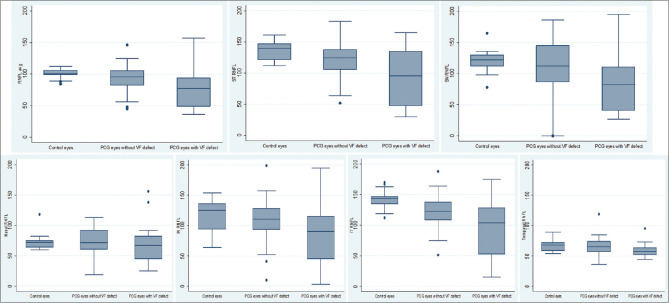
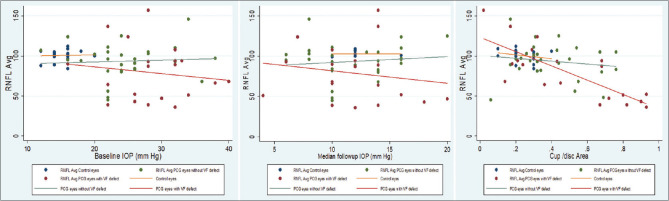
Forty-eight eyes of 34 children operated for PCG and 19 eyes of 17 controls were analyzed. A statistically significant thinner average RNFL thickness of 87.2 ± 28 μm was noted in PCG eyes as compared to controls with 100.6 ± 7.2 μm (P = 0.04). The mean cup-disc area ratio on OCT in PCG eyes was 0.43 ± 0.2 (0.02-0.93) and in control eyes was 0.23 ± 0.07 (0.1-0.4) (P < 0.001). On RNFL OCT, there was significant focal RNFL loss in temporal superior (P = 0.003), nasal inferior (P = 0.037) and temporal inferior (P < 0.001) quadrants compared to controls. Among PCG eyes, 20/48 eyes (41.7%), had definitive, reproducible glaucomatous VF defects. Mean baseline IOP in PCG eyes with VF defect was 28.7 ± 5.7 mmHg and in eyes with normal VF was 24.6 ± 5.9 mmHg (P = 0.03). On univariate regression analysis, higher baseline IOP was significantly associated with both RNFL loss (odds ratio (OR): -2.17) and VF defects (OR: 3.35). Fluctuation in follow-up IOP (OR: 3.33) was also significantly associated with the presence of VF defects. On multivariable regression analysis maximum, IOP was significantly associated with RNFL loss and VF defects.
Peripapillary RNFL thickness could be used to identify PCG eyes having visual field loss and possibly poor visual function from PCG eyes without visual field defects. Baseline and follow-up IOP, significantly correlated with RNFL thickness in PCG eyes.
利用光学相干断层扫描(OCT)评估原发性先天性青光眼(PCG)的视野变化与视网膜神经纤维层(RNFL)厚度的关系。
这是一项横断面、观察性研究,连续纳入了接受小梁切开联合小梁切除术且定期随访的 PCG 患儿。所有患者年龄均大于 4 岁,且能够配合完成 RNFL-OCT 和视野检查。使用 Humphrey 视野分析仪(HVF)的 30-2 和 10-2 SITA 标准算法进行视野检查(如果无法进行可靠的自动视野检查,则进行动态视野检查)。在基线和每次随访时记录以下内容:年龄、性别、视力、眼内压(IOP)、杯盘比(CDR)、角膜直径、屈光度、任何局部抗青光眼药物、手术、手术年龄和手术至最后一次检查的时间间隔。
共分析了 34 名患儿的 48 只眼和 17 名对照者的 19 只眼。与对照组的 100.6 ± 7.2 μm 相比,PCG 眼的平均 RNFL 厚度明显更薄,为 87.2 ± 28 μm(P = 0.04)。PCG 眼 OCT 上的平均杯盘面积比为 0.43 ± 0.2(0.02-0.93),对照组为 0.23 ± 0.07(0.1-0.4)(P < 0.001)。在 RNFL-OCT 上,与对照组相比,颞上(P = 0.003)、鼻下(P = 0.037)和颞下(P < 0.001)象限存在明显的局灶性 RNFL 丢失。在 48 只 PCG 眼中,20 只(41.7%)眼有明确、可重复的青光眼视野缺损。有视野缺损的 PCG 眼的平均基线 IOP 为 28.7 ± 5.7 mmHg,无视野缺损的眼为 24.6 ± 5.9 mmHg(P = 0.03)。单因素回归分析显示,较高的基线 IOP 与 RNFL 丢失(比值比(OR):-2.17)和视野缺损(OR:3.35)均显著相关。随访中 IOP 的波动(OR:3.33)也与视野缺损的存在显著相关。多变量回归分析显示,最大 IOP 与 RNFL 丢失和视野缺损显著相关。
视盘周围 RNFL 厚度可用于识别具有视野丧失且可能存在视力障碍的 PCG 眼,以及不伴有视野缺损的 PCG 眼。PCG 眼中的基线和随访 IOP 与 RNFL 厚度显著相关。