Takahashi Miwako, Akamatsu Go, Iwao Yuma, Tashima Hideaki, Yoshida Eiji, Yamaya Taiga
Department of Advanced Nuclear Medicine Sciences, Institute for Quantum Medical Science, National Institutes for Quantum Science and Technology (QST), 4-9-1 Anagawa, Inage-ku, Chiba, 263-8555, Japan.
EJNMMI Phys. 2022 Oct 8;9(1):69. doi: 10.1186/s40658-022-00498-4.
To confirm the performance of the first hemispherical positron emission tomography (PET) for the brain (Vrain) that we developed to visualise the small nuclei in the deep brain area, we compared F-fluorodeoxyglucose (FDG) brain images with whole-body PET images.
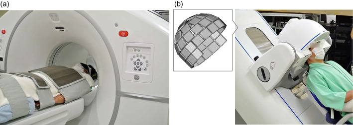
Ten healthy male volunteers (aged 22-45 years) underwent a representative clinical whole-body PET, followed by Vrain each for 10 min. These two scans were initiated 30 min and 45 min after FDG injection (4.1 ± 0.5 MBq/kg), respectively. First, we visually identified the small nuclei and then compared their standardised uptake values (SUVs) with the participants' age. Next, the SUVs of each brain region, which were determined by applying a volume-of-interest template for anatomically normalised PET images, were compared between the brain images with the Vrain and those with the whole-body PET images.
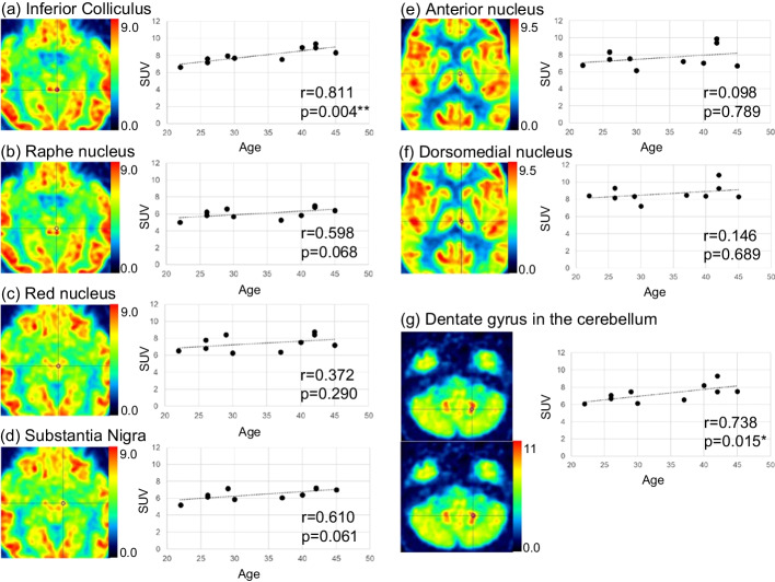
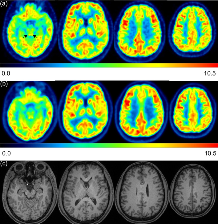
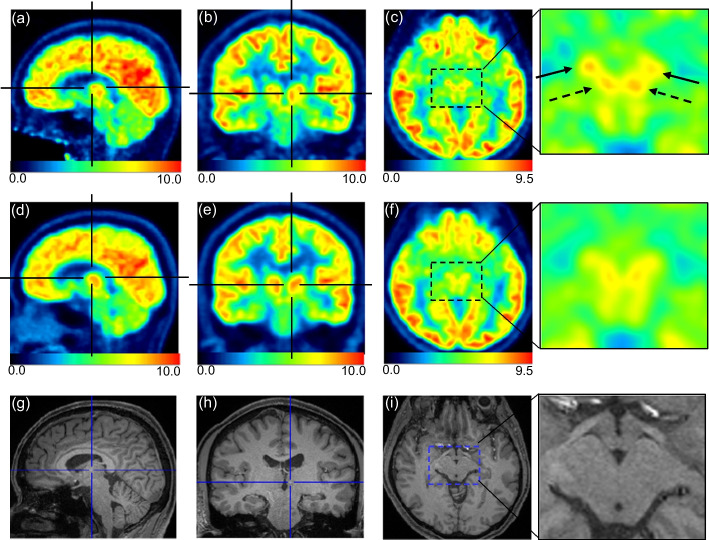
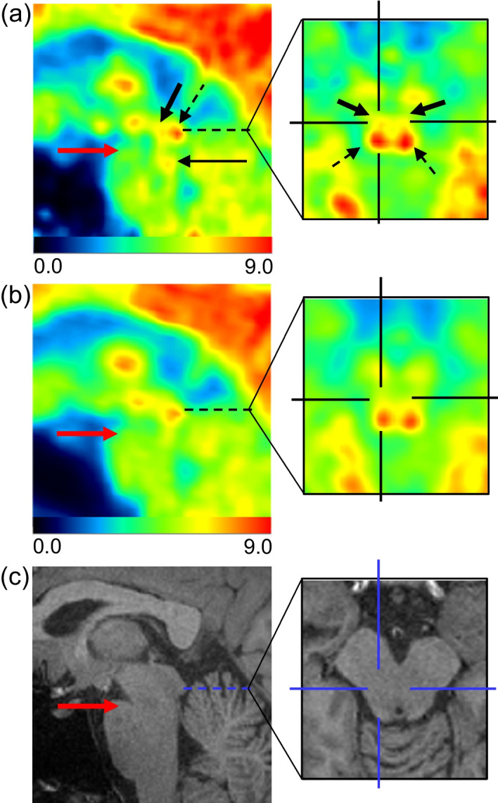
Small nuclei, such as the inferior colliculus, red nucleus, and substantia nigra, were more clearly visualised in Vrain than in whole-body PET. The anterior nucleus and dorsomedial nucleus in the thalamus and raphe nucleus in the brainstem were identified in Vrain but not in whole-body PET. The SUVs of the inferior colliculus and dentate gyrus in the cerebellum positively correlated with age (Spearman's correlation coefficient r = 0.811, p = 0.004; r = 0.738, p = 0.015, respectively). The SUVs of Vrain were slightly higher in the mesial temporal and medial parietal lobes than those in whole-body PET.
This was the first time that the raphe nuclei, anterior nuclei, and dorsomedial nuclei were successfully visualised using the first hemispherical brain PET. TRIAL REGISTRATION : Japan Registry of Clinical Trials, jRCTs032210086, Registered 13 May 2021, https://jrct.niph.go.jp/latest-detail/jRCTs032210086 .
为了确认我们开发的用于可视化深部脑区小核团的首款半球形脑正电子发射断层扫描(PET)(Vrain)的性能,我们将氟代脱氧葡萄糖(FDG)脑图像与全身PET图像进行了比较。
10名健康男性志愿者(年龄22 - 45岁)先进行了一次具有代表性的临床全身PET检查,随后进行Vrain检查,每次检查持续10分钟。这两次扫描分别在注射FDG(4.1±0.5MBq/kg)后30分钟和45分钟开始。首先,我们通过视觉识别小核团,然后将其标准化摄取值(SUVs)与参与者的年龄进行比较。接下来,通过对解剖学标准化PET图像应用感兴趣体积模板来确定每个脑区的SUVs,并在Vrain脑图像和全身PET脑图像之间进行比较。
诸如下丘、红核和黑质等小核团在Vrain中比在全身PET中显示得更清晰。在Vrain中识别出了丘脑的前核和背内侧核以及脑干中的中缝核,但在全身PET中未识别出。小脑下丘和齿状回的SUVs与年龄呈正相关(Spearman相关系数r分别为0.811,p = 0.004;r = 0.738,p = 0.015)。Vrain在颞叶内侧和顶叶内侧的SUVs略高于全身PET。
这是首次使用首款半球形脑PET成功可视化中缝核、前核和背内侧核。试验注册:日本临床试验注册中心,jRCTs032210086,于2021年5月13日注册,https://jrct.niph.go.jp/latest-detail/jRCTs032210086 。