• 文献检索
  • 文档翻译
  • 深度研究
  • 学术资讯
  • Suppr Zotero 插件Zotero 插件
  • 邀请有礼
  • 套餐&价格
  • 历史记录
应用&插件
Suppr Zotero 插件Zotero 插件浏览器插件Mac 客户端Windows 客户端微信小程序
定价
高级版会员购买积分包购买API积分包
服务
文献检索文档翻译深度研究API 文档MCP 服务
关于我们
关于 Suppr公司介绍联系我们用户协议隐私条款
关注我们

Suppr 超能文献

核心技术专利:CN118964589B侵权必究
粤ICP备2023148730 号-1Suppr @ 2026

文献检索

告别复杂PubMed语法,用中文像聊天一样搜索,搜遍4000万医学文献。AI智能推荐,让科研检索更轻松。

立即免费搜索

文件翻译

保留排版,准确专业,支持PDF/Word/PPT等文件格式,支持 12+语言互译。

免费翻译文档

深度研究

AI帮你快速写综述,25分钟生成高质量综述,智能提取关键信息,辅助科研写作。

立即免费体验

在 Transwell 细胞培养小室内分离、培养和冷冻切片原代猪视网膜色素上皮细胞。

Isolation, culture, and cryosectioning of primary porcine retinal pigment epithelium on transwell cell culture inserts.

机构信息

Cell Biology, Neurobiology, and Anatomy, Medical College of Wisconsin, Milwaukee, WI, USA; Ophthalmology and Visual Sciences, Medical College of Wisconsin, Milwaukee, WI, USA.

Department of Ophthalmology and Visual Sciences, Heersink School of Medicine, University of Alabama at Birmingham, Birmingham, AL, USA.

出版信息

STAR Protoc. 2022 Dec 16;3(4):101758. doi: 10.1016/j.xpro.2022.101758. Epub 2022 Oct 12.

DOI:10.1016/j.xpro.2022.101758
PMID:36227744
原文链接:https://pmc.ncbi.nlm.nih.gov/articles/PMC9576630/
Abstract

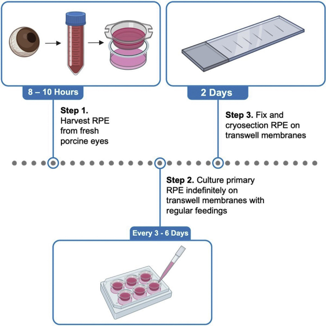
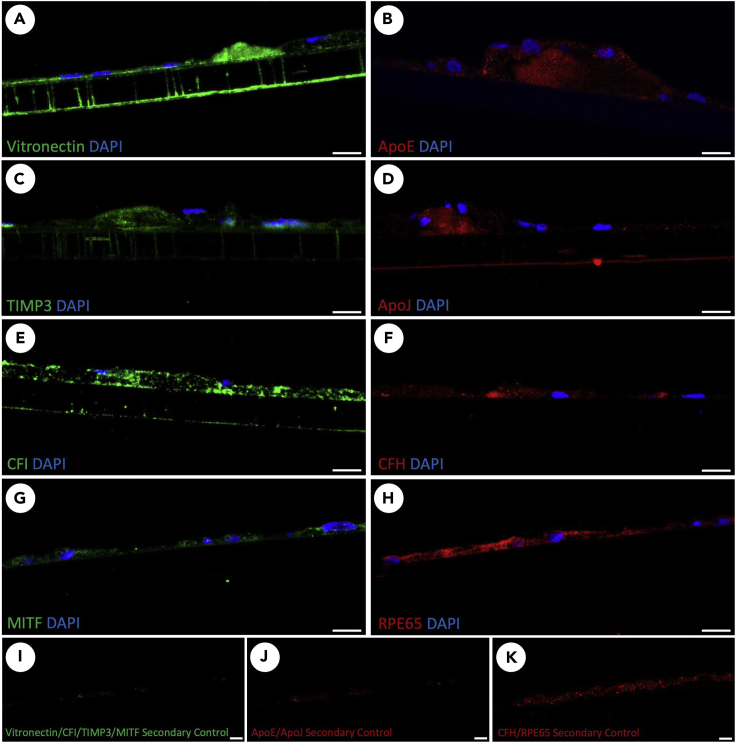
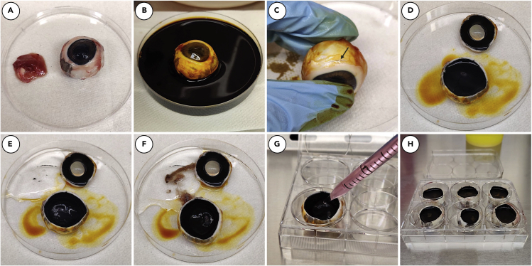
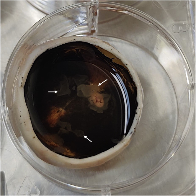
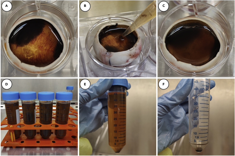
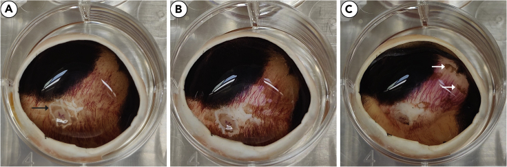
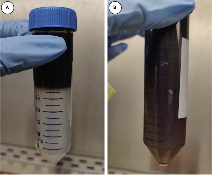
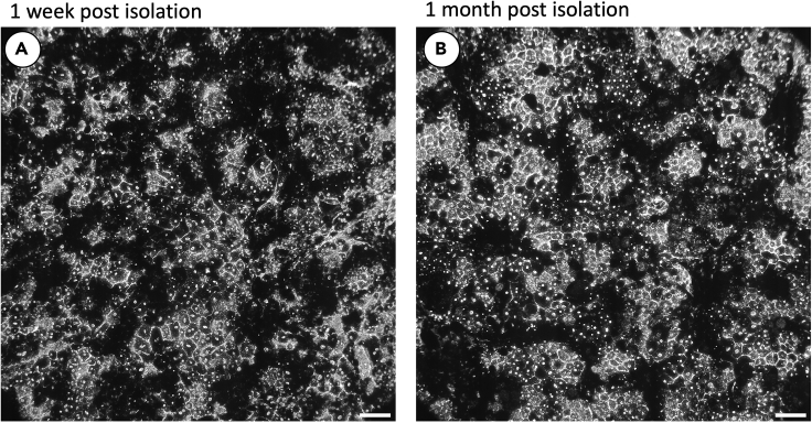
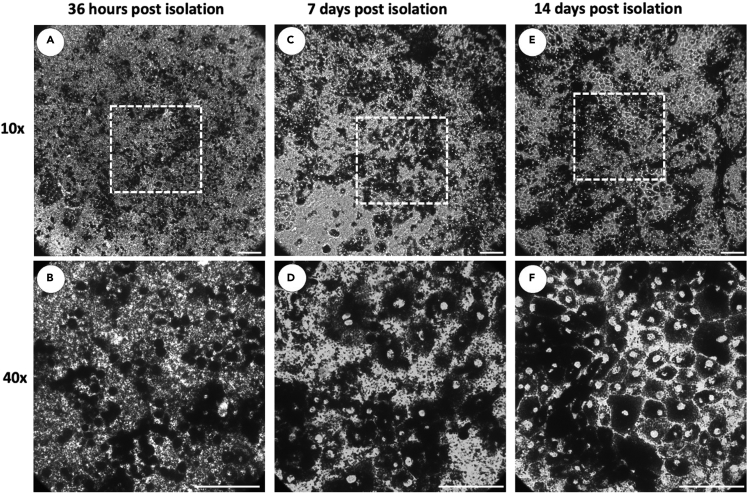
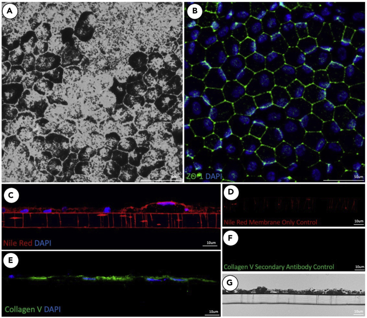
Primary culture and long-term maintenance of primary retinal pigment epithelium (RPE) is a useful model system for the study of ocular pathologies such as age-related macular degeneration. Here, we detail the steps for the isolation and long-term culture of primary porcine RPE. We also describe steps for cryoprotecting, cryosectioning, and interrogating with immunofluorescence and histochemistry RPE cells grown on transwell membranes. These techniques can be used in histological studies to detect sub-RPE deposits. For complete details on the use and execution of this protocol, please refer to Pilgrim et al., (2017).

摘要

原代培养和长期维持原代视网膜色素上皮(RPE)是研究眼部疾病(如年龄相关性黄斑变性)的有用模型系统。在这里,我们详细介绍了分离和长期培养原代猪 RPE 的步骤。我们还描述了在 Transwell 膜上生长的 RPE 细胞进行冷冻保护、冷冻切片和免疫荧光及组织化学检测的步骤。这些技术可用于组织学研究,以检测 RPE 下沉积物。有关此方案的使用和执行的完整详细信息,请参阅 Pilgrim 等人(2017 年)。

https://cdn.ncbi.nlm.nih.gov/pmc/blobs/b3cc/9576630/8ff102f0faa7/gr12.jpg
https://cdn.ncbi.nlm.nih.gov/pmc/blobs/b3cc/9576630/69fdfe8c0561/fx1.jpg
https://cdn.ncbi.nlm.nih.gov/pmc/blobs/b3cc/9576630/5775d21a11f3/gr1.jpg
https://cdn.ncbi.nlm.nih.gov/pmc/blobs/b3cc/9576630/85cc7f4d23be/gr2.jpg
https://cdn.ncbi.nlm.nih.gov/pmc/blobs/b3cc/9576630/5e52bc21406f/gr3.jpg
https://cdn.ncbi.nlm.nih.gov/pmc/blobs/b3cc/9576630/66601821c727/gr4.jpg
https://cdn.ncbi.nlm.nih.gov/pmc/blobs/b3cc/9576630/59cfcabffd68/gr5.jpg
https://cdn.ncbi.nlm.nih.gov/pmc/blobs/b3cc/9576630/6103bfb26c42/gr6.jpg
https://cdn.ncbi.nlm.nih.gov/pmc/blobs/b3cc/9576630/a2faf42d13bd/gr7.jpg
https://cdn.ncbi.nlm.nih.gov/pmc/blobs/b3cc/9576630/1602b931abed/gr8.jpg
https://cdn.ncbi.nlm.nih.gov/pmc/blobs/b3cc/9576630/de95cc5422c5/gr9.jpg
https://cdn.ncbi.nlm.nih.gov/pmc/blobs/b3cc/9576630/dc6a6c4c2c16/gr10.jpg
https://cdn.ncbi.nlm.nih.gov/pmc/blobs/b3cc/9576630/31a0992fe46e/gr11.jpg
https://cdn.ncbi.nlm.nih.gov/pmc/blobs/b3cc/9576630/8ff102f0faa7/gr12.jpg

相似文献

1
Isolation, culture, and cryosectioning of primary porcine retinal pigment epithelium on transwell cell culture inserts.在 Transwell 细胞培养小室内分离、培养和冷冻切片原代猪视网膜色素上皮细胞。
STAR Protoc. 2022 Dec 16;3(4):101758. doi: 10.1016/j.xpro.2022.101758. Epub 2022 Oct 12.
2
Modulation of Sub-RPE deposits in vitro: a potential model for age-related macular degeneration.体外视网膜色素上皮下沉积物的调节:年龄相关性黄斑变性的潜在模型
Invest Ophthalmol Vis Sci. 2004 May;45(5):1281-8. doi: 10.1167/iovs.03-0671.
3
A convenient protocol for establishing a human cell culture model of the outer retina.一种用于建立视网膜外层人类细胞培养模型的简便方案。
F1000Res. 2018 Jul 18;7:1107. doi: 10.12688/f1000research.15409.1. eCollection 2018.
4
Characterization of the effects of retinal pigment epithelium-conditioned media on porcine and aged human retina.描述视网膜色素上皮细胞条件培养基对猪眼和老年人眼视网膜的影响。
Graefes Arch Clin Exp Ophthalmol. 2013 Jun;251(6):1515-28. doi: 10.1007/s00417-013-2326-3. Epub 2013 Apr 11.
5
Triphasic developmentally guided protocol to generate retinal pigment epithelium from induced pluripotent stem cells.诱导多能干细胞生成视网膜色素上皮细胞的三相发育指导方案。
STAR Protoc. 2022 Jul 20;3(3):101582. doi: 10.1016/j.xpro.2022.101582. eCollection 2022 Sep 16.
6
An improved protocol for generation and characterization of human-induced pluripotent stem cell-derived retinal pigment epithelium cells.一种改良的人诱导多能干细胞源性视网膜色素上皮细胞的生成和鉴定方案。
STAR Protoc. 2022 Nov 4;3(4):101803. doi: 10.1016/j.xpro.2022.101803. eCollection 2022 Dec 16.
7
Interaction of inflammatorily activated retinal pigment epithelium with retinal microglia and neuronal cells.炎症激活的视网膜色素上皮细胞与视网膜小胶质细胞和神经元细胞的相互作用。
Exp Eye Res. 2020 Oct;199:108167. doi: 10.1016/j.exer.2020.108167. Epub 2020 Jul 28.
8
Unstimulated, Serum-free Cultures of Retinal Pigment Epithelium Excrete Large Mounds of Drusen-like Deposits.未受刺激的视网膜色素上皮无血清培养物会分泌大量类玻璃膜疣沉积物。
Curr Eye Res. 2020 Nov;45(11):1390-1394. doi: 10.1080/02713683.2020.1740744. Epub 2020 Mar 30.
9
Oxidative stress protection by exogenous delivery of rhHsp70 chaperone to the retinal pigment epithelium (RPE), a possible therapeutic strategy against RPE degeneration.通过向视网膜色素上皮(RPE)外源性递送重组人热休克蛋白70(rhHsp70)伴侣蛋白实现氧化应激保护,这是一种针对RPE变性的可能治疗策略。
Pharm Res. 2015 Jan;32(1):211-21. doi: 10.1007/s11095-014-1456-6. Epub 2014 Jul 17.
10
Optimization of an in vitro bilayer model for studying the functional interplay between human primary retinal pigment epithelial and choroidal endothelial cells isolated from donor eyes.优化用于研究从供体眼中分离出的人原代视网膜色素上皮细胞和脉络膜内皮细胞之间功能相互作用的体外双层模型。
BMC Res Notes. 2019 May 30;12(1):307. doi: 10.1186/s13104-019-4333-x.

引用本文的文献

1
Differentiation and histological analysis of primary human bronchial epithelial cells cultured on transwell membranes using air-liquid interface culture.使用气液界面培养法对在Transwell膜上培养的原代人支气管上皮细胞进行分化和组织学分析。
MethodsX. 2025 Jul 7;15:103492. doi: 10.1016/j.mex.2025.103492. eCollection 2025 Dec.
2
Porcine Sub-Retinal Pigment Epithelium Deposits: A Model for Dry Age-Related Macular Degeneration With Comparison to Human Drusen.猪视网膜色素上皮下沉积物:一种干性年龄相关性黄斑变性模型,并与人类玻璃膜疣进行比较。
Invest Ophthalmol Vis Sci. 2025 Mar 3;66(3):18. doi: 10.1167/iovs.66.3.18.
3
Evaluation of mesenchymal stem cells as an model for inherited retinal diseases.
间充质干细胞作为遗传性视网膜疾病模型的评估。
Front Cell Dev Biol. 2024 Nov 15;12:1455140. doi: 10.3389/fcell.2024.1455140. eCollection 2024.
4
Characterization of drusen formation in a primary porcine tissue culture model of dry AMD.干性年龄相关性黄斑变性的原代猪组织培养模型中玻璃膜疣形成的特征分析
Mol Ther Methods Clin Dev. 2024 Aug 31;32(4):101331. doi: 10.1016/j.omtm.2024.101331. eCollection 2024 Dec 12.
5
A cell culture system for RPE hypoxia, a physiologic stressor relevant to AMD deposit formation.一种用于视网膜色素上皮细胞缺氧的细胞培养系统,视网膜色素上皮细胞缺氧是一种与年龄相关性黄斑变性沉积物形成相关的生理应激源。
Eye (Lond). 2025 Mar;39(4):611-612. doi: 10.1038/s41433-024-03166-2. Epub 2024 Jun 13.
6
A Simplified Method for Isolation and Culture of Retinal Pigment Epithelial Cells from Adult Mice.一种从成年小鼠中分离和培养视网膜色素上皮细胞的简化方法。
J Vis Exp. 2024 May 24(207). doi: 10.3791/66921.
7
Age-Related Macular Degeneration, a Mathematically Tractable Disease.年龄相关性黄斑变性,一种可用数学方法处理的疾病。
Invest Ophthalmol Vis Sci. 2024 Mar 5;65(3):4. doi: 10.1167/iovs.65.3.4.
8
Co-cultivation of primary porcine RPE cells and neuroretina induces inflammation: a potential inflammatory AMD-model.原代猪 RPE 细胞和神经视网膜的共培养诱导炎症:一种潜在的炎症性 AMD 模型。
Sci Rep. 2023 Nov 7;13(1):19345. doi: 10.1038/s41598-023-46029-8.
9
INFLAMMATORY CELL ACTIVITY IN TREATED NEOVASCULAR AGE-RELATED MACULAR DEGENERATION: A Histologic Case Study.治疗新生血管年龄相关性黄斑变性中的炎症细胞活性:一项组织病理学案例研究。
Retina. 2023 Nov 1;43(11):1904-1913. doi: 10.1097/IAE.0000000000003881.