• 文献检索
  • 文档翻译
  • 深度研究
  • 学术资讯
  • Suppr Zotero 插件Zotero 插件
  • 邀请有礼
  • 套餐&价格
  • 历史记录
应用&插件
Suppr Zotero 插件Zotero 插件浏览器插件Mac 客户端Windows 客户端微信小程序
定价
高级版会员购买积分包购买API积分包
服务
文献检索文档翻译深度研究API 文档MCP 服务
关于我们
关于 Suppr公司介绍联系我们用户协议隐私条款
关注我们

Suppr 超能文献

核心技术专利:CN118964589B侵权必究
粤ICP备2023148730 号-1Suppr @ 2026

文献检索

告别复杂PubMed语法,用中文像聊天一样搜索,搜遍4000万医学文献。AI智能推荐,让科研检索更轻松。

立即免费搜索

文件翻译

保留排版,准确专业,支持PDF/Word/PPT等文件格式,支持 12+语言互译。

免费翻译文档

深度研究

AI帮你快速写综述,25分钟生成高质量综述,智能提取关键信息,辅助科研写作。

立即免费体验

治疗新生血管年龄相关性黄斑变性中的炎症细胞活性:一项组织病理学案例研究。

INFLAMMATORY CELL ACTIVITY IN TREATED NEOVASCULAR AGE-RELATED MACULAR DEGENERATION: A Histologic Case Study.

机构信息

Department of Ophthalmology and Visual Sciences, Heersink School of Medicine, University of Alabama at Birmingham, Birmingham, Alabama.

Department of Ophthalmology, University Hospital Würzburg, Würzburg, Germany.

出版信息

Retina. 2023 Nov 1;43(11):1904-1913. doi: 10.1097/IAE.0000000000003881.

DOI:10.1097/IAE.0000000000003881
PMID:37871271
原文链接:https://pmc.ncbi.nlm.nih.gov/articles/PMC10801910/
Abstract

BACKGROUND

Imaging indicators of macular neovascularization risk can help determine patient eligibility for new treatments for geographic atrophy secondary to age-related macular degeneration. Because type 1 macular neovascularization includes inflammation, we assessed by histology the distribution of cells with inflammatory potential in two fellow eyes with age-related macular degeneration.

METHODS

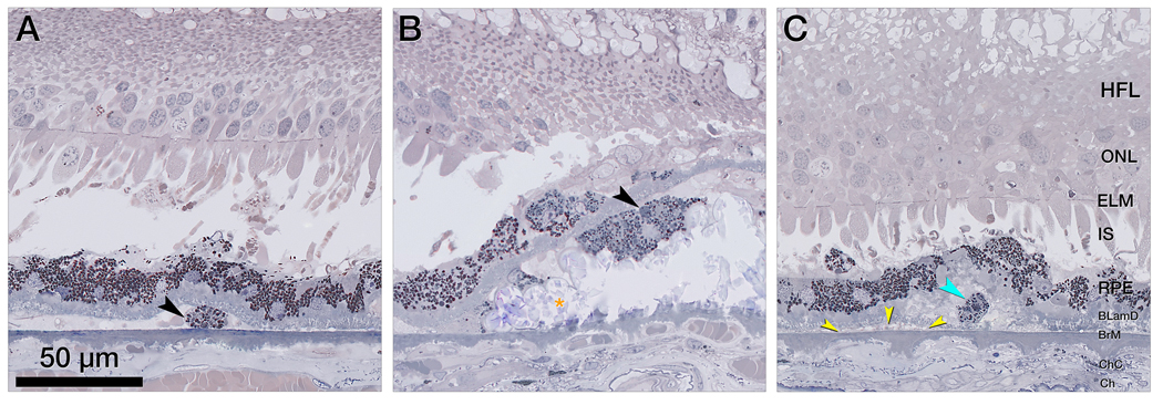
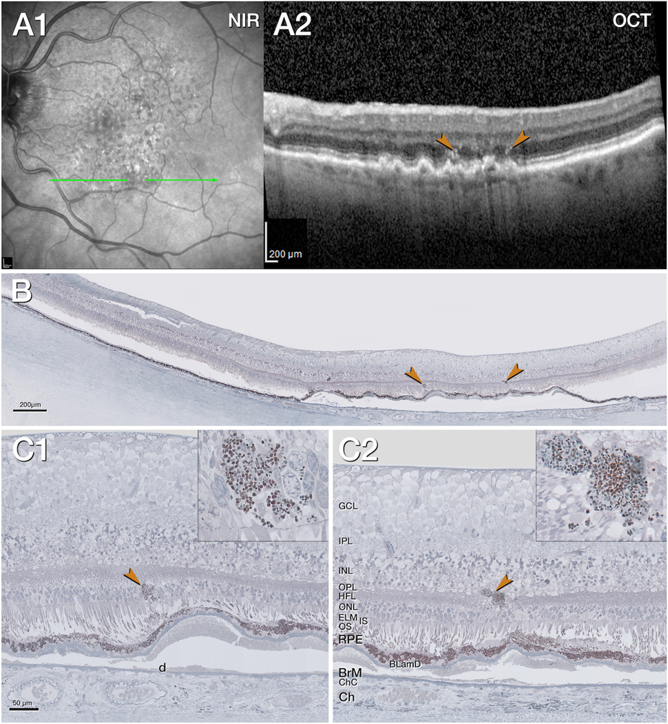
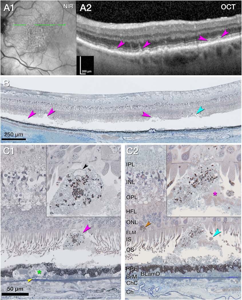
Two eyes of a White woman in her 90's with type 3 macular neovascularization treated with antivascular endothelial growth factor were prepared for high-resolution histology. Eye-tracked spectral domain optical coherence tomography applied to the preserved donor eyes linked in vivo imaging to histology. Cells were enumerated in the intraretinal, subretinal, and subretinal retinal pigment epithelium (RPE)-basal lamina compartments on 199 glass slides. Cells with numerous organelles were considered to RPE-derived; cells with sparse RPE organelles were considered non-RPE phagocytes.

RESULTS

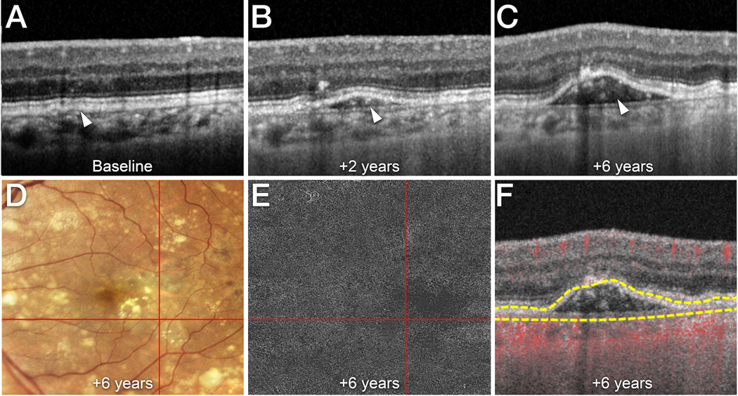
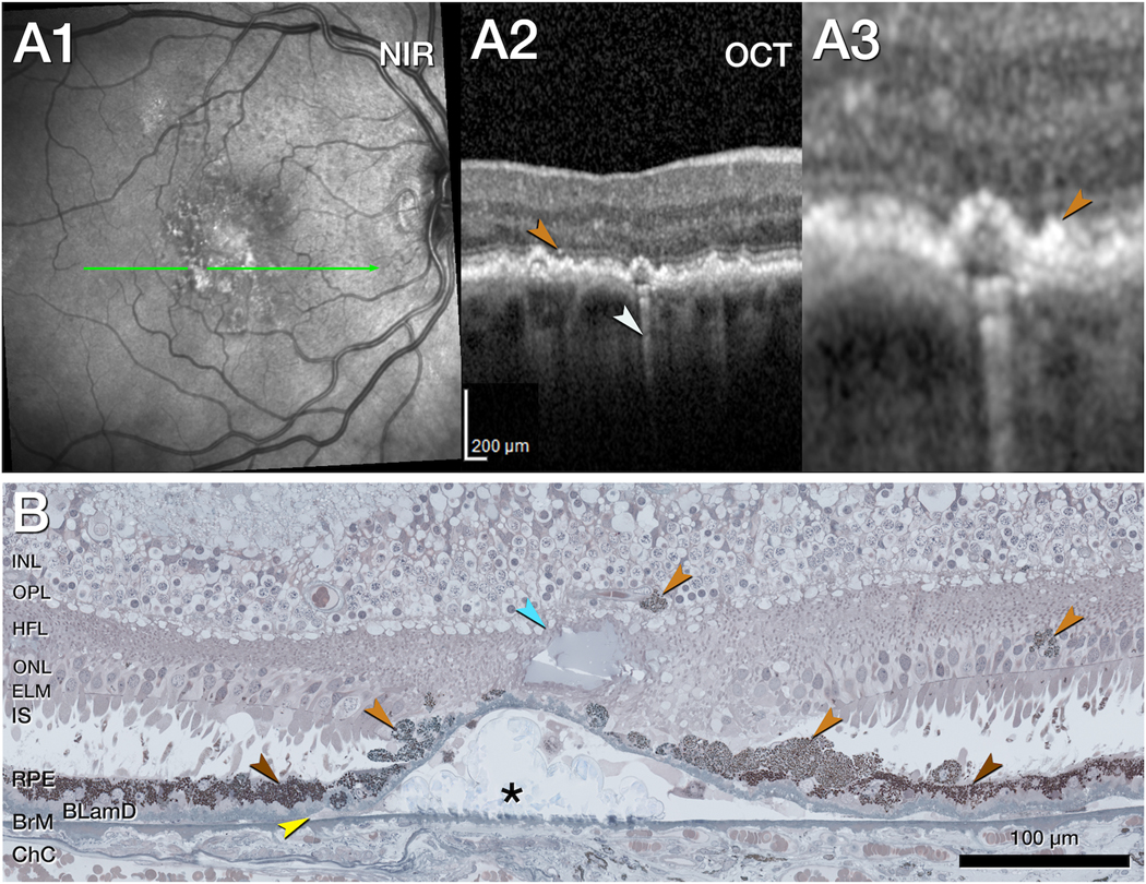
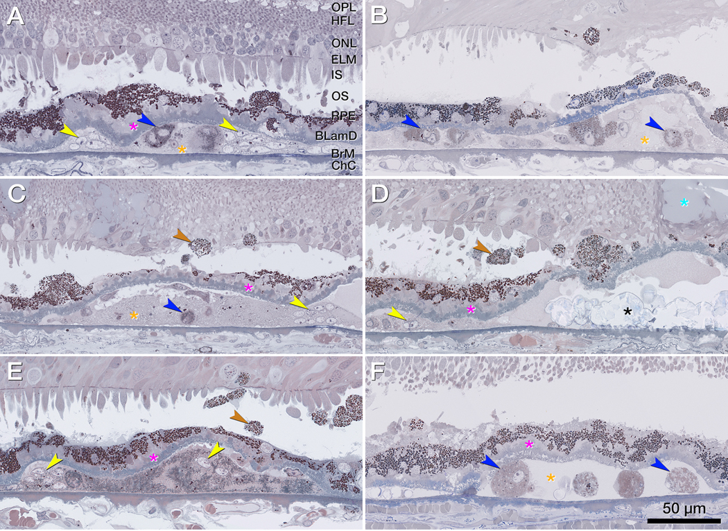
Both eyes had soft drusen and abundant subretinal drusenoid deposit. In the retina and subretinal space, RPE-derived cells, including hyperreflective foci, were common (n = 125 and 73, respectively). Non-RPE phagocytes were infrequent (n = 5 in both). Over drusen, RPE morphology transitioned smoothly from the age-normal layer toward the top, suggesting transdifferentiation. The sub-RPE-basal lamina space had RPE-derived cells (n = 87) and non-RPE phagocytes (n = 49), including macrophages and giant cells.

CONCLUSION

Numerous sub-RPE-basal lamina cells of several types are consistent with the documented presence of proinflammatory lipids in drusen and aged Bruch's membrane. The relatively compartmentalized abundance of infiltrating cells suggests that drusen contents are more inflammatory than subretinal drusenoid deposit, perhaps reflecting their environments. Ectopic RPE occurs frequently. Some manifest as hyperreflective foci. More cells may be visible as optical coherence tomography technologies evolve.

摘要

背景

有助于确定黄斑新生血管风险的影像学指标,可帮助确定是否适合接受新的治疗方法来治疗年龄相关性黄斑变性引起的地图状萎缩。由于 1 型黄斑新生血管包括炎症,我们通过组织学评估了 2 例年龄相关性黄斑变性伴 3 型黄斑新生血管患者的两只同眼具有炎症潜能的细胞分布。

方法

对 1 例 90 多岁白人女性的 2 只眼进行了 3 型黄斑新生血管的抗血管内皮生长因子治疗,并用高分辨率组织学准备。将保存在供体眼中的眼跟踪谱域光学相干断层扫描应用于活体成像与组织学联系起来。在 199 张载玻片上对视网膜内、视网膜下和视网膜色素上皮(RPE)-基底膜腔室中的细胞进行计数。具有大量细胞器的细胞被认为是 RPE 衍生的;细胞器稀疏的细胞被认为是非 RPE 吞噬细胞。

结果

两只眼均有软玻璃膜疣和大量的视网膜下玻璃膜疣沉积物。在视网膜和视网膜下空间中,RPE 衍生细胞(包括高反射焦点)很常见(分别为 125 个和 73 个)。非 RPE 吞噬细胞很少(各 5 个)。在玻璃膜疣上,RPE 形态从正常年龄层平滑过渡到顶部,提示转分化。RPE 下基底膜腔室中有 RPE 衍生细胞(n = 87)和非 RPE 吞噬细胞(n = 49),包括巨噬细胞和巨细胞。

结论

大量的 RPE 下基底膜腔室中的多种类型细胞与文献报道的玻璃膜疣和老化 Bruch 膜中存在的促炎脂质一致。浸润细胞相对分隔的丰富程度表明,玻璃膜疣内容物比视网膜下玻璃膜疣沉积物更具炎症性,这可能反映了它们的环境。异位 RPE 很常见。有些表现为高反射焦点。随着光学相干断层扫描技术的发展,可能会有更多的细胞可见。

https://cdn.ncbi.nlm.nih.gov/pmc/blobs/5cd5/10801910/4f82cd58b249/nihms-1913232-f0006.jpg
https://cdn.ncbi.nlm.nih.gov/pmc/blobs/5cd5/10801910/72ed5c500471/nihms-1913232-f0001.jpg
https://cdn.ncbi.nlm.nih.gov/pmc/blobs/5cd5/10801910/50c64f53e6a3/nihms-1913232-f0002.jpg
https://cdn.ncbi.nlm.nih.gov/pmc/blobs/5cd5/10801910/7368200df574/nihms-1913232-f0003.jpg
https://cdn.ncbi.nlm.nih.gov/pmc/blobs/5cd5/10801910/0b4ba2e7c222/nihms-1913232-f0004.jpg
https://cdn.ncbi.nlm.nih.gov/pmc/blobs/5cd5/10801910/c63ce4a7bc0d/nihms-1913232-f0005.jpg
https://cdn.ncbi.nlm.nih.gov/pmc/blobs/5cd5/10801910/4f82cd58b249/nihms-1913232-f0006.jpg

相似文献

1
INFLAMMATORY CELL ACTIVITY IN TREATED NEOVASCULAR AGE-RELATED MACULAR DEGENERATION: A Histologic Case Study.治疗新生血管年龄相关性黄斑变性中的炎症细胞活性:一项组织病理学案例研究。
Retina. 2023 Nov 1;43(11):1904-1913. doi: 10.1097/IAE.0000000000003881.
2
Histologic and Optical Coherence Tomographic Correlates in Drusenoid Pigment Epithelium Detachment in Age-Related Macular Degeneration.年龄相关性黄斑变性中玻璃膜疣样色素上皮脱离的组织学与光学相干断层扫描相关性
Ophthalmology. 2017 May;124(5):644-656. doi: 10.1016/j.ophtha.2016.12.034. Epub 2017 Jan 30.
3
SUBRETINAL DRUSENOID DEPOSIT IN AGE-RELATED MACULAR DEGENERATION: Histologic Insights Into Initiation, Progression to Atrophy, and Imaging.年龄相关性黄斑变性的视网膜下硬性脂褐质沉积:发病机制、向萎缩进展及影像学研究的组织学观察。
Retina. 2020 Apr;40(4):618-631. doi: 10.1097/IAE.0000000000002657.
4
Chorioretinal thickness and retinal pigment epithelial degeneration of fellow eyes in patients with unilateral neovascular age-related macular degeneration with subretinal drusenoid deposits.单侧伴脉络膜视网膜结节样沉积物的年龄相关性黄斑变性患者对侧眼的脉络膜视网膜厚度和视网膜色素上皮变性。
BMC Ophthalmol. 2022 Jul 14;22(1):304. doi: 10.1186/s12886-022-02518-4.
5
Activated Retinal Pigment Epithelium, an Optical Coherence Tomography Biomarker for Progression in Age-Related Macular Degeneration.活化视网膜色素上皮,一种用于年龄相关性黄斑变性进展的光学相干断层扫描生物标志物。
Invest Ophthalmol Vis Sci. 2017 May 1;58(6):BIO211-BIO226. doi: 10.1167/iovs.17-21872.
6
Clinicopathologic Correlation of Anti-Vascular Endothelial Growth Factor-Treated Type 3 Neovascularization in Age-Related Macular Degeneration.抗血管内皮生长因子治疗与年龄相关性黄斑变性 3 型新生血管的临床病理相关性。
Ophthalmology. 2018 Feb;125(2):276-287. doi: 10.1016/j.ophtha.2017.08.019. Epub 2017 Sep 28.
7
Optical Coherence Tomography Predictors of Risk for Progression to Non-Neovascular Atrophic Age-Related Macular Degeneration.光学相干断层扫描预测进展为非新生血管性萎缩性年龄相关性黄斑变性风险的因素
Ophthalmology. 2017 Dec;124(12):1764-1777. doi: 10.1016/j.ophtha.2017.06.032. Epub 2017 Aug 26.
8
Imaging of the retinal pigment epithelium in age-related macular degeneration using polarization-sensitive optical coherence tomography.利用偏振敏感光学相干断层扫描技术对年龄相关性黄斑变性的视网膜色素上皮进行成像。
Invest Ophthalmol Vis Sci. 2010 Apr;51(4):2149-57. doi: 10.1167/iovs.09-3817. Epub 2009 Sep 24.
9
Stages of Drusen-Associated Atrophy in Age-Related Macular Degeneration Visible via Histologically Validated Fundus Autofluorescence.通过经组织学验证的眼底自发荧光观察与年龄相关性黄斑变性相关的玻璃膜疣相关萎缩的阶段。
Ophthalmol Retina. 2021 Aug;5(8):730-742. doi: 10.1016/j.oret.2020.11.006. Epub 2020 Nov 18.
10
CLINICOPATHOLOGIC CORRELATION OF GEOGRAPHIC ATROPHY SECONDARY TO AGE-RELATED MACULAR DEGENERATION.年龄相关性黄斑变性所致地图状萎缩的临床病理相关性。
Retina. 2019 Apr;39(4):802-816. doi: 10.1097/IAE.0000000000002461.

引用本文的文献

1
Photoreceptor Function and Structure in Retinal Areas With Intraretinal Hyperreflective Foci in Age-Related Macular Degeneration.年龄相关性黄斑变性中视网膜内高反射灶区域的光感受器功能与结构
Invest Ophthalmol Vis Sci. 2025 Feb 3;66(2):27. doi: 10.1167/iovs.66.2.27.
2
Spatial Distribution of Hyperreflective Choroidal Foci in the Macula of Normal Eyes.正常眼黄斑区高反射性脉络膜病灶的空间分布。
Transl Vis Sci Technol. 2024 Aug 1;13(8):35. doi: 10.1167/tvst.13.8.35.

本文引用的文献

1
Histology of Type 3 Macular Neovascularization and Microvascular Anomalies in Treated Age-Related Macular Degeneration: A Case Study.治疗后的年龄相关性黄斑变性中3型黄斑新生血管形成和微血管异常的组织学:一项病例研究
Ophthalmol Sci. 2023 Feb 10;3(3):100280. doi: 10.1016/j.xops.2023.100280. eCollection 2023 Sep.
2
Melanophages give rise to hyperreflective foci in AMD, a disease-progression marker.黑色素细胞在 AMD 中产生高反射性病灶,这是疾病进展的标志。
J Neuroinflammation. 2023 Feb 8;20(1):28. doi: 10.1186/s12974-023-02699-9.
3
Isolation, culture, and cryosectioning of primary porcine retinal pigment epithelium on transwell cell culture inserts.
在 Transwell 细胞培养小室内分离、培养和冷冻切片原代猪视网膜色素上皮细胞。
STAR Protoc. 2022 Dec 16;3(4):101758. doi: 10.1016/j.xpro.2022.101758. Epub 2022 Oct 12.
4
Correlation of Optical Coherence Tomography Angiography of Type 3 Macular Neovascularization With Corresponding Histology.3 型黄斑新生血管的光学相干断层扫描血管造影与相应组织病理学的相关性。
JAMA Ophthalmol. 2022 Jun 1;140(6):628-633. doi: 10.1001/jamaophthalmol.2022.0890.
5
Histology and clinical imaging lifecycle of black pigment in fibrosis secondary to neovascular age-related macular degeneration.纤维化继发于新生血管年龄相关性黄斑变性的黑色色素的组织学和临床成像生命周期。
Exp Eye Res. 2022 Jan;214:108882. doi: 10.1016/j.exer.2021.108882. Epub 2021 Dec 8.
6
Role of inflammasome activation in neovascular age-related macular degeneration.炎症小体激活在新生血管性年龄相关性黄斑变性中的作用。
FEBS J. 2023 Jan;290(1):28-36. doi: 10.1111/febs.16278. Epub 2021 Dec 4.
7
Hyperreflective Foci, Optical Coherence Tomography Progression Indicators in Age-Related Macular Degeneration, Include Transdifferentiated Retinal Pigment Epithelium.老年黄斑变性的光学相干断层扫描高反射病灶的进展指标包括转分化的视网膜色素上皮。
Invest Ophthalmol Vis Sci. 2021 Aug 2;62(10):34. doi: 10.1167/iovs.62.10.34.
8
The RAP study, report 4: morphological and topographical characteristics of multifocal macular neovascularization type 3.RAP 研究,报告 4:多灶性黄斑新生血管病变 3 型的形态学和解剖学特征。
Graefes Arch Clin Exp Ophthalmol. 2022 Jan;260(1):141-147. doi: 10.1007/s00417-021-05332-8. Epub 2021 Aug 26.
9
Mitochondrial Reactive Oxygen Species and Their Contribution in Chronic Kidney Disease Progression Through Oxidative Stress.线粒体活性氧及其通过氧化应激在慢性肾脏病进展中的作用
Front Physiol. 2021 Apr 23;12:627837. doi: 10.3389/fphys.2021.627837. eCollection 2021.
10
Low frequency mitochondrial DNA heteroplasmy SNPs in blood, retina, and [RPE+choroid] of age-related macular degeneration subjects.年龄相关性黄斑变性患者血液、视网膜和 [RPE+脉络膜] 中的低频线粒体 DNA 异质性 SNPs。
PLoS One. 2021 Jan 29;16(1):e0246114. doi: 10.1371/journal.pone.0246114. eCollection 2021.