Ohe Rintaro, Kaneko Yutaka, Namba Hiroyuki, Nishi Katsuhiro, Goto Jun-Ichi, Futakuchi Mitsuru, Nishitsuka Koichi
Department of Pathology, Yamagata University Faculty of Medicine, Yamagata, Japan.
Department of Ophthalmology and Visual Sciences, Yamagata University Faculty of Medicine, Yamagata, Japan.
Clin Ophthalmol. 2022 Oct 10;16:3289-3296. doi: 10.2147/OPTH.S376141. eCollection 2022.
The eyes are one of the most frequently involved organs in sarcoidosis in Asia, including Japan. Sarcoid uveitis is the major complaint of ocular sarcoidosis. The detection of epithelioid granuloma (EG) requires histological biopsy of the uvea for the precise diagnosis of sarcoid uveitis, because it is challenging to diagnose sarcoid uveitis without a history of systemic sarcoidosis. To diagnose sarcoid uveitis, we have established novel methods.
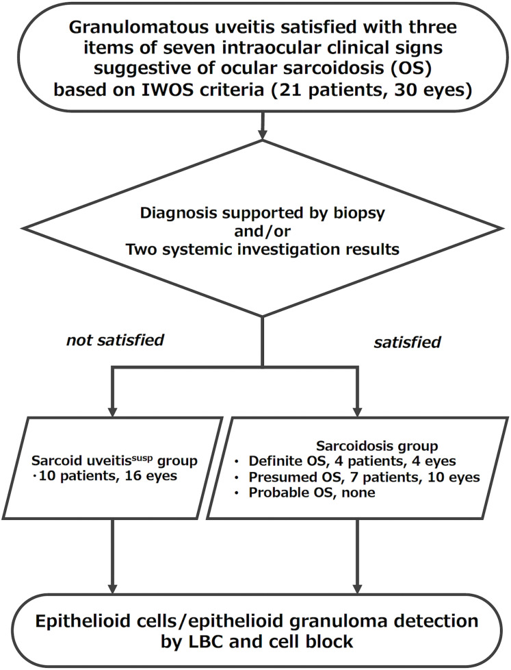
In this study, we included 30 eyes of 21 patients with granulomatous uveitis diagnosed via slit-lamp examinations, gonioscopy, fundus photography, and fluorescein angiography. Vitrectomy was performed to remove the vitreous opacity with vision loss. To examine vitreous cell components, we used liquid-based cytology (LBC). To detect EG in an intraocular irrigating solution, we collected vitreous cell components, and then the cell pellets were embedded in the cell block procedure.
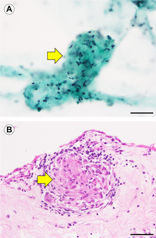
Here, we demonstrated the usefulness of the histological detection of EG and epithelioid cells (ECs) in LBC from vitreous body specimens and in the cell block procedure from vitreous cell components in an intraocular irrigating solution. Our results showed that the detection rates of EG were 6.3% (1/16) in LBC and 9.1% (1/11) in the cell block procedure in the sarcoid uveitis-suspected group and 7.7% (1/13) in LBC and 28.6% (2/7) in the cell block procedure in the sarcoidosis group. We would discuss the specificity of the EG/EC detection of ocular sarcoidosis.
Our methods are helpful in the precise diagnosis of ocular sarcoidosis and the control of the development of systemic sarcoidosis.
在亚洲,包括日本,眼睛是结节病中最常受累的器官之一。结节性葡萄膜炎是眼部结节病的主要症状。上皮样肉芽肿(EG)的检测需要对葡萄膜进行组织活检以精确诊断结节性葡萄膜炎,因为在没有全身性结节病病史的情况下诊断结节性葡萄膜炎具有挑战性。为了诊断结节性葡萄膜炎,我们建立了新的方法。
在本研究中,我们纳入了21例经裂隙灯检查、前房角镜检查、眼底照相和荧光素血管造影诊断为肉芽肿性葡萄膜炎的患者的30只眼睛。进行玻璃体切除术以清除导致视力丧失的玻璃体混浊。为了检查玻璃体细胞成分,我们使用了液基细胞学(LBC)。为了检测眼内灌洗溶液中的EG,我们收集了玻璃体细胞成分,然后将细胞沉淀包埋在细胞块制作过程中。
在此,我们证明了在玻璃体标本的LBC中以及在眼内灌洗溶液中玻璃体细胞成分的细胞块制作过程中,组织学检测EG和上皮样细胞(ECs)的有用性。我们的结果表明,在疑似结节性葡萄膜炎组中,LBC中EG的检出率为6.3%(1/16),细胞块制作过程中为9.1%(1/11);在结节病组中,LBC中为7.7%(1/13),细胞块制作过程中为28.6%(2/7)。我们将讨论眼部结节病EG/EC检测的特异性。
我们的方法有助于精确诊断眼部结节病并控制全身性结节病的发展。