Department of Anesthesiology, The Second Affiliated Hospital of Chongqing Medical University, Chongqing 400010, China.
Department of Rehabilitation, the Fifth People's Hospital of Chongqing, Chinese Academy of Sciences, Chongqing 400062, China.
Oxid Med Cell Longev. 2022 Sep 30;2022:7958542. doi: 10.1155/2022/7958542. eCollection 2022.
This study is aimed at identifying the potential diagnostic markers for circulating endothelial cells (CECs) for acute myocardial ischemia (AMI) and exploring the regulatory mechanisms of the selected biomarker in mitochondrial oxidative damage and vascular inflammation in AMI pathology.
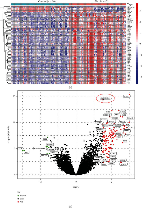
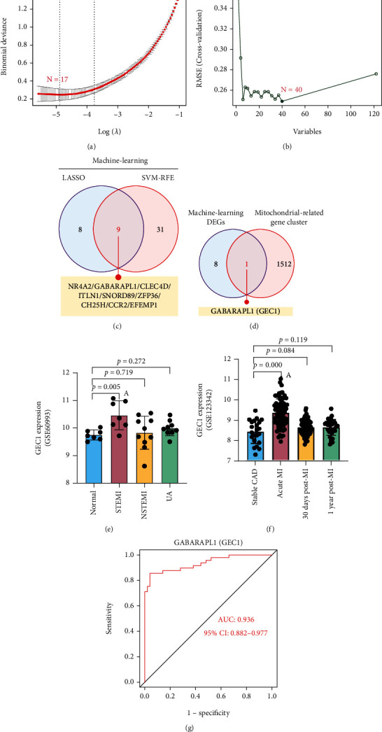
Utilizing the Gene Expression Omnibus dataset GSE66360, we scanned for differentially expressed genes (DEGs) in 49 AMI patients and 50 healthy subjects. To discover possible biomarkers, LASSO regression and support vector machine recursive feature elimination examinations were conducted. Using the GSE60993 and GSE123342 datasets and AMI rat models, the expression levels and diagnostic accuracy of the biomarkers in AMI were thoroughly verified. CIBERSORT was employed to evaluate the compositional patterns of 22 distinct immunological cell percentages in AMI according to combined cohorts. The oxidative-damaged mitochondria were detected by confocal microscopy observation of MitoTracker, ROS-DCFH-DA, and mCherry-GFP-LC3B.
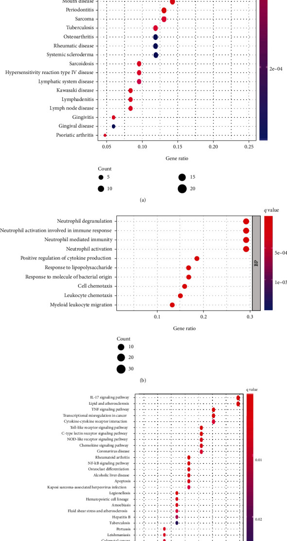
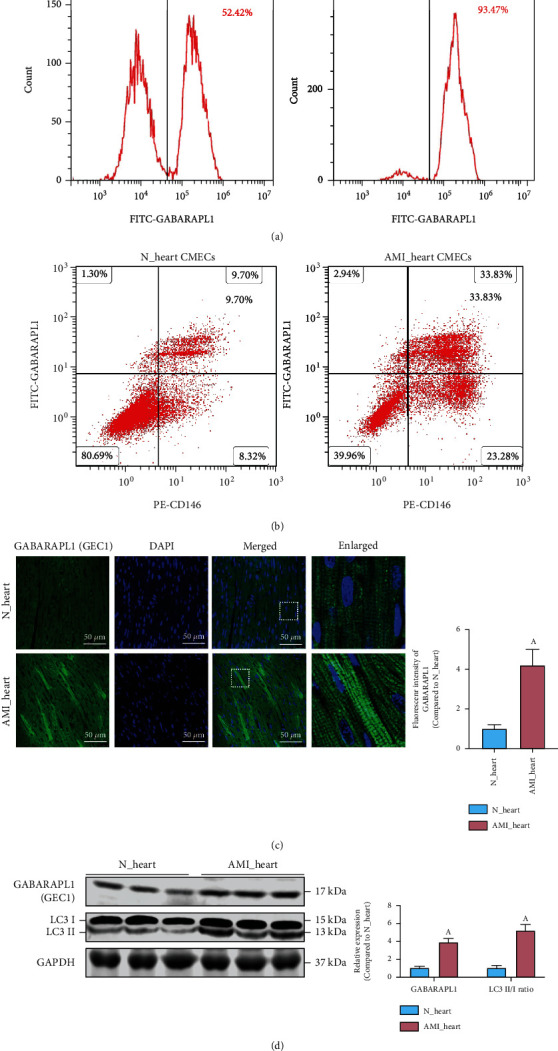
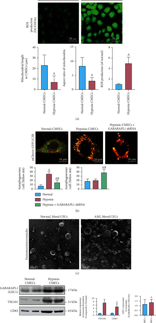
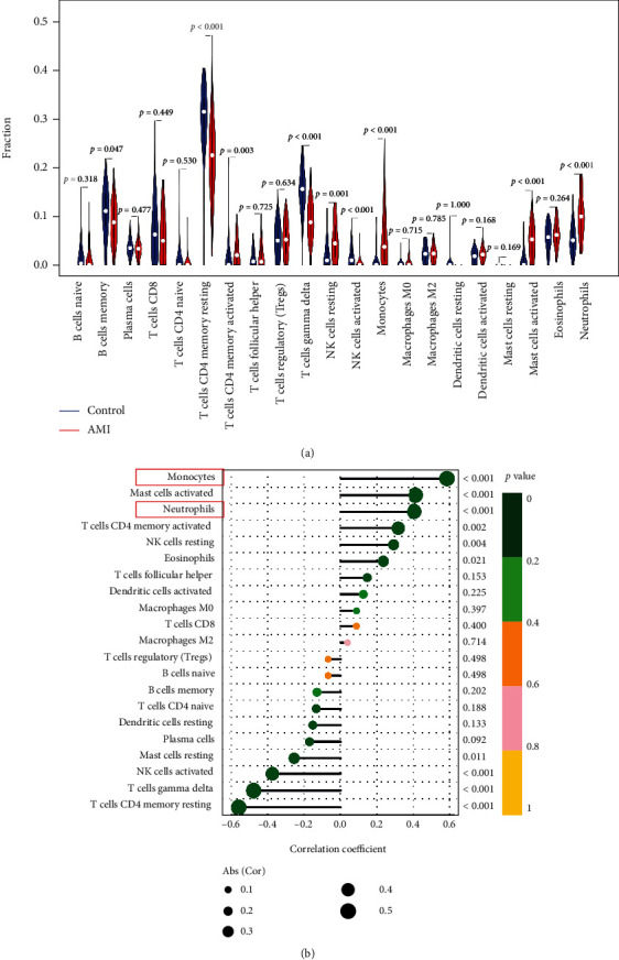
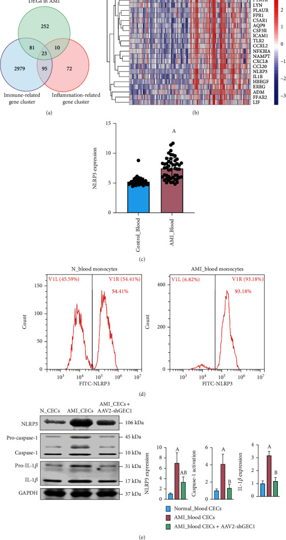
In total, 122 genes were identified. The identified DEGs primarily contributed in arteriosclerosis, arteriosclerotic cardiovascular disorders, bacterial infectious disorder, coronary artery disease, and myocardial infarction. Nine features (NR4A2, GABARAPL1 (GEC1), CLEC4D, ITLN1, SNORD89, ZFP36, CH25H, CCR2, and EFEMP1) of the DEGs were shared by two algorithms, and GABARAPL1 (GEC1) was identified and verified as a diagnostic mitochondrial biomarker for AMI. Confocal results showed that there existed mitochondrial damage and oxidative stress in cardiac CMECs after AMI, and the blocked autophagy flux could be released by exosome burst in cardiac CMECs and blood CECs. Immune cell infiltration testing declared that elevated GEC1 expression in blood CECs was linked to the rise of monocytes and neutrophils. Functional tests revealed that high GEC1 expression in CMECs and CECs could activate the vascular inflammatory response by stimulating NLRP3 inflammasome production after AMI.
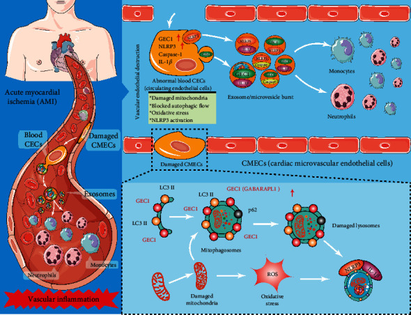
Oxidative-damaged mitochondria in cardiac CMECs activate GEC1-mediated autophagosomes but block autophagy flux after AMI. The exfoliated cardiac CMECs evolve into abnormal blood CECs, and the undegraded GEC1 autophagosomes produce a large number of NLRP3 inflammasomes by exosome burst, stimulating the increase in monocytes and neutrophils and ultimately triggering vascular inflammation after AMI. Therefore, GEC1 in blood CECs is a highly specific diagnostic mitochondrial biomarker for AMI.
本研究旨在鉴定循环内皮细胞(CEC)在急性心肌缺血(AMI)中的潜在诊断标志物,并探讨所选生物标志物在 AMI 病理中线粒体氧化损伤和血管炎症中的调控机制。
利用基因表达综合数据库 GSE66360,我们对 49 例 AMI 患者和 50 例健康对照者的差异表达基因(DEG)进行扫描。为了发现可能的生物标志物,我们进行了 LASSO 回归和支持向量机递归特征消除检查。利用 GSE60993 和 GSE123342 数据集和 AMI 大鼠模型,全面验证了生物标志物在 AMI 中的表达水平和诊断准确性。根据联合队列,采用 CIBERSORT 评估 22 种不同免疫细胞比例的组成模式。通过共聚焦显微镜观察 MitoTracker、ROS-DCFH-DA 和 mCherry-GFP-LC3B 检测氧化损伤的线粒体。
共鉴定出 122 个基因。这些差异表达基因主要参与动脉粥样硬化、动脉粥样硬化性心血管疾病、细菌性传染病、冠状动脉疾病和心肌梗死。两种算法共共享了 9 个特征(NR4A2、GABARAPL1(GEC1)、CLEC4D、ITLN1、SNORD89、ZFP36、CH25H、CCR2 和 EFEMP1),并鉴定和验证 GABARAPL1(GEC1)是 AMI 的诊断性线粒体生物标志物。共聚焦结果显示,AMI 后心肌 CMEC 中存在线粒体损伤和氧化应激,心肌 CMEC 和血 CEC 中的外泌体爆发可以释放阻断的自噬流。免疫细胞浸润试验表明,血 CEC 中 GEC1 表达升高与单核细胞和中性粒细胞的升高有关。功能试验表明,AMI 后,CMEC 和 CEC 中高表达的 GEC1 通过刺激 NLRP3 炎性小体的产生,激活血管炎症反应。
AMI 后,心肌 CMEC 中的氧化损伤线粒体激活 GEC1 介导的自噬体,但阻断自噬流。脱落的心肌 CMEC 演变成异常的血 CEC,未降解的 GEC1 自噬体通过外泌体爆发产生大量 NLRP3 炎性小体,刺激单核细胞和中性粒细胞增加,最终触发 AMI 后的血管炎症。因此,血 CEC 中的 GEC1 是 AMI 高度特异性的诊断性线粒体生物标志物。