Department of 2nd Oncology, Guangdong Second Provincial General Hospital, Guangzhou, 510317 Guangdong, China.
Institute of Pathology, University Medical Center, Göttingen, Germany.
Oxid Med Cell Longev. 2022 Oct 6;2022:4320809. doi: 10.1155/2022/4320809. eCollection 2022.
Cancer-associated fibroblasts (CAFs) within the tumor microenvironment are key players in tumorigenesis and tumor development. Nevertheless, the regulatory mechanisms of CAFs on lung squamous cell carcinoma- (LUSC-) associated remain poorly elucidated.
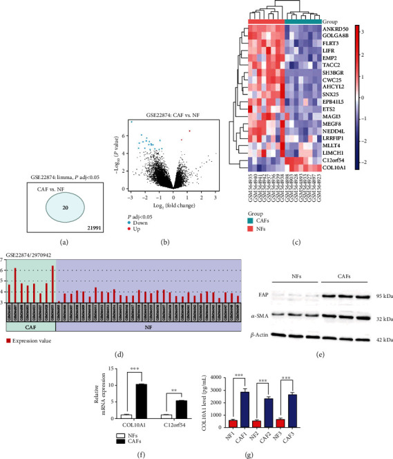
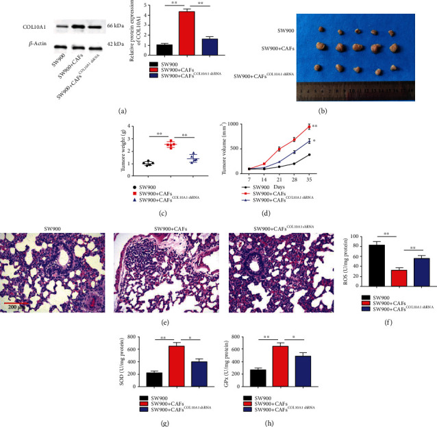
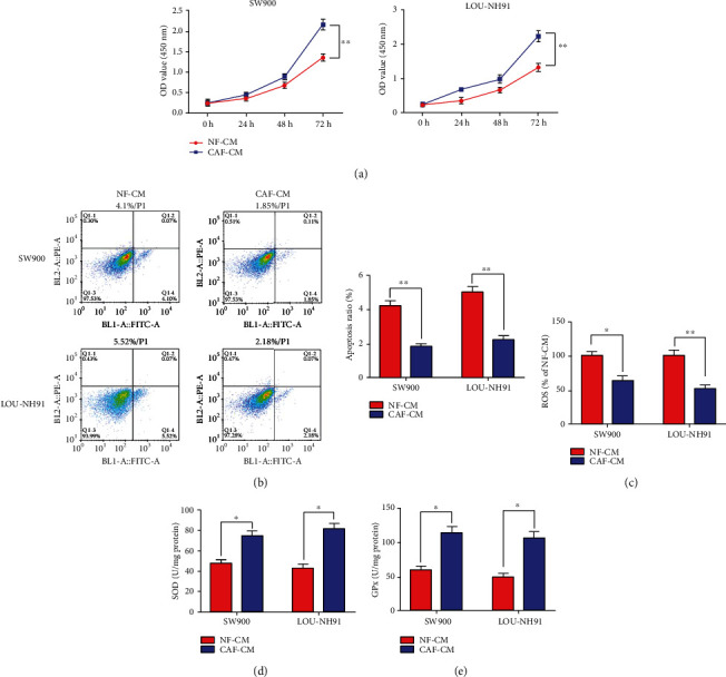
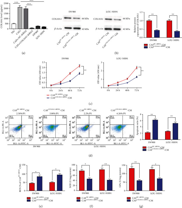
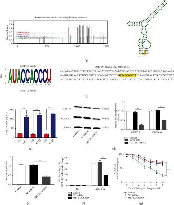
The microarray dataset GSE22874, containing 30 specimens of primary culture of normal fibroblasts (NFs) and 8 specimens of cancer-associated fibroblasts (CAFs) samples derived from LUSC, was retrieved from the Gene Expression Omnibus (GEO) database and then calculated by using the R language (limma package) to identify differentially expressed genes (DEGs). CAF-conditioned medium (CAF-CM) was collected and used to culture LUSC cells, followed by assessment of cell proliferation, apoptosis, and oxidative stress levels by using CCK-8, annexin V-FITC/PI double staining and ELISA assays. Subsequently, COL10A1 was knocked down in CAFs to assess the role of COL10A1 in CAF regulation of LUSC behavior. Bioinformatics online analysis and MeRIP were applied to predict and test the mA modification of COL10A1 mRNA and the regulatory relationship with METTL3. Rescue experiments were next performed to explore the effects of METTL3 and COL10A1 in CAFs on LUSC cell proliferation, apoptosis, and oxidative stress. LUSC tumor cells with or without (COL10A1-silenced) CAFs were subcutaneously inoculated in nude mice to evaluate the effect of COL10A1 in CAFs on LUSC tumor growth.
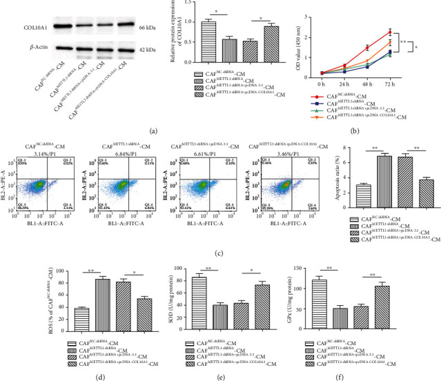
Elevated expression of COL10A1 was found in LUSC-derived CAFs by GSE22874 dataset analysis. We discovered that COL10A1 and METTL3 was expressed in both LUSC cells and matched CAFs, while COL10A1 expression was prominently higher in CAFs than in LUSC cells. CAF-CM memorably encouraged LUSC cell proliferation and suppressed apoptosis-induced oxidative stress, which was reversed by interfering with COL10A1 expression in CAFs, suggesting that COL10A1 might be secreted by CAFs into the culture medium to exert its effects inside LUSC cells. Global mA modification was decreased in METTL3 knocked down CAFs. MA modification, expression levels, and stability of COL10A1 mRNA were impaired upon METTL3 knockdown in CAFs. Overexpression of COL10A1 in CAFs partially reversed the effect of METTL3 knockdown on the malignant behavior of LUSC cells. studies confirmed that CAFs accelerated LUSC tumor growth, and this effect was counteracted by COL10A1 silencing.
COL10A1 secreted by CAFs could facilitate LUSC cell proliferation and repress apoptosis-induced oxidative stress, and the mechanism was due to elevated expression mediated by METTL3 promoting its mRNA mA modification, thereby accelerating tumor growth.
肿瘤微环境中的癌症相关成纤维细胞(CAFs)是肿瘤发生和发展的关键因素。然而,CAFs 对肺鳞状细胞癌(LUSC)相关的调控机制仍未得到充分阐明。
从基因表达综合数据库(GEO)中检索到包含 30 例原发性正常成纤维细胞(NFs)和 8 例 LUSC 来源的癌症相关成纤维细胞(CAFs)样本的微阵列数据集 GSE22874,然后使用 R 语言(limma 包)进行计算,以鉴定差异表达基因(DEGs)。收集 CAF 条件培养基(CAF-CM)并用于培养 LUSC 细胞,然后使用 CCK-8、 Annexin V-FITC/PI 双重染色和 ELISA 测定评估细胞增殖、凋亡和氧化应激水平。随后,在 CAFs 中敲低 COL10A1,以评估 COL10A1 在 CAF 调节 LUSC 行为中的作用。应用生物信息学在线分析和 MeRIP 预测和测试 COL10A1 mRNA 的 mA 修饰及其与 METTL3 的调控关系。接下来进行挽救实验,以探讨 CAFs 中 METTL3 和 COL10A1 对 LUSC 细胞增殖、凋亡和氧化应激的影响。将有或没有(COL10A1 沉默)CAFs 的 LUSC 肿瘤细胞皮下接种于裸鼠,以评估 CAFs 中 COL10A1 对 LUSC 肿瘤生长的影响。
通过 GSE22874 数据集分析发现,COL10A1 在 LUSC 衍生的 CAFs 中表达升高。我们发现 COL10A1 和 METTL3 在 LUSC 细胞和匹配的 CAFs 中均有表达,而 CAFs 中 COL10A1 的表达明显高于 LUSC 细胞。CAF-CM 显著促进 LUSC 细胞增殖并抑制凋亡诱导的氧化应激,而 CAFs 中 COL10A1 表达的干扰则逆转了这种作用,提示 COL10A1 可能由 CAFs 分泌到培养基中,在 LUSC 细胞内发挥作用。METTL3 敲低的 CAFs 中全局 mA 修饰减少。METTL3 敲低后 CAFs 中 COL10A1 mRNA 的表达水平和稳定性受损。CAFs 中 COL10A1 的过表达部分逆转了 METTL3 敲低对 LUSC 细胞恶性行为的影响。体内研究证实,CAFs 加速了 LUSC 肿瘤的生长,而 COL10A1 沉默则拮抗了这种作用。
CAFs 分泌的 COL10A1 可促进 LUSC 细胞增殖并抑制凋亡诱导的氧化应激,其机制是通过升高表达的 METTL3 促进其 mRNA mA 修饰,从而加速肿瘤生长。