• 文献检索
  • 文档翻译
  • 深度研究
  • 学术资讯
  • Suppr Zotero 插件Zotero 插件
  • 邀请有礼
  • 套餐&价格
  • 历史记录
应用&插件
Suppr Zotero 插件Zotero 插件浏览器插件Mac 客户端Windows 客户端微信小程序
定价
高级版会员购买积分包购买API积分包
服务
文献检索文档翻译深度研究API 文档MCP 服务
关于我们
关于 Suppr公司介绍联系我们用户协议隐私条款
关注我们

Suppr 超能文献

核心技术专利:CN118964589B侵权必究
粤ICP备2023148730 号-1Suppr @ 2026

文献检索

告别复杂PubMed语法,用中文像聊天一样搜索,搜遍4000万医学文献。AI智能推荐,让科研检索更轻松。

立即免费搜索

文件翻译

保留排版,准确专业,支持PDF/Word/PPT等文件格式,支持 12+语言互译。

免费翻译文档

深度研究

AI帮你快速写综述,25分钟生成高质量综述,智能提取关键信息,辅助科研写作。

立即免费体验

心肌纤维化的检测:我们目前的进展

Detection of myocardial fibrosis: Where we stand.

作者信息

Zhu Leyi, Wang Yining, Zhao Shihua, Lu Minjie

机构信息

State Key Laboratory of Cardiovascular Disease, Department of Magnetic Resonance Imaging, National Center for Cardiovascular Diseases, Fuwai Hospital, Beijing, China.

Chinese Academy of Medical Sciences and Peking Union Medical College, Beijing, China.

出版信息

Front Cardiovasc Med. 2022 Sep 29;9:926378. doi: 10.3389/fcvm.2022.926378. eCollection 2022.

DOI:10.3389/fcvm.2022.926378
PMID:36247487
原文链接:https://pmc.ncbi.nlm.nih.gov/articles/PMC9557071/
Abstract

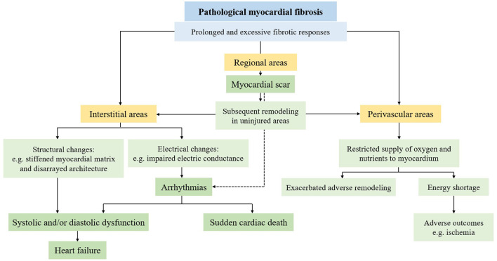
Myocardial fibrosis, resulting from the disturbance of extracellular matrix homeostasis in response to different insults, is a common and important pathological remodeling process that is associated with adverse clinical outcomes, including arrhythmia, heart failure, or even sudden cardiac death. Over the past decades, multiple non-invasive detection methods have been developed. Laboratory biomarkers can aid in both detection and risk stratification by reflecting cellular and even molecular changes in fibrotic processes, yet more evidence that validates their detection accuracy is still warranted. Different non-invasive imaging techniques have been demonstrated to not only detect myocardial fibrosis but also provide information on prognosis and management. Cardiovascular magnetic resonance (CMR) is considered as the gold standard imaging technique to non-invasively identify and quantify myocardial fibrosis with its natural ability for tissue characterization. This review summarizes the current understanding of the non-invasive detection methods of myocardial fibrosis, with the focus on different techniques and clinical applications of CMR.

摘要

心肌纤维化是一种常见且重要的病理重塑过程,它是由于细胞外基质稳态受到不同损伤的干扰而产生的,与不良临床结局相关,包括心律失常、心力衰竭,甚至心源性猝死。在过去几十年中,已经开发出多种非侵入性检测方法。实验室生物标志物可以通过反映纤维化过程中的细胞甚至分子变化来辅助检测和风险分层,但仍需要更多证据来验证其检测准确性。不同的非侵入性成像技术已被证明不仅可以检测心肌纤维化,还能提供有关预后和治疗的信息。心血管磁共振(CMR)因其具有组织特征的天然能力,被认为是用于非侵入性识别和量化心肌纤维化的金标准成像技术。本综述总结了目前对心肌纤维化非侵入性检测方法的理解,重点关注CMR的不同技术和临床应用。

https://cdn.ncbi.nlm.nih.gov/pmc/blobs/5c5f/9557071/0e2affed3bea/fcvm-09-926378-g0002.jpg
https://cdn.ncbi.nlm.nih.gov/pmc/blobs/5c5f/9557071/6b879e56dea9/fcvm-09-926378-g0001.jpg
https://cdn.ncbi.nlm.nih.gov/pmc/blobs/5c5f/9557071/0e2affed3bea/fcvm-09-926378-g0002.jpg
https://cdn.ncbi.nlm.nih.gov/pmc/blobs/5c5f/9557071/6b879e56dea9/fcvm-09-926378-g0001.jpg
https://cdn.ncbi.nlm.nih.gov/pmc/blobs/5c5f/9557071/0e2affed3bea/fcvm-09-926378-g0002.jpg

相似文献

1
Detection of myocardial fibrosis: Where we stand.心肌纤维化的检测:我们目前的进展
Front Cardiovasc Med. 2022 Sep 29;9:926378. doi: 10.3389/fcvm.2022.926378. eCollection 2022.
2
Recent Progresses in the Multimodality Imaging Assessment of Myocardial Fibrosis.心肌纤维化多模态成像评估的最新进展
Rev Cardiovasc Med. 2024 Jan 8;25(1):5. doi: 10.31083/j.rcm2501005. eCollection 2024 Jan.
3
T1 mapping: characterisation of myocardial interstitial space.T1 映射:心肌间质空间的特征描述
Insights Imaging. 2015 Apr;6(2):189-202. doi: 10.1007/s13244-014-0366-9. Epub 2014 Nov 26.
4
Imaging Interstitial Fibrosis, Left Ventricular Remodeling, and Function in Stage A and B Heart Failure.A期和B期心力衰竭患者的间质纤维化、左心室重构及功能的影像学表现
JACC Cardiovasc Imaging. 2021 May;14(5):1038-1052. doi: 10.1016/j.jcmg.2020.05.036. Epub 2020 Aug 19.
5
Magnetic resonance imaging (MRI) for the assessment of myocardial viability: an evidence-based analysis.用于评估心肌存活性的磁共振成像(MRI):一项基于证据的分析。
Ont Health Technol Assess Ser. 2010;10(15):1-45. Epub 2010 Jul 1.
6
Myocardial Fibrosis in Heart Failure: Anti-Fibrotic Therapies and the Role of Cardiovascular Magnetic Resonance in Drug Trials.心力衰竭中的心肌纤维化:抗纤维化治疗及心血管磁共振在药物试验中的作用
Cardiol Ther. 2020 Dec;9(2):363-376. doi: 10.1007/s40119-020-00199-y. Epub 2020 Aug 30.
7
Myocardial fibrosis detected by cardiac magnetic resonance imaging in heart failure: impact on remodeling, diastolic function and BNP levels.心脏磁共振成像检测到的心力衰竭患者心肌纤维化:对重塑、舒张功能及脑钠肽水平的影响
Anadolu Kardiyol Derg. 2011 Feb;11(1):71-6. doi: 10.5152/akd.2011.013. Epub 2011 Jan 11.
8
Noninvasive imaging of myocardial extracellular matrix for assessment of fibrosis.心肌细胞外基质的无创成像用于纤维化评估。
Curr Opin Cardiol. 2013 May;28(3):282-9. doi: 10.1097/HCO.0b013e32835f5a2b.
9
A prospective study examining the role of myocardial Fibrosis in outcome following mitral valve repair IN DEgenerative mitral Regurgitation: rationale and design of the mitral FINDER study.一项前瞻性研究,探讨心肌纤维化在退行性二尖瓣反流二尖瓣修复术后预后中的作用:二尖瓣FINDER研究的原理与设计
BMC Cardiovasc Disord. 2017 Nov 22;17(1):282. doi: 10.1186/s12872-017-0715-y.
10
Multimodality imaging evaluation of Chagas disease: an expert consensus of Brazilian Cardiovascular Imaging Department (DIC) and the European Association of Cardiovascular Imaging (EACVI).恰加斯病的多模态成像评估:巴西心血管成像部(DIC)与欧洲心血管成像协会(EACVI)的专家共识
Eur Heart J Cardiovasc Imaging. 2018 Apr 1;19(4):459-460n. doi: 10.1093/ehjci/jex154.

引用本文的文献

1
Cardiac toxicity and intervention strategies during thoracic cancer radiotherapy.胸段癌放疗期间的心脏毒性及干预策略
Front Oncol. 2025 Aug 4;15:1638035. doi: 10.3389/fonc.2025.1638035. eCollection 2025.
2
Left ventricle strain and T1 mapping evaluation in a mouse model with myocardial infarction.心肌梗死小鼠模型中的左心室应变和T1映射评估
Sci Rep. 2025 Aug 12;15(1):29579. doi: 10.1038/s41598-025-09699-0.
3
The small molecule LOXL2 inhibitor SNT-5382 reduces cardiac fibrosis and achieves strong clinical target engagement.

本文引用的文献

1
2022 AHA/ACC/HFSA Guideline for the Management of Heart Failure: A Report of the American College of Cardiology/American Heart Association Joint Committee on Clinical Practice Guidelines.2022年美国心脏协会/美国心脏病学会/美国心力衰竭学会心力衰竭管理指南:美国心脏病学会/美国心脏协会临床实践指南联合委员会报告
Circulation. 2022 May 3;145(18):e895-e1032. doi: 10.1161/CIR.0000000000001063. Epub 2022 Apr 1.
2
Synthetic Extracellular Volume in Cardiac Magnetic Resonance Without Blood Sampling: a Reliable Tool to Replace Conventional Extracellular Volume.心脏磁共振无采血的合成细胞外容积:替代常规细胞外容积的可靠工具。
Circ Cardiovasc Imaging. 2022 Apr;15(4):e013745. doi: 10.1161/CIRCIMAGING.121.013745. Epub 2022 Apr 1.
3
小分子赖氨酰氧化酶样蛋白2(LOXL2)抑制剂SNT-5382可减轻心脏纤维化并实现强大的临床靶点结合。
Sci Rep. 2025 Jul 2;15(1):22653. doi: 10.1038/s41598-025-06312-2.
4
Cardiac T1 mapping in non-ST-segment elevation myocardial infarction: temporal changes in myocardial fibrosis.非ST段抬高型心肌梗死中的心脏T1映射:心肌纤维化的时间变化
Front Cardiovasc Med. 2025 May 23;12:1563368. doi: 10.3389/fcvm.2025.1563368. eCollection 2025.
5
Diagnostic performance of cardiovascular magnetic resonance parametric mapping as per modified Lake Louise Criteria in acute myocarditis: an updated systematic review and meta-analysis.根据改良的路易斯湖标准,心血管磁共振参数成像在急性心肌炎中的诊断性能:一项更新的系统评价和荟萃分析。
J Cardiovasc Imaging. 2025 Jun 3;33(1):5. doi: 10.1186/s44348-025-00048-3.
6
Angiotensin II-induced cardiac fibrosis and dysfunction are exacerbated by deletion of cGKI in periostin+ myofibroblasts.在骨膜素阳性肌成纤维细胞中,cGKI基因缺失会加剧血管紧张素II诱导的心脏纤维化和功能障碍。
Clin Sci (Lond). 2025 Apr 23;139(11):507-26. doi: 10.1042/CS20241204.
7
The Prevalence, Pathophysiological Role and Determinants of Mitral Annular Disjunction Among Patients with Mitral Valve Prolapse: A Systematic Review.二尖瓣脱垂患者二尖瓣环分离的患病率、病理生理作用及决定因素:一项系统评价
J Clin Med. 2025 Feb 20;14(5):1423. doi: 10.3390/jcm14051423.
8
Risk stratification for future cardiac arrest after COVID-19 vaccination.新冠病毒疫苗接种后未来发生心脏骤停的风险分层
World J Cardiol. 2025 Feb 26;17(2):103909. doi: 10.4330/wjc.v17.i2.103909.
9
Baricitinib represses the myocardial fibrosis via blocking JAK/STAT and TGF-β1 pathways in vivo and in vitro.巴瑞替尼在体内和体外通过阻断JAK/STAT和TGF-β1信号通路来抑制心肌纤维化。
BMC Cardiovasc Disord. 2025 Jan 31;25(1):65. doi: 10.1186/s12872-025-04517-x.
10
Explainable AI associates ECG aging effects with increased cardiovascular risk in a longitudinal population study.在一项纵向人群研究中,可解释人工智能将心电图老化效应与心血管风险增加联系起来。
NPJ Digit Med. 2025 Jan 13;8(1):25. doi: 10.1038/s41746-024-01428-7.
Myocardial Work Brings New Insights into Left Ventricular Remodelling in Cardio-Oncology Patients.
心肌做功为心血管肿瘤患者左心室重构带来新见解。
Int J Environ Res Public Health. 2022 Feb 28;19(5):2826. doi: 10.3390/ijerph19052826.
4
Predictive Value of Echocardiographic Strain for Myocardial Fibrosis and Adverse Outcomes in Autoimmune Diseases.超声心动图应变对自身免疫性疾病中心肌纤维化及不良结局的预测价值
Front Cardiovasc Med. 2022 Feb 21;9:836942. doi: 10.3389/fcvm.2022.836942. eCollection 2022.
5
Reduced myocardial septal function assessed by cardiac magnetic resonance feature tracking in patients with hypertrophic obstructive cardiomyopathy: associated with histological myocardial fibrosis and ventricular arrhythmias.心脏磁共振斑点追踪技术评估肥厚型梗阻性心肌病患者的心肌室间隔功能:与组织学心肌纤维化和室性心律失常相关。
Eur Heart J Cardiovasc Imaging. 2022 Jul 21;23(8):1006-1015. doi: 10.1093/ehjci/jeac032.
6
Investigation of Left Ventricular Strain and Its Morphological Basis During Different Stages of Diastolic and Systolic Dysfunction in Spontaneously Hypertensive Rat.自发性高血压大鼠舒张和收缩功能障碍不同阶段左心室应变及其形态学基础的研究。
Am J Hypertens. 2022 May 10;35(5):423-432. doi: 10.1093/ajh/hpac008.
7
Detection of myocardial fibrosis by speckle-tracking echocardiography: from prediction to clinical applications.斑点追踪超声心动图检测心肌纤维化:从预测到临床应用。
Heart Fail Rev. 2022 Sep;27(5):1857-1867. doi: 10.1007/s10741-022-10214-0. Epub 2022 Jan 19.
8
Fibroblast activation protein imaging in reperfused ST-elevation myocardial infarction: comparison with cardiac magnetic resonance imaging.再灌注治疗的 ST 段抬高型心肌梗死中的成纤维细胞激活蛋白成像:与心脏磁共振成像的比较。
Eur J Nucl Med Mol Imaging. 2022 Jul;49(8):2786-2797. doi: 10.1007/s00259-021-05674-9. Epub 2022 Jan 5.
9
Association Between 2D- and 3D-Speckle-Tracking Longitudinal Strain and Cardiovascular Magnetic Resonance Evidence of Diffuse Myocardial Fibrosis in Heart Transplant Recipients.心脏移植受者二维和三维斑点追踪纵向应变与心血管磁共振弥散性心肌纤维化证据之间的关联
Front Cardiovasc Med. 2021 Nov 30;8:727745. doi: 10.3389/fcvm.2021.727745. eCollection 2021.
10
Latest Advances in Multimodality Imaging of Aortic Stenosis.主动脉瓣狭窄的多模态成像最新进展。
J Nucl Med. 2022 Mar;63(3):353-358. doi: 10.2967/jnumed.121.262304. Epub 2021 Dec 9.