• 文献检索
  • 文档翻译
  • 深度研究
  • 学术资讯
  • Suppr Zotero 插件Zotero 插件
  • 邀请有礼
  • 套餐&价格
  • 历史记录
应用&插件
Suppr Zotero 插件Zotero 插件浏览器插件Mac 客户端Windows 客户端微信小程序
定价
高级版会员购买积分包购买API积分包
服务
文献检索文档翻译深度研究API 文档MCP 服务
关于我们
关于 Suppr公司介绍联系我们用户协议隐私条款
关注我们

Suppr 超能文献

核心技术专利:CN118964589B侵权必究
粤ICP备2023148730 号-1Suppr @ 2026

文献检索

告别复杂PubMed语法,用中文像聊天一样搜索,搜遍4000万医学文献。AI智能推荐,让科研检索更轻松。

立即免费搜索

文件翻译

保留排版,准确专业,支持PDF/Word/PPT等文件格式,支持 12+语言互译。

免费翻译文档

深度研究

AI帮你快速写综述,25分钟生成高质量综述,智能提取关键信息,辅助科研写作。

立即免费体验

基于负重计算机断层扫描的膝关节骨与关节间隙宽度的多参数三维分析

Multiparametric 3-D analysis of bone and joint space width at the knee from weight bearing computed tomography.

作者信息

Turmezei Tom D, Low Samantha B, Rupret Simon, Treece Graham M, Gee Andrew H, MacKay James W, Lynch John A, Poole Kenneth Es, Segal Neil A

机构信息

Norfolk and Norwich University Hospital NHS Foundation Trust, Colney Lane, Norwich, UK.

University of East Anglia, Norwich Research Park, Norwich, UK.

出版信息

Osteoarthr Imaging. 2022 Jun;2(2). doi: 10.1016/j.ostima.2022.100069. Epub 2022 Jun 17.

DOI:10.1016/j.ostima.2022.100069
PMID:36249485
原文链接:https://pmc.ncbi.nlm.nih.gov/articles/PMC9559750/
Abstract

OBJECTIVE

Computed tomography (CT) can deliver multiple parameters relevant to osteoarthritis. In this study we demonstrate that a 3-D multiparametric approach at the weight bearing knee with cone beam CT is feasible, can include multiple parameters from across the joint space, and can reveal stronger relationships with disease status in combination.

DESIGN

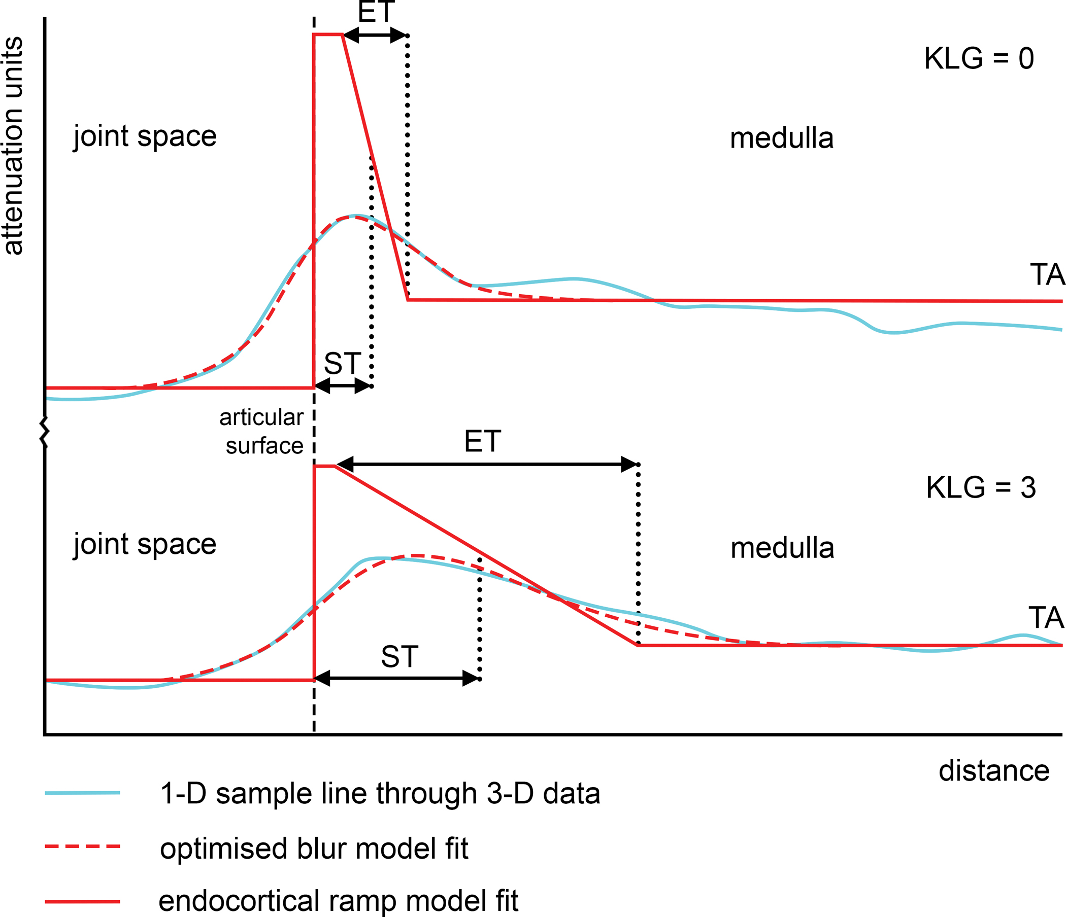
33 participants with knee weight bearing CT (WBCT) were analysed with joint space mapping and cortical bone mapping to deliver joint space width (JSW), subchondral bone plate thickness, endocortical thickness, and trabecular attenuation at both sides of the joint. All data were co-localised to the same canonical surface. Statistical parametric mapping (SPM) was applied in uni- and multivariate models to demonstrate significant dependence of parameters on Kellgren & Lawrence grade (KLG). Correlation between JSW and bony parameters and 2-week test-retest repeatability were also calculated.

RESULTS

SPM revealed that the central-to-posterior medial tibiofemoral joint space was significantly narrowed by up to 0.5 mm with significantly higher tibial trabecular attenuation up to 50 units for each increment in KLG as single features, and in a wider distribution when combined (p<0.05). These were also more strongly correlated with worsening KLG grade category. Test-retest repeatability was subvoxel (0.37 mm) for nearly all thickness parameters.

CONCLUSIONS

3-D JSW and tibial trabecular attenuation are repeatable and significantly dependent on radiographic disease severity at the weight bearing knee joint not just alone, but more strongly in combination. A quantitative multiparametric approach with WBCT may have potential for more sensitive investigation of disease progression in osteoarthritis.

摘要

目的

计算机断层扫描(CT)能够提供与骨关节炎相关的多个参数。在本研究中,我们证明了采用锥形束CT对负重膝关节进行三维多参数分析方法是可行的,该方法可以纳入整个关节间隙的多个参数,并且联合使用时能揭示与疾病状态更强的相关性。

设计

对33名进行膝关节负重CT(WBCT)检查的参与者进行关节间隙映射和皮质骨映射分析,以得出关节两侧的关节间隙宽度(JSW)、软骨下骨板厚度、皮质内厚度和小梁衰减。所有数据均共定位到同一个标准表面。在单变量和多变量模型中应用统计参数映射(SPM),以证明参数对凯尔格伦&劳伦斯分级(KLG)的显著依赖性。还计算了JSW与骨参数之间的相关性以及两周重测的重复性。

结果

SPM显示,随着KLG每增加一级,作为单一特征,胫股关节中后内侧关节间隙显著变窄达0.5毫米,胫骨小梁衰减显著增加达50个单位,联合使用时分布更广泛(p<0.05)。这些变化也与KLG分级恶化更密切相关。几乎所有厚度参数的重测重复性均为亚体素(0.37毫米)。

结论

三维JSW和胫骨小梁衰减具有可重复性,并且不仅单独显著依赖于负重膝关节的影像学疾病严重程度,联合使用时相关性更强。采用WBCT的定量多参数分析方法可能对骨关节炎疾病进展进行更敏感的研究具有潜力。

https://cdn.ncbi.nlm.nih.gov/pmc/blobs/fac6/9559750/2f2ae111e290/nihms-1831155-f0004.jpg
https://cdn.ncbi.nlm.nih.gov/pmc/blobs/fac6/9559750/a6be2fba20dd/nihms-1831155-f0001.jpg
https://cdn.ncbi.nlm.nih.gov/pmc/blobs/fac6/9559750/b509683d53dc/nihms-1831155-f0002.jpg
https://cdn.ncbi.nlm.nih.gov/pmc/blobs/fac6/9559750/55db420c4ec4/nihms-1831155-f0003.jpg
https://cdn.ncbi.nlm.nih.gov/pmc/blobs/fac6/9559750/2f2ae111e290/nihms-1831155-f0004.jpg

相似文献

1
Multiparametric 3-D analysis of bone and joint space width at the knee from weight bearing computed tomography.基于负重计算机断层扫描的膝关节骨与关节间隙宽度的多参数三维分析
Osteoarthr Imaging. 2022 Jun;2(2). doi: 10.1016/j.ostima.2022.100069. Epub 2022 Jun 17.
2
Quantitative Three-dimensional Assessment of Knee Joint Space Width from Weight-bearing CT.基于负重 CT 的膝关节间隙宽度的定量三维评估
Radiology. 2021 Jun;299(3):649-659. doi: 10.1148/radiol.2021203928. Epub 2021 Apr 13.
3
3-D joint space mapping at the ankle from weight-bearing CT: reproducibility, repeatability, and challenges for standardisation.踝关节负重 CT 三维关节间隙成像:可重复性、再现性及标准化挑战。
Eur Radiol. 2023 Nov;33(11):8333-8342. doi: 10.1007/s00330-023-09718-6. Epub 2023 May 31.
4
Three-dimensional analysis for quantification of knee joint space width with weight-bearing CT: comparison with non-weight-bearing CT and weight-bearing radiography.采用负重CT对膝关节间隙宽度进行定量分析的三维研究:与非负重CT及负重X线摄影的比较
Osteoarthritis Cartilage. 2022 May;30(5):671-680. doi: 10.1016/j.joca.2021.11.019. Epub 2021 Dec 7.
5
Improved responsiveness to change in joint space width over 24-month follow-up: comparison of 3D JSW on weight-bearing CT vs 2D JSW on radiographs in the MOST study.24 个月随访时关节间隙宽度变化的反应性改善:负重 CT 的 3D JSW 与 MOST 研究中 X 线的 2D JSW 的比较。
Osteoarthritis Cartilage. 2023 Mar;31(3):406-413. doi: 10.1016/j.joca.2022.12.002. Epub 2022 Dec 13.
6
In vivo analysis of subchondral trabecular bone in patients with osteoarthritis of the knee using second-generation high-resolution peripheral quantitative computed tomography (HR-pQCT).采用第二代高分辨率外周定量计算机断层扫描(HR-pQCT)对膝关节骨关节炎患者的软骨下骨小梁进行体内分析。
Bone. 2020 Mar;132:115155. doi: 10.1016/j.bone.2019.115155. Epub 2019 Nov 14.
7
Quantitative analysis of subchondral sclerosis of the tibia by bone texture parameters in knee radiographs: site-specific relationships with joint space width.膝关节 X 线片中骨纹理参数对胫骨软骨下硬化的定量分析:与关节间隙宽度的特定部位关系。
Osteoarthritis Cartilage. 2009 Nov;17(11):1453-60. doi: 10.1016/j.joca.2009.05.004. Epub 2009 May 18.
8
Comparison between 2D radiographic weight-bearing joint space width and 3D MRI non-weight-bearing cartilage thickness measures in the knee using non-weight-bearing 2D and 3D CT as an intermediary.以非负重二维和三维CT作为中介,比较膝关节二维负重X线摄影关节间隙宽度与三维MRI非负重软骨厚度测量值。
Ther Adv Chronic Dis. 2021 Aug 21;12:20406223211037868. doi: 10.1177/20406223211037868. eCollection 2021.
9
Correlation of 3D Joint Space Width From Weightbearing CT With Outcomes After Intra-articular Calcaneal Fracture.跟骨关节内骨折术后负重 CT 三维关节间隙宽度与疗效的相关性。
Foot Ankle Int. 2020 Sep;41(9):1106-1116. doi: 10.1177/1071100720933891. Epub 2020 Jul 10.
10
Quantifying the tibiofemoral joint space using x-ray tomosynthesis.使用 X 射线断层合成术定量测量胫股关节间隙。
Med Phys. 2011 Dec;38(12):6672-82. doi: 10.1118/1.3662891.

引用本文的文献

1
Effects of a multicomponent exercise regimen on subchondral bone and cartilage in postmenopausal women with knee osteoarthritis: protocol for a randomized controlled trial.多组分运动方案对绝经后膝骨关节炎女性软骨下骨和软骨的影响:一项随机对照试验的方案
Trials. 2025 Jun 23;26(1):222. doi: 10.1186/s13063-025-08928-1.
2
Trends in the Use of Weightbearing Computed Tomography.负重计算机断层扫描的使用趋势
J Clin Med. 2024 Sep 18;13(18):5519. doi: 10.3390/jcm13185519.
3
Imaging of joint response to exercise with MRI and PET.MRI 和 PET 检查对运动后关节反应的影像学评估。

本文引用的文献

1
Quantitative Three-dimensional Assessment of Knee Joint Space Width from Weight-bearing CT.基于负重 CT 的膝关节间隙宽度的定量三维评估
Radiology. 2021 Jun;299(3):649-659. doi: 10.1148/radiol.2021203928. Epub 2021 Apr 13.
2
Three-Dimensional Surface-Based Analysis of Cartilage MRI Data in Knee Osteoarthritis: Validation and Initial Clinical Application.膝关节骨关节炎软骨MRI数据的基于三维表面的分析:验证与初步临床应用
J Magn Reson Imaging. 2020 Oct;52(4):1139-1151. doi: 10.1002/jmri.27193. Epub 2020 May 24.
3
Quantifying Subresolution 3D Morphology of Bone with Clinical Computed Tomography.
Skeletal Radiol. 2023 Nov;52(11):2159-2183. doi: 10.1007/s00256-022-04271-7. Epub 2023 Jan 17.
用临床计算机断层扫描定量骨的亚分辨率 3D 形态。
Ann Biomed Eng. 2020 Feb;48(2):595-605. doi: 10.1007/s10439-019-02374-2. Epub 2019 Oct 3.
4
Statistical shape modelling reveals large and distinct subchondral bony differences in osteoarthritic knees.统计形状建模揭示了骨关节炎膝关节中大而明显的软骨下骨差异。
J Biomech. 2019 Aug 27;93:177-184. doi: 10.1016/j.jbiomech.2019.07.003. Epub 2019 Jul 10.
5
Relationships between cartilage thickness and subchondral bone mineral density in non-osteoarthritic and severely osteoarthritic knees: In vivo concomitant 3D analysis using CT arthrography.非骨关节炎和严重骨关节炎膝关节软骨厚度与软骨下骨骨密度的关系:关节 CT 造影术的体内同时 3D 分析。
Osteoarthritis Cartilage. 2019 Apr;27(4):621-629. doi: 10.1016/j.joca.2018.12.014. Epub 2019 Jan 3.
6
Cortical Bone Mapping: Measurement and Statistical Analysis of Localised Skeletal Changes.皮质骨绘图:局部骨骼变化的测量和统计分析。
Curr Osteoporos Rep. 2018 Oct;16(5):617-625. doi: 10.1007/s11914-018-0475-3.
7
Concurrent Assessment of Cartilage Morphology and Bone Microarchitecture in the Human Knee Using Contrast-Enhanced HR-pQCT Imaging.采用对比增强高分辨率 CT 成像技术对人体膝关节软骨形态和骨微结构进行同步评估。
J Clin Densitom. 2019 Jan-Mar;22(1):74-85. doi: 10.1016/j.jocd.2018.07.002. Epub 2018 Jul 19.
8
A new quantitative 3D approach to imaging of structural joint disease.一种新的定量 3D 成像方法,用于结构性关节疾病的成像。
Sci Rep. 2018 Jun 18;8(1):9280. doi: 10.1038/s41598-018-27486-y.
9
Differences in subchondral bone plate and cartilage thickness between women with anterior cruciate ligament reconstructions and uninjured controls.女性前交叉韧带重建患者与未受伤对照者的软骨下骨板和软骨厚度的差异。
Osteoarthritis Cartilage. 2018 Jul;26(7):929-939. doi: 10.1016/j.joca.2018.04.006. Epub 2018 Apr 17.
10
Measurement of the bone endocortical region using clinical CT.临床 CT 测量骨内皮质区
Med Image Anal. 2018 Feb;44:28-40. doi: 10.1016/j.media.2017.11.006. Epub 2017 Nov 16.