Lang Jonas B, Buck Michèle C, Rivière Jennifer, Stambouli Oumaima, Sachenbacher Ken, Choudhary Purva, Dietz Hendrik, Giebel Bernd, Bassermann Florian, Oostendorp Robert A J, Götze Katharina S, Hecker Judith S
Department of Medicine III, Technical University of Munich (TUM), Klinikum rechts der Isar, Munich, Germany.
Institute for Transfusion Medicine, University Hospital Essen, Essen, Germany.
Front Oncol. 2022 Oct 3;12:949261. doi: 10.3389/fonc.2022.949261. eCollection 2022.
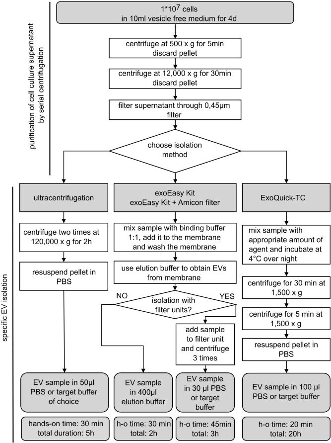
Cellular crosstalk between hematopoietic stem/progenitor cells and the bone marrow (BM) niche is vital for the development and maintenance of myeloid malignancies. These compartments can communicate bidirectional transfer of extracellular vesicles (EVs). EV trafficking in acute myeloid leukemia (AML) plays a crucial role in shaping the BM microenvironment into a leukemia-permissive niche. Although several EV isolation methods have been developed, it remains a major challenge to define the most accurate and reliable procedure. Here, we tested the efficacy and functional assay compatibility of four different EV isolation methods in leukemia-derived EVs: (1) membrane affinity-based: exoEasy Kit alone and (2) in combination with Amicon filtration; (3) precipitation: ExoQuick-TC; and (4) ultracentrifugation (UC). Western blot analysis of EV fractions showed the highest enrichment of EV marker expression (e.g., CD63, HSP70, and TSG101) by precipitation with removal of overabundant soluble proteins [e.g., bovine serum albumin (BSA)], which were not discarded using UC. Besides the presence of damaged EVs after UC, intact EVs were successfully isolated with all methods as evidenced by highly maintained spherical- and cup-shaped vesicles in transmission electron microscopy. Nanoparticle tracking analysis of EV particle size and concentration revealed significant differences in EV isolation efficacy, with exoEasy Kit providing the highest EV yield recovery. Of note, functional assays with exoEasy Kit-isolated EVs showed significant toxicity towards treated target cells [e.g., mesenchymal stromal cells (MSCs)], which was abrogated when combining exoEasy Kit with Amicon filtration. Additionally, MSC treated with green fluorescent protein (GFP)-tagged exoEasy Kit-isolated EVs did not show any EV uptake, while EV isolation by precipitation demonstrated efficient EV internalization. Taken together, the choice of EV isolation procedure significantly impacts the yield and potential functionality of leukemia-derived EVs. The cheapest method (UC) resulted in contaminated and destructed EV fractions, while the isolation method with the highest EV yield (exoEasy Kit) appeared to be incompatible with functional assays. We identified two methods (precipitation-based ExoQuick-TC and membrane affinity-based exoEasy Kit combined with Amicon filtration) yielding pure and intact EVs, also suitable for application in functional assays. This study highlights the importance of selecting the right EV isolation method depending on the desired experimental design.
造血干/祖细胞与骨髓(BM)微环境之间的细胞间串扰对于髓系恶性肿瘤的发生和维持至关重要。这些区室可通过细胞外囊泡(EVs)进行双向传递。急性髓系白血病(AML)中的EV运输在将BM微环境塑造成白血病允许的生态位方面起着关键作用。尽管已经开发了几种EV分离方法,但确定最准确和可靠的程序仍然是一项重大挑战。在这里,我们测试了四种不同的EV分离方法对白血病来源的EVs的功效和功能分析兼容性:(1)基于膜亲和力的:单独使用exoEasy试剂盒以及(2)与Amicon过滤相结合;(3)沉淀法:ExoQuick-TC;以及(4)超速离心(UC)。对EV级分的蛋白质印迹分析表明,通过沉淀去除过量的可溶性蛋白质(例如牛血清白蛋白(BSA)),EV标记物表达(例如CD63、HSP70和TSG101)的富集程度最高,而超速离心未丢弃这些蛋白质。除了超速离心后存在受损的EVs外,所有方法均成功分离出完整的EVs,这在透射电子显微镜下表现为高度保持的球形和杯形囊泡。对EV粒径和浓度的纳米颗粒跟踪分析揭示了EV分离功效的显著差异,exoEasy试剂盒的EV产量回收率最高。值得注意的是,用exoEasy试剂盒分离的EVs进行的功能分析显示对处理的靶细胞[例如间充质基质细胞(MSCs)]具有显著毒性,而将exoEasy试剂盒与Amicon过滤结合时这种毒性被消除。此外,用绿色荧光蛋白(GFP)标记的exoEasy试剂盒分离的EVs处理的MSC未显示任何EV摄取,而通过沉淀法分离EVs则显示出有效的EV内化。综上所述,EV分离程序的选择显著影响白血病来源的EVs的产量和潜在功能。最便宜的方法(超速离心)导致EV级分受到污染和破坏,而EV产量最高的分离方法(exoEasy试剂盒)似乎与功能分析不兼容。我们确定了两种方法(基于沉淀的ExoQuick-TC和基于膜亲和力的exoEasy试剂盒与Amicon过滤相结合)可产生纯净且完整的EVs,也适用于功能分析。这项研究强调了根据所需实验设计选择正确的EV分离方法的重要性。