• 文献检索
  • 文档翻译
  • 深度研究
  • 学术资讯
  • Suppr Zotero 插件Zotero 插件
  • 邀请有礼
  • 套餐&价格
  • 历史记录
应用&插件
Suppr Zotero 插件Zotero 插件浏览器插件Mac 客户端Windows 客户端微信小程序
定价
高级版会员购买积分包购买API积分包
服务
文献检索文档翻译深度研究API 文档MCP 服务
关于我们
关于 Suppr公司介绍联系我们用户协议隐私条款
关注我们

Suppr 超能文献

核心技术专利:CN118964589B侵权必究
粤ICP备2023148730 号-1Suppr @ 2026

文献检索

告别复杂PubMed语法,用中文像聊天一样搜索,搜遍4000万医学文献。AI智能推荐,让科研检索更轻松。

立即免费搜索

文件翻译

保留排版,准确专业,支持PDF/Word/PPT等文件格式,支持 12+语言互译。

免费翻译文档

深度研究

AI帮你快速写综述,25分钟生成高质量综述,智能提取关键信息,辅助科研写作。

立即免费体验

基于深度学习的脑出血计算机断层扫描图像分割与体积测量

Deep learning-based computed tomography image segmentation and volume measurement of intracerebral hemorrhage.

作者信息

Peng Qi, Chen Xingcai, Zhang Chao, Li Wenyan, Liu Jingjing, Shi Tingxin, Wu Yi, Feng Hua, Nian Yongjian, Hu Rong

机构信息

Department of Digital Medicine, School of Biomedical Engineering and Imaging Medicine, Army Medical University, Third Military Medical University, Chongqing, China.

Department of Neurosurgery, First Affiliated Hospital,Southwest Hospital, Army Medical University, Third Military Medical University, Chongqing, China.

出版信息

Front Neurosci. 2022 Oct 3;16:965680. doi: 10.3389/fnins.2022.965680. eCollection 2022.

DOI:10.3389/fnins.2022.965680
PMID:36263364
原文链接:https://pmc.ncbi.nlm.nih.gov/articles/PMC9575984/
Abstract

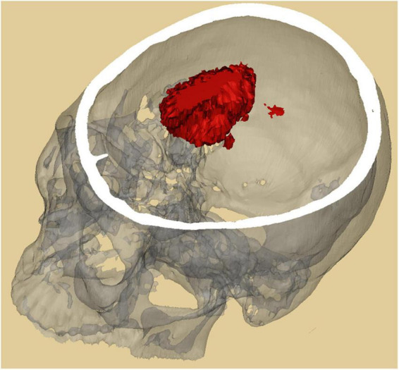
The study aims to enhance the accuracy and practicability of CT image segmentation and volume measurement of ICH by using deep learning technology. A dataset including the brain CT images and clinical data of 1,027 patients with spontaneous ICHs treated from January 2010 to December 2020 were retrospectively analyzed, and a deep segmentation network (AttFocusNet) integrating the focus structure and the attention gate (AG) mechanism is proposed to enable automatic, accurate CT image segmentation and volume measurement of ICHs. In internal validation set, experimental results showed that AttFocusNet achieved a Dice coefficient of 0.908, an intersection-over-union (IoU) of 0.874, a sensitivity of 0.913, a positive predictive value (PPV) of 0.957, and a 95% Hausdorff distance (HD95) (mm) of 5.960. The intraclass correlation coefficient (ICC) of the ICH volume measurement between AttFocusNet and the ground truth was 0.997. The average time of per case achieved by AttFocusNet, Coniglobus formula and manual segmentation is 5.6, 47.7, and 170.1 s. In the two external validation sets, AttFocusNet achieved a Dice coefficient of 0.889 and 0.911, respectively, an IoU of 0.800 and 0.836, respectively, a sensitivity of 0.817 and 0.849, respectively, a PPV of 0.976 and 0.981, respectively, and a HD95 of 5.331 and 4.220, respectively. The ICC of the ICH volume measurement between AttFocusNet and the ground truth were 0.939 and 0.956, respectively. The proposed segmentation network AttFocusNet significantly outperforms the Coniglobus formula in terms of ICH segmentation and volume measurement by acquiring measurement results closer to the true ICH volume and significantly reducing the clinical workload.

摘要

本研究旨在利用深度学习技术提高脑出血(ICH)CT图像分割及体积测量的准确性和实用性。回顾性分析了2010年1月至2020年12月期间接受治疗的1027例自发性ICH患者的脑CT图像及临床数据,并提出了一种整合焦点结构和注意力门控(AG)机制的深度分割网络(AttFocusNet),以实现ICH的CT图像自动、准确分割及体积测量。在内验证集中,实验结果显示AttFocusNet的Dice系数为0.908,交并比(IoU)为0.874,敏感度为0.913,阳性预测值(PPV)为0.957,95%豪斯多夫距离(HD95)(mm)为5.960。AttFocusNet与真实值之间ICH体积测量的组内相关系数(ICC)为0.997。AttFocusNet、Coniglobus公式和手动分割每例的平均时间分别为5.6、47.7和170.1秒。在两个外验证集中,AttFocusNet的Dice系数分别为0.889和0.911,IoU分别为0.800和0.836,敏感度分别为0.817和0.849,PPV分别为0.976和0.981,HD95分别为5.331和4.220。AttFocusNet与真实值之间ICH体积测量的ICC分别为0.939和0.956。所提出的分割网络AttFocusNet在ICH分割和体积测量方面显著优于Coniglobus公式,获得了更接近真实ICH体积的测量结果,并显著减少了临床工作量。

https://cdn.ncbi.nlm.nih.gov/pmc/blobs/04d8/9575984/6ed6611a88d8/fnins-16-965680-g009.jpg
https://cdn.ncbi.nlm.nih.gov/pmc/blobs/04d8/9575984/ecb9d270831f/fnins-16-965680-g001.jpg
https://cdn.ncbi.nlm.nih.gov/pmc/blobs/04d8/9575984/634b20e94cc6/fnins-16-965680-g002.jpg
https://cdn.ncbi.nlm.nih.gov/pmc/blobs/04d8/9575984/c013f59d8a71/fnins-16-965680-g003.jpg
https://cdn.ncbi.nlm.nih.gov/pmc/blobs/04d8/9575984/8236e67928b7/fnins-16-965680-g004.jpg
https://cdn.ncbi.nlm.nih.gov/pmc/blobs/04d8/9575984/c95d7685204f/fnins-16-965680-g005.jpg
https://cdn.ncbi.nlm.nih.gov/pmc/blobs/04d8/9575984/d4cc464527ad/fnins-16-965680-g006.jpg
https://cdn.ncbi.nlm.nih.gov/pmc/blobs/04d8/9575984/c03f233ebd44/fnins-16-965680-g007.jpg
https://cdn.ncbi.nlm.nih.gov/pmc/blobs/04d8/9575984/4d4d0cab8557/fnins-16-965680-g008.jpg
https://cdn.ncbi.nlm.nih.gov/pmc/blobs/04d8/9575984/6ed6611a88d8/fnins-16-965680-g009.jpg

相似文献

1
Deep learning-based computed tomography image segmentation and volume measurement of intracerebral hemorrhage.基于深度学习的脑出血计算机断层扫描图像分割与体积测量
Front Neurosci. 2022 Oct 3;16:965680. doi: 10.3389/fnins.2022.965680. eCollection 2022.
2
A Robust Deep Learning Segmentation Method for Hematoma Volumetric Detection in Intracerebral Hemorrhage.一种用于脑出血血肿容量检测的稳健深度学习分割方法。
Stroke. 2022 Jan;53(1):167-176. doi: 10.1161/STROKEAHA.120.032243. Epub 2021 Oct 4.
3
Deep learning shows good reliability for automatic segmentation and volume measurement of brain hemorrhage, intraventricular extension, and peripheral edema.深度学习在脑出血、脑室内延伸和周围水肿的自动分割和体积测量方面具有较好的可靠性。
Eur Radiol. 2021 Jul;31(7):5012-5020. doi: 10.1007/s00330-020-07558-2. Epub 2021 Jan 6.
4
Deep learning models for separate segmentations of intracerebral and intraventricular hemorrhage on head CT and segmentation quality assessment.用于头部CT上脑内和脑室内出血单独分割的深度学习模型及分割质量评估
Med Phys. 2024 Nov;51(11):8317-8333. doi: 10.1002/mp.17343. Epub 2024 Aug 12.
5
Deep Network for the Automatic Segmentation and Quantification of Intracranial Hemorrhage on CT.用于CT图像上颅内出血自动分割与量化的深度网络
Front Neurosci. 2021 Jan 11;14:541817. doi: 10.3389/fnins.2020.541817. eCollection 2020.
6
A Deep Learning-Based Automatic Segmentation and 3D Visualization Technique for Intracranial Hemorrhage Detection Using Computed Tomography Images.一种基于深度学习的自动分割与三维可视化技术,用于利用计算机断层扫描图像检测颅内出血。
Diagnostics (Basel). 2023 Jul 31;13(15):2537. doi: 10.3390/diagnostics13152537.
7
Deep-learning measurement of intracerebral haemorrhage with mixed precision training: a coarse-to-fine study.基于混合精度训练的深度学习脑内出血测量:一项从粗到细的研究。
Clin Radiol. 2023 Apr;78(4):e328-e335. doi: 10.1016/j.crad.2022.12.019. Epub 2023 Jan 22.
8
Improved automatic segmentation of brain metastasis gross tumor volume in computed tomography images for radiotherapy: a position attention module for U-Net architecture.用于放疗的计算机断层扫描图像中脑转移瘤大体肿瘤体积的改进自动分割:一种用于U-Net架构的位置注意力模块
Quant Imaging Med Surg. 2024 Jul 1;14(7):4475-4489. doi: 10.21037/qims-23-1627. Epub 2024 May 24.
9
Accuracy of automated segmentation and volumetry of acute intracerebral hemorrhage following minimally invasive surgery using a patch-based convolutional neural network in a small dataset.基于小块卷积神经网络的微创手术后急性脑出血自动分割与体积测量在小数据集中的准确性
Neuroradiology. 2024 Apr;66(4):601-608. doi: 10.1007/s00234-024-03311-4. Epub 2024 Feb 17.
10
Efficiency of a deep learning-based artificial intelligence diagnostic system in spontaneous intracerebral hemorrhage volume measurement.基于深度学习的人工智能诊断系统在自发性脑出血容量测量中的效率。
BMC Med Imaging. 2021 Aug 13;21(1):125. doi: 10.1186/s12880-021-00657-6.

引用本文的文献

1
A Machine Learning Pipeline for Automated Bolus Segmentation and Area Measurement in Swallowing Videofluoroscopy Images of an Infant Pig Model.一种用于在幼猪模型吞咽荧光透视图像中自动进行团注分割和面积测量的机器学习管道。
Dysphagia. 2025 Apr 28. doi: 10.1007/s00455-025-10829-z.
2
Optimizing Automated Hematoma Expansion Classification from Baseline and Follow-Up Head Computed Tomography.通过基线和随访头部计算机断层扫描优化自动血肿扩大分类
Appl Sci (Basel). 2025 Jan;15(1). doi: 10.3390/app15010111. Epub 2024 Dec 27.
3
A Hybrid Transformer-Convolutional Neural Network for Segmentation of Intracerebral Hemorrhage and Perihematomal Edema on Non-Contrast Head Computed Tomography (CT) with Uncertainty Quantification to Improve Confidence.

本文引用的文献

1
A Clinical Predictive Nomogram for Traumatic Brain Parenchyma Hematoma Progression.创伤性脑实质血肿进展的临床预测列线图
Neurol Ther. 2022 Mar;11(1):185-203. doi: 10.1007/s40120-021-00306-8. Epub 2021 Dec 2.
2
Hematoma Evacuation via Image-Guided Para-Corticospinal Tract Approach in Patients with Spontaneous Intracerebral Hemorrhage.影像引导下经皮质脊髓束旁入路治疗自发性脑出血的血肿清除术
Neurol Ther. 2021 Dec;10(2):1001-1013. doi: 10.1007/s40120-021-00279-8. Epub 2021 Sep 12.
3
Construction of a Machine Learning Dataset through Collaboration: The RSNA 2019 Brain CT Hemorrhage Challenge.
一种用于非增强头部计算机断层扫描(CT)上脑内出血和血肿周围水肿分割的混合Transformer-卷积神经网络,具有不确定性量化以提高可信度。
Bioengineering (Basel). 2024 Dec 15;11(12):1274. doi: 10.3390/bioengineering11121274.
4
The Role of ICP Monitoring in Minimally Invasive Surgery for the Management of Intracerebral Hemorrhage.颅内压监测在微创治疗脑出血手术中的作用
Transl Stroke Res. 2025 Apr;16(2):547-556. doi: 10.1007/s12975-023-01219-4. Epub 2023 Dec 29.
5
Role of artificial intelligence and machine learning in the diagnosis of cerebrovascular disease.人工智能和机器学习在脑血管疾病诊断中的作用。
Front Hum Neurosci. 2023 Sep 7;17:1254417. doi: 10.3389/fnhum.2023.1254417. eCollection 2023.
6
Validation of a deep learning model for traumatic brain injury detection and NIRIS grading on non-contrast CT: a multi-reader study with promising results and opportunities for improvement.深度学习模型在非对比 CT 上检测创伤性脑损伤和 NIRIS 分级的验证:一项多读者研究,结果有前景,有改进的机会。
Neuroradiology. 2023 Nov;65(11):1605-1617. doi: 10.1007/s00234-023-03170-5. Epub 2023 Jun 3.
通过合作构建机器学习数据集:2019年RSNA脑CT出血挑战赛
Radiol Artif Intell. 2020 Apr 29;2(3):e190211. doi: 10.1148/ryai.2020190211. eCollection 2020 May.
4
Assessment of an Artificial Intelligence Algorithm for Detection of Intracranial Hemorrhage.颅内出血检测人工智能算法的评估。
World Neurosurg. 2021 Jun;150:e209-e217. doi: 10.1016/j.wneu.2021.02.134. Epub 2021 Mar 5.
5
Deep Network for the Automatic Segmentation and Quantification of Intracranial Hemorrhage on CT.用于CT图像上颅内出血自动分割与量化的深度网络
Front Neurosci. 2021 Jan 11;14:541817. doi: 10.3389/fnins.2020.541817. eCollection 2020.
6
Deep learning shows good reliability for automatic segmentation and volume measurement of brain hemorrhage, intraventricular extension, and peripheral edema.深度学习在脑出血、脑室内延伸和周围水肿的自动分割和体积测量方面具有较好的可靠性。
Eur Radiol. 2021 Jul;31(7):5012-5020. doi: 10.1007/s00330-020-07558-2. Epub 2021 Jan 6.
7
UNet++: Redesigning Skip Connections to Exploit Multiscale Features in Image Segmentation.UNet++:重新设计跳过连接以利用图像分割中的多尺度特征。
IEEE Trans Med Imaging. 2020 Jun;39(6):1856-1867. doi: 10.1109/TMI.2019.2959609. Epub 2019 Dec 13.
8
Expert-level detection of acute intracranial hemorrhage on head computed tomography using deep learning.使用深度学习技术对头 CT 进行急性颅内出血的专家级检测。
Proc Natl Acad Sci U S A. 2019 Nov 5;116(45):22737-22745. doi: 10.1073/pnas.1908021116. Epub 2019 Oct 21.
9
Deep Learning and Neurology: A Systematic Review.深度学习与神经病学:一项系统综述。
Neurol Ther. 2019 Dec;8(2):351-365. doi: 10.1007/s40120-019-00153-8. Epub 2019 Aug 21.
10
Advanced machine learning in action: identification of intracranial hemorrhage on computed tomography scans of the head with clinical workflow integration.实用的高级机器学习:通过整合临床工作流程,在头部计算机断层扫描中识别颅内出血。
NPJ Digit Med. 2018 Apr 4;1:9. doi: 10.1038/s41746-017-0015-z. eCollection 2018.