• 文献检索
  • 文档翻译
  • 深度研究
  • 学术资讯
  • Suppr Zotero 插件Zotero 插件
  • 邀请有礼
  • 套餐&价格
  • 历史记录
应用&插件
Suppr Zotero 插件Zotero 插件浏览器插件Mac 客户端Windows 客户端微信小程序
定价
高级版会员购买积分包购买API积分包
服务
文献检索文档翻译深度研究API 文档MCP 服务
关于我们
关于 Suppr公司介绍联系我们用户协议隐私条款
关注我们

Suppr 超能文献

核心技术专利:CN118964589B侵权必究
粤ICP备2023148730 号-1Suppr @ 2026

文献检索

告别复杂PubMed语法,用中文像聊天一样搜索,搜遍4000万医学文献。AI智能推荐,让科研检索更轻松。

立即免费搜索

文件翻译

保留排版,准确专业,支持PDF/Word/PPT等文件格式,支持 12+语言互译。

免费翻译文档

深度研究

AI帮你快速写综述,25分钟生成高质量综述,智能提取关键信息,辅助科研写作。

立即免费体验

定量核组织形态计量学可预测髓母细胞瘤的分子亚型和临床结局:初步研究结果。

Quantitative Nuclear Histomorphometry Predicts Molecular Subtype and Clinical Outcome in Medulloblastomas: Preliminary Findings.

作者信息

Whitney Jon, Dollinger Liisa, Tamrazi Benita, Hawes Debra, Couce Marta, Marcheque Julia, Judkins Alexander, Margol Ashley, Madabhushi Anant

机构信息

Department of Biomedical Engineering, Cleveland, OH, USA.

Case Western Reserve University School of Medicine, Cleveland, OH, USA.

出版信息

J Pathol Inform. 2022 Feb 17;13:100090. doi: 10.1016/j.jpi.2022.100090. eCollection 2022.

DOI:10.1016/j.jpi.2022.100090
PMID:36268104
原文链接:https://pmc.ncbi.nlm.nih.gov/articles/PMC9576985/
Abstract

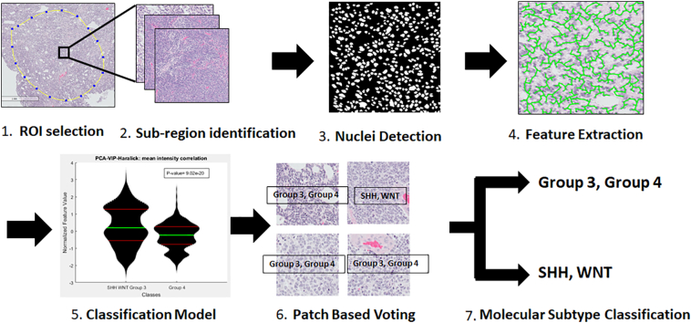
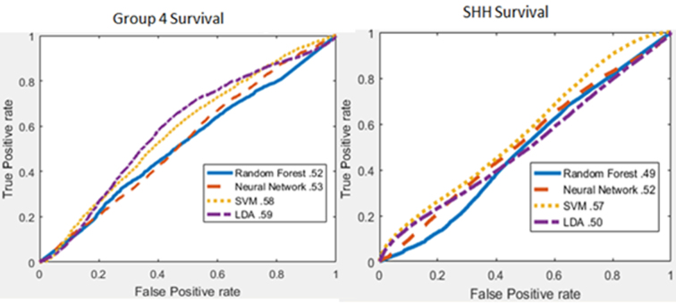
Molecular subtypes of medulloblastoma [Sonic Hedgehog (SHH), Wingless/INT (WNT), Group 3, and Group 4] are defined by common patterns of gene expression. These differential gene expression patterns appear to result in different histomorphology and prognosis. Quantitative histomorphometry is a well-known method of computer-aided pathology image analysis. The hypotheses we sought to examine in this preliminary proof of concept study were whether computer extracted nuclear morphological features of medulloblastomas from digitized tissue slide images could independently: (1) distinguish between molecularly determined subgroups and (2) identify patterns within these subgroups that correspond with clinical outcome. Our dataset was composed of 46 medulloblastoma patients: 16 SHH (5 dead, 11 survived), 3 WNT (0 dead, 3 survived), 12 Group 3 (4 dead, 8 survived), and 15 were Group 4 (5 dead, 10 survived). A watershed-based thresholding scheme was used to automatically identify individual nuclei within digitized whole slide hematoxylin and eosin tissue images. Quantitative histomorphometric features corresponding to the texture (variation in pixel intensity), shape (variations in size, roundness), and architectural rearrangement (distances between, and number of connected neighbors) of nuclei were subsequently extracted. These features were ranked using feature selection schemes and these top-ranked features were then used to train machine-learning classifiers via threefold cross-validation to separate patients based on: (1) molecular subtype and (2) disease-specific outcomes within the individual molecular subtype groups. SHH and WNT tumors were separated from Groups 3 and 4 tumors with a maximum area under the receiver operating characteristic curve (AUC) of 0.7, survival within Group 3 tumors was predicted with an AUC of 0.92, and Group 3 and 4 patients were separated into high- and low-risk groups with = 0.002. Model prediction was quantitatively compared with age, stage, and histological subtype using univariate and multivariate Cox hazard ratio models. Age was the most statistically significant variable for predicting survival in Group 3 and 4 tumors, but model predictions had the highest hazard ratio value. Quantitative nuclear histomorphometry can be used to study medulloblastoma genetic expression phenotypes as it may distinguish meaningful features of disease pathology.

摘要

髓母细胞瘤的分子亚型[音猬因子(SHH)、无翅/整合(WNT)、3组和4组]由常见的基因表达模式定义。这些不同的基因表达模式似乎导致了不同的组织形态学和预后。定量组织形态计量学是一种众所周知的计算机辅助病理图像分析方法。在这项初步概念验证研究中,我们试图检验的假设是,从数字化组织切片图像中计算机提取的髓母细胞瘤核形态特征是否能够独立:(1)区分分子确定的亚组;(2)识别这些亚组内与临床结果相关的模式。我们的数据集由46例髓母细胞瘤患者组成:16例SHH型(5例死亡,11例存活),3例WNT型(0例死亡,3例存活),12例3组(4例死亡,8例存活),15例4组(5例死亡,10例存活)。基于分水岭的阈值化方案用于自动识别数字化全切片苏木精和伊红组织图像中的单个细胞核。随后提取与细胞核的纹理(像素强度变化)、形状(大小、圆度变化)和结构重排(相邻细胞核之间的距离和连接邻居的数量)相对应的定量组织形态计量学特征。使用特征选择方案对这些特征进行排序,然后使用这些排名靠前的特征通过三重交叉验证来训练机器学习分类器,以根据以下因素对患者进行分类:(1)分子亚型;(2)各个分子亚型组内的疾病特异性结果。SHH和WNT肿瘤与3组和4组肿瘤分离,受试者操作特征曲线(AUC)下的最大面积为0.7,3组肿瘤内的生存情况预测AUC为0.92,3组和4组患者被分为高风险和低风险组,P = 0.002。使用单变量和多变量Cox风险比模型将模型预测与年龄、分期和组织学亚型进行定量比较。年龄是预测3组和4组肿瘤生存的最具统计学意义的变量,但模型预测的风险比值最高。定量细胞核组织形态计量学可用于研究髓母细胞瘤的基因表达表型,因为它可能区分疾病病理学的有意义特征。

https://cdn.ncbi.nlm.nih.gov/pmc/blobs/3c9f/9576985/6ca26cda3ecd/mmc8.jpg
https://cdn.ncbi.nlm.nih.gov/pmc/blobs/3c9f/9576985/e1092c6c245c/gr1.jpg
https://cdn.ncbi.nlm.nih.gov/pmc/blobs/3c9f/9576985/5c9dfa4ae672/gr2.jpg
https://cdn.ncbi.nlm.nih.gov/pmc/blobs/3c9f/9576985/c577be7fb033/gr3.jpg
https://cdn.ncbi.nlm.nih.gov/pmc/blobs/3c9f/9576985/e63a9aee1e55/gr4.jpg
https://cdn.ncbi.nlm.nih.gov/pmc/blobs/3c9f/9576985/94232510e7b7/gr5.jpg
https://cdn.ncbi.nlm.nih.gov/pmc/blobs/3c9f/9576985/3b3c8f7ef774/gr6.jpg
https://cdn.ncbi.nlm.nih.gov/pmc/blobs/3c9f/9576985/5eabe5b08a6f/mmc1.jpg
https://cdn.ncbi.nlm.nih.gov/pmc/blobs/3c9f/9576985/e677edb3ecc9/mmc3.jpg
https://cdn.ncbi.nlm.nih.gov/pmc/blobs/3c9f/9576985/f16f53e8e3ed/mmc4.jpg
https://cdn.ncbi.nlm.nih.gov/pmc/blobs/3c9f/9576985/b87cf63ee48f/mmc5.jpg
https://cdn.ncbi.nlm.nih.gov/pmc/blobs/3c9f/9576985/78c14718a746/mmc6.jpg
https://cdn.ncbi.nlm.nih.gov/pmc/blobs/3c9f/9576985/d71290552ca9/mmc7.jpg
https://cdn.ncbi.nlm.nih.gov/pmc/blobs/3c9f/9576985/6ca26cda3ecd/mmc8.jpg

相似文献

1
Quantitative Nuclear Histomorphometry Predicts Molecular Subtype and Clinical Outcome in Medulloblastomas: Preliminary Findings.定量核组织形态计量学可预测髓母细胞瘤的分子亚型和临床结局:初步研究结果。
J Pathol Inform. 2022 Feb 17;13:100090. doi: 10.1016/j.jpi.2022.100090. eCollection 2022.
2
Quantitative nuclear histomorphometry predicts oncotype DX risk categories for early stage ER+ breast cancer.定量核组织形态计量学可预测早期 ER+ 乳腺癌的 Oncotype DX 风险类别。
BMC Cancer. 2018 May 30;18(1):610. doi: 10.1186/s12885-018-4448-9.
3
Quantitative nuclear histomorphometric features are predictive of Oncotype DX risk categories in ductal carcinoma in situ: preliminary findings.定量核组织形态计量学特征可预测导管原位癌的 Oncotype DX 风险类别:初步研究结果。
Breast Cancer Res. 2019 Oct 17;21(1):114. doi: 10.1186/s13058-019-1200-6.
4
MR Imaging-Based Radiomic Signatures of Distinct Molecular Subgroups of Medulloblastoma.基于磁共振成像的髓母细胞瘤不同分子亚群的放射组学特征。
AJNR Am J Neuroradiol. 2019 Jan;40(1):154-161. doi: 10.3174/ajnr.A5899. Epub 2018 Dec 6.
5
Texture Analysis of T1-Weighted Contrast-Enhanced Magnetic Resonance Imaging Potentially Predicts Outcomes of Patients with Non-Wingless-Type/Non-Sonic Hedgehog Medulloblastoma.T1 加权对比增强磁共振成像纹理分析可能预测非 Wnt 型/非 Sonic Hedgehog 型髓母细胞瘤患者的结局。
World Neurosurg. 2020 May;137:e27-e33. doi: 10.1016/j.wneu.2019.09.142. Epub 2019 Oct 4.
6
Medulloblastoma: clinicopathological correlates of SHH, WNT, and non-SHH/WNT molecular subgroups.髓母细胞瘤:SHH、WNT 和非 SHH/WNT 分子亚群的临床病理相关性。
Acta Neuropathol. 2011 Mar;121(3):381-96. doi: 10.1007/s00401-011-0800-8. Epub 2011 Jan 26.
7
Computer-extracted features of nuclear morphology in hematoxylin and eosin images distinguish stage II and IV colon tumors.苏木精和伊红图像中细胞核形态的计算机提取特征可区分 II 期和 IV 期结肠肿瘤。
J Pathol. 2022 May;257(1):17-28. doi: 10.1002/path.5864. Epub 2022 Feb 22.
8
Prognostic effect of whole chromosomal aberration signatures in standard-risk, non-WNT/non-SHH medulloblastoma: a retrospective, molecular analysis of the HIT-SIOP PNET 4 trial.标准风险、非 WNT/非 SHH 髓母细胞瘤中全染色体畸变特征的预后影响:HIT-SIOP PNET 4 试验的回顾性、分子分析。
Lancet Oncol. 2018 Dec;19(12):1602-1616. doi: 10.1016/S1470-2045(18)30532-1. Epub 2018 Nov 1.
9
Subgroup-specific prognostic implications of TP53 mutation in medulloblastoma.髓母细胞瘤中 TP53 突变的亚组特异性预后意义。
J Clin Oncol. 2013 Aug 10;31(23):2927-35. doi: 10.1200/JCO.2012.48.5052. Epub 2013 Jul 8.
10
Differentiation of fat-poor angiomyolipoma from clear cell renal cell carcinoma in contrast-enhanced MDCT images using quantitative feature classification.基于定量特征分类的 MDCT 增强图像鉴别乏脂性血管平滑肌脂肪瘤与透明细胞肾细胞癌
Med Phys. 2017 Jul;44(7):3604-3614. doi: 10.1002/mp.12258. Epub 2017 Jun 9.

引用本文的文献

1
Machine learning-based models for prediction of survival in medulloblastoma: a systematic review and meta-analysis.基于机器学习的髓母细胞瘤生存预测模型:一项系统综述和荟萃分析。
Neurol Sci. 2025 Feb;46(2):689-696. doi: 10.1007/s10072-024-07879-w. Epub 2024 Nov 12.
2
Radio-pathomic approaches in pediatric neuro-oncology: Opportunities and challenges.儿科神经肿瘤学中的放射组学方法:机遇与挑战。
Neurooncol Adv. 2023 Sep 13;5(1):vdad119. doi: 10.1093/noajnl/vdad119. eCollection 2023 Jan-Dec.

本文引用的文献

1
Pathology, diagnostics, and classification of medulloblastoma.髓母细胞瘤的病理学、诊断和分类。
Brain Pathol. 2020 May;30(3):664-678. doi: 10.1111/bpa.12837.
2
Medulloblastoma in the age of molecular subgroups: a review.分子亚组时代的髓母细胞瘤:综述
J Neurosurg Pediatr. 2019 Oct 1;24(4):353-363. doi: 10.3171/2019.5.PEDS18381.
3
DNA-methylation profiling discloses significant advantages over NanoString method for molecular classification of medulloblastoma.DNA甲基化谱分析在髓母细胞瘤分子分类方面比NanoString方法具有显著优势。
Acta Neuropathol. 2017 Dec;134(6):965-967. doi: 10.1007/s00401-017-1776-9. Epub 2017 Oct 13.
4
A deep learning based strategy for identifying and associating mitotic activity with gene expression derived risk categories in estrogen receptor positive breast cancers.一种基于深度学习的策略,用于识别雌激素受体阳性乳腺癌中的有丝分裂活性并将其与基因表达衍生的风险类别相关联。
Cytometry A. 2017 Jun;91(6):566-573. doi: 10.1002/cyto.a.23065. Epub 2017 Feb 13.
5
Integrative Analysis of Cellular Morphometric Context Reveals Clinically Relevant Signatures in Lower Grade Glioma.细胞形态测量背景的综合分析揭示了低级别胶质瘤中与临床相关的特征。
Med Image Comput Comput Assist Interv. 2016 Oct;9900:72-80. doi: 10.1007/978-3-319-46720-7_9. Epub 2016 Oct 2.
6
Automated Tubule Nuclei Quantification and Correlation with Oncotype DX risk categories in ER+ Breast Cancer Whole Slide Images.自动肾小管核定量与 ER+乳腺癌全 slides 图像中 Oncotype DX 风险分类的相关性。
Sci Rep. 2016 Sep 7;6:32706. doi: 10.1038/srep32706.
7
Active medulloblastoma enhancers reveal subgroup-specific cellular origins.活跃的髓母细胞瘤增强子揭示了亚组特异性细胞起源。
Nature. 2016 Feb 4;530(7588):57-62. doi: 10.1038/nature16546. Epub 2016 Jan 27.
8
Augmented expression of MYC and/or MYCN protein defines highly aggressive MYC-driven neuroblastoma: a Children's Oncology Group study.MYC和/或MYCN蛋白的过表达定义了高度侵袭性的MYC驱动的神经母细胞瘤:一项儿童肿瘤学组的研究。
Br J Cancer. 2015 Jun 30;113(1):57-63. doi: 10.1038/bjc.2015.188. Epub 2015 Jun 2.
9
Novel PCA-VIP scheme for ranking MRI protocols and identifying computer-extracted MRI measurements associated with central gland and peripheral zone prostate tumors.用于对MRI协议进行排名并识别与中央腺体和外周带前列腺肿瘤相关的计算机提取MRI测量值的新型PCA-VIP方案。
J Magn Reson Imaging. 2015 May;41(5):1383-93. doi: 10.1002/jmri.24676. Epub 2014 Jun 18.
10
Cell orientation entropy (COrE): predicting biochemical recurrence from prostate cancer tissue microarrays.细胞取向熵(COrE):从前列腺癌组织微阵列预测生化复发
Med Image Comput Comput Assist Interv. 2013;16(Pt 3):396-403. doi: 10.1007/978-3-642-40760-4_50.