Jambhulkar Mohit, Bhatia Jasvinder K, Singh Samresh K
Department of Pathology, Command Hospital Eastern Command, Kolkata, West Bengal, India.
J Cytol. 2022 Jul-Sep;39(3):91-97. doi: 10.4103/joc.joc_80_21. Epub 2022 Jul 30.
Fine-needle aspiration cytology is considered the gold standard screening test in the evaluation of a thyroid nodule. We studied whether cell block cytology can be used in addition to conventional smears for the evaluation of tissue from fine-needle aspirations or fluid aspirations and also compared it with histopathological diagnosis.
The primary aim of this study was to know the utility of cell blocks in the diagnosis of thyroid lesions.
This was a prospective observational study conducted from June 2018 to September 2020 at a tertiary Care Hospital in Eastern India. Ethical approval was obtained from the Ethics Committee of the institution. Patients above 18 years who presented with goiter were included in the study. Thirty patients were enrolled in the study after informed consent.
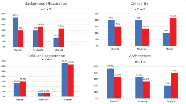
Smears prepared from the aspirates were stained with Leishman-Giemsa (LG) and Pap stain. The remnant from the needle hub was transferred to a sterile container. Cell blocks were prepared from the remnants. Smears were scored based on cell obscuration by blood, cellularity, cell degeneration, and cell architecture. The results were compared with histopathology.
Data were recorded using Microsoft Excel. Descriptive statistics, frequency, and proportion were used to describe demographic variables.
The majority of the patients (23.3%) were in their third decade of life, followed by 16.7% of the patients in their fourth and fifth decades. The patient age ranged from 25 to 80 years, with a mean age of 50.83 years and a standard deviation of 16.72. The largest number of patients were females accounting for 80% (24/30) of the total participants. The majority of the patients (36.7%) (11/30) had thyroid gland enlargement for a period of 15 days to three months. 14% of the participants were not able to recall its duration. The majority (60%) (18/30) had left lobe lesions, followed by 33.3% (10/30) who had right lobe lesions, and 6.7% (2/30) who had bilateral lobe swelling. The mean size of the lesion was 2.84 cm. 50% were found to be Bethesda II lesions, while 13.3% were Bethesda IV, and 36.7% were found to be Bethesda VI lesions. The cell block score (7) was found to be better compared to Fine Needle Aspiration Cytology (FNAC) (4.7). Tissue Coagulum Clot and Clot Scrape methods were found to yield better results compared to the Cytocentrifuge method. The value was found to be significant (<0.001).
Cell blocks were found to improve the cell morphology compared to FNAC alone and can be used as an adjunct to FNAC in the diagnosis of various thyroid lesions.
细针穿刺细胞学检查被认为是评估甲状腺结节的金标准筛查试验。我们研究了细胞块细胞学检查是否可用于辅助传统涂片,以评估细针穿刺或液体抽吸获得的组织,并将其与组织病理学诊断进行比较。
本研究的主要目的是了解细胞块在甲状腺病变诊断中的效用。
这是一项前瞻性观察性研究,于2018年6月至2020年9月在印度东部的一家三级护理医院进行。获得了该机构伦理委员会的伦理批准。纳入研究的患者为18岁以上出现甲状腺肿的患者。30名患者在获得知情同意后被纳入研究。
将抽吸物制备的涂片用利什曼-吉姆萨(LG)和巴氏染色。针座残留的样本转移至无菌容器中。从残留样本制备细胞块。根据涂片上血液造成的细胞模糊、细胞数量、细胞变性和细胞结构对涂片进行评分。将结果与组织病理学进行比较。
使用Microsoft Excel记录数据。描述性统计、频率和比例用于描述人口统计学变量。
大多数患者(23.3%)处于第三个十年年龄段,其次是16.7%的患者处于第四和第五个十年年龄段。患者年龄范围为25至80岁,平均年龄为50.83岁,标准差为16.72。女性患者数量最多,占总参与者的80%(24/30)。大多数患者(36.7%)(11/30)甲状腺肿大持续15天至3个月。14%的参与者无法回忆起其持续时间。大多数(60%)(18/30)患者有左叶病变,其次是33.3%(10/30)有右叶病变,6.7%(2/30)有双侧叶肿大。病变的平均大小为2.84厘米。50%被发现为贝塞斯达II类病变,而13.3%为贝塞斯达IV类,36.7%被发现为贝塞斯达VI类病变。发现细胞块评分(7分)优于细针穿刺细胞学检查(FNAC)(4.7分)。与细胞离心法相比,组织凝块和凝块刮取法的效果更好。发现P值具有显著性(<0.001)。
与单独的FNAC相比,细胞块可改善细胞形态,可作为FNAC的辅助手段用于诊断各种甲状腺病变。