Department of Medical Pharmacology and Physiology, School of Medicine, University of Missouri, Columbia, Missouri.
Department of Physiology and Cell Biology, The Ohio State University, Columbus, Ohio.
Am J Physiol Cell Physiol. 2022 Dec 1;323(6):C1728-C1739. doi: 10.1152/ajpcell.00101.2022. Epub 2022 Oct 24.
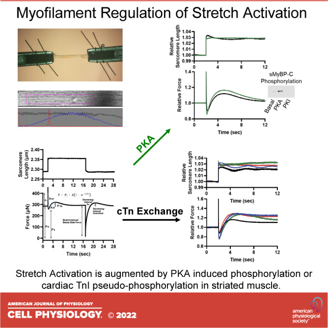
Stretch activation is defined as a delayed increase in force after rapid stretches. Although there is considerable evidence for stretch activation in isolated cardiac myofibrillar preparations, few studies have measured mechanisms of stretch activation in mammalian skeletal muscle fibers. We measured stretch activation following rapid step stretches [∼1%-4% sarcomere length (SL)] during submaximal Ca activations of rat permeabilized slow-twitch skeletal muscle fibers before and after protein kinase A (PKA), which phosphorylates slow myosin binding protein-C. PKA significantly increased stretch activation during low (∼25%) Ca activation and accelerated rates of delayed force development () during both low and half-maximal Ca activation. Following the step stretches and subsequent force development, fibers were rapidly shortened to original sarcomere length, which often elicited a shortening-induced transient force overshoot. After PKA, step shortening-induced transient force overshoot increased ∼10-fold following an ∼4% SL shortening during low Ca activation levels. following step shortening also increased after PKA during low and half-maximal Ca activations. We next investigated thin filament regulation of stretch activation. We tested the interplay between cardiac troponin I (cTnI) phosphorylation at the canonical PKA and novel tyrosine kinase sites on stretch activation. Native slow-skeletal Tn complexes were exchanged with recombinant human cTn complex with different human cTnI N-terminal pseudo-phosphorylation molecules: ) nonphosphorylated wild type (WT), ) the canonical S22/23D PKA sites, ) the tyrosine kinase Y26E site, and ) the combinatorial S22/23D + Y26E cTnI. All three pseudo-phosphorylated cTnIs elicited greater stretch activation than WT. Following stretch activation, a new, elevated stretch-induced steady-state force was reached with pseudo-phosphorylated cTnI. Combinatorial S22/23D + Y26E pseudo-phosphorylated cTnI increased . These results suggest that slow-skeletal yosin inding rotein- (sMyBP-C) phosphorylation modulates stretch activation by a combination of cross-bridge recruitment and faster cycling kinetics, whereas cTnI phosphorylation regulates stretch activation by both redundant and synergistic mechanisms; and, taken together, these sarcomere phosphoproteins offer precision targets for enhanced contractility.
伸展激活被定义为快速伸展后力的延迟增加。尽管在分离的心肌肌原纤维制剂中存在大量伸展激活的证据,但很少有研究测量哺乳动物骨骼肌纤维中伸展激活的机制。我们在大鼠通透慢肌纤维的亚最大 Ca 激活期间测量了快速阶跃伸展[1%-4%肌节长度(SL)]后的伸展激活,然后在蛋白激酶 A(PKA)前后测量,PKA 磷酸化慢肌球蛋白结合蛋白-C。PKA 在低(25%)Ca 激活期间显著增加伸展激活,并在低和半最大 Ca 激活期间加速延迟力发展的速率()。在阶跃伸展和随后的力发展之后,纤维被快速缩短到原始肌节长度,这通常会引起缩短诱导的瞬态力过冲。在 PKA 之后,在低 Ca 激活水平下进行~4%SL 缩短后,阶跃缩短诱导的瞬态力过冲增加了约 10 倍。在低和半最大 Ca 激活期间,PKA 后阶跃缩短也增加了。接下来,我们研究了薄丝蛋白对伸展激活的调节。我们测试了心脏肌钙蛋白 I(cTnI)在经典 PKA 和新的酪氨酸激酶位点的磷酸化与伸展激活之间的相互作用。用重组人 cTn 复合物交换天然慢肌 Tn 复合物,该复合物具有不同的人 cTnI N 端假磷酸化分子:)非磷酸化野生型(WT),)经典 S22/23D PKA 位点,)酪氨酸激酶 Y26E 位点,以及)S22/23D+Y26E cTnI 组合。所有三种假磷酸化的 cTnI 都比 WT 产生更大的伸展激活。伸展激活后,新的、升高的伸展诱导稳态力达到假磷酸化 cTnI。组合 S22/23D+Y26E 假磷酸化 cTnI 增加了。这些结果表明,慢肌肌球蛋白结合蛋白-C(sMyBP-C)的磷酸化通过横桥募集和更快的循环动力学的组合来调节伸展激活,而 cTnI 磷酸化通过冗余和协同机制来调节伸展激活;并且,综合起来,这些肌节磷酸蛋白为增强收缩力提供了精确的靶点。