• 文献检索
  • 文档翻译
  • 深度研究
  • 学术资讯
  • Suppr Zotero 插件Zotero 插件
  • 邀请有礼
  • 套餐&价格
  • 历史记录
应用&插件
Suppr Zotero 插件Zotero 插件浏览器插件Mac 客户端Windows 客户端微信小程序
定价
高级版会员购买积分包购买API积分包
服务
文献检索文档翻译深度研究API 文档MCP 服务
关于我们
关于 Suppr公司介绍联系我们用户协议隐私条款
关注我们

Suppr 超能文献

核心技术专利:CN118964589B侵权必究
粤ICP备2023148730 号-1Suppr @ 2026

文献检索

告别复杂PubMed语法,用中文像聊天一样搜索,搜遍4000万医学文献。AI智能推荐,让科研检索更轻松。

立即免费搜索

文件翻译

保留排版,准确专业,支持PDF/Word/PPT等文件格式,支持 12+语言互译。

免费翻译文档

深度研究

AI帮你快速写综述,25分钟生成高质量综述,智能提取关键信息,辅助科研写作。

立即免费体验

MRI 扫描在伴有病变(既往缺氧缺血性损伤)儿童中对 3D 打印脑的保真度。

Fidelity of 3D Printed Brains from MRI Scan in Children with Pathology (Prior Hypoxic Ischemic Injury).

机构信息

School of Clinical Sciences, Faculty of Medicine, University of Bristol, Bristol, BS28DX, UK.

Department of Radiology, Children's Hospital of Philadelphia, Philadelphia, PA, USA.

出版信息

J Digit Imaging. 2023 Feb;36(1):17-28. doi: 10.1007/s10278-022-00723-7. Epub 2022 Oct 24.

DOI:10.1007/s10278-022-00723-7
PMID:36280655
原文链接:https://pmc.ncbi.nlm.nih.gov/articles/PMC9984578/
Abstract

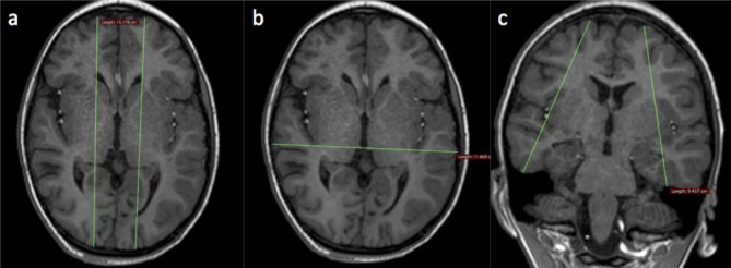
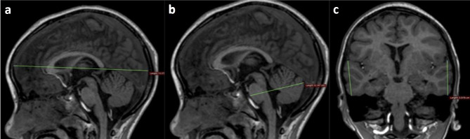
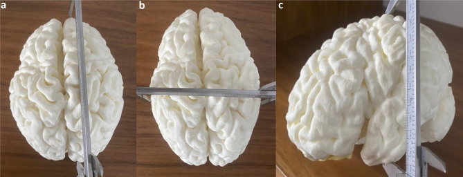
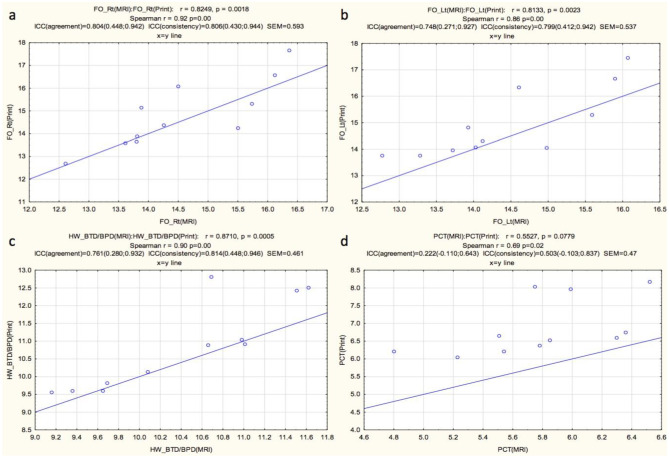
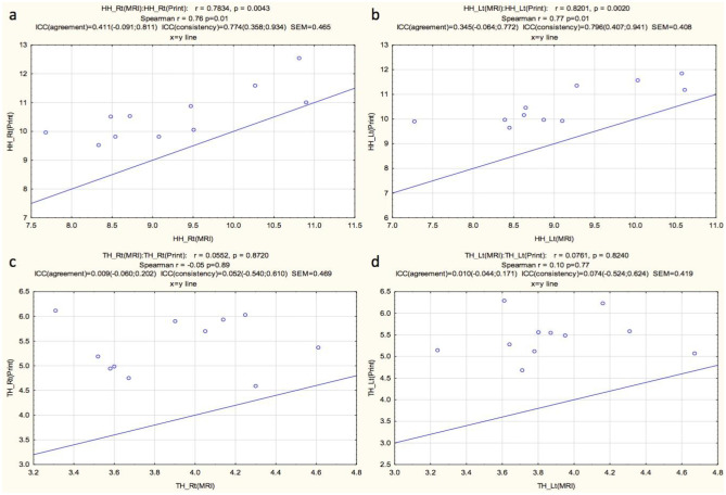
Cortical injury on the surface of the brain in children with hypoxic ischemic injury (HII) can be difficult to demonstrate to non-radiologists and lay people using brain images alone. Three-dimensional (3D) printing is helpful to communicate the volume loss and pathology due to HII in children's brains. 3D printed models represent the brain to scale and can be held up against models of normal brains for appreciation of volume loss. If 3D printed brains are to be used for formal communication, e.g., with medical colleagues or in court, they should have high fidelity of reproduction of the actual size of patients' brains. Here, we evaluate the size fidelity of 3D printed models from MRI scans of the brain, in children with prior HII. Twelve 3D prints of the brain were created from MRI scans of children with HII and selected to represent a variety of cortical pathologies. Specific predetermined measures of the 3D prints were made and compared to measures in matched planes on MRI. Fronto-occipital length (FOL) and bi-temporal/bi-parietal diameters (BTD/BPD) demonstrated high interclass correlations (ICC). Correlations were moderate to weak for hemispheric height, temporal height, and pons-cerebellar thickness. The average standard error of measurement (SEM) was 0.48 cm. Our results demonstrate high correlations in overall measurements of each 3D printed model derived from brain MRI scans versus the original MRI, evidenced by high ICC values for FOL and BTD/BPD. Measures with low correlation values can be explained by variability in matching the plane of measurement to the MRI slice orientation.

摘要

对于缺氧缺血性损伤(HII)患儿,大脑表面的皮质损伤单凭脑图像很难向非放射科医生和非专业人士解释清楚。三维(3D)打印有助于沟通儿童大脑因 HII 导致的容积损失和病变。3D 打印模型按比例代表大脑,可以与正常大脑的模型进行对比,以了解容积损失情况。如果要将 3D 打印的大脑用于正式交流,例如与医疗同事交流或在法庭上使用,那么它们应该高度忠实地再现患者大脑的实际大小。在这里,我们评估了先前患有 HII 的儿童的脑 MRI 扫描的 3D 打印模型的大小准确性。从患有 HII 的儿童的 MRI 扫描中创建了 12 个大脑 3D 打印模型,以代表各种皮质病变。对 3D 打印模型的特定预定测量值进行了测量,并与 MRI 上匹配的平面进行了比较。额枕长度(FOL)和双侧颞叶/顶叶直径(BTD/BPD)表现出较高的组内相关系数(ICC)。半球高度、颞叶高度和桥脑小脑厚度的相关性为中度至弱。平均测量标准误差(SEM)为 0.48cm。我们的结果表明,从脑 MRI 扫描中获得的每个 3D 打印模型的总体测量值与原始 MRI 之间具有高度相关性,FOL 和 BTD/BPD 的 ICC 值较高证明了这一点。具有低相关值的测量值可以通过将测量平面与 MRI 切片方向匹配的可变性来解释。

https://cdn.ncbi.nlm.nih.gov/pmc/blobs/e233/9984578/2623aff2ef47/10278_2022_723_Fig8_HTML.jpg
https://cdn.ncbi.nlm.nih.gov/pmc/blobs/e233/9984578/a4850201962a/10278_2022_723_Fig1_HTML.jpg
https://cdn.ncbi.nlm.nih.gov/pmc/blobs/e233/9984578/8a4144383ed7/10278_2022_723_Fig2_HTML.jpg
https://cdn.ncbi.nlm.nih.gov/pmc/blobs/e233/9984578/c011710a5fd1/10278_2022_723_Fig3_HTML.jpg
https://cdn.ncbi.nlm.nih.gov/pmc/blobs/e233/9984578/38264225f7eb/10278_2022_723_Fig4_HTML.jpg
https://cdn.ncbi.nlm.nih.gov/pmc/blobs/e233/9984578/ea79be15017d/10278_2022_723_Fig5_HTML.jpg
https://cdn.ncbi.nlm.nih.gov/pmc/blobs/e233/9984578/0c22a2cd14aa/10278_2022_723_Fig6_HTML.jpg
https://cdn.ncbi.nlm.nih.gov/pmc/blobs/e233/9984578/c49c9247e777/10278_2022_723_Fig7_HTML.jpg
https://cdn.ncbi.nlm.nih.gov/pmc/blobs/e233/9984578/2623aff2ef47/10278_2022_723_Fig8_HTML.jpg

相似文献

1
Fidelity of 3D Printed Brains from MRI Scan in Children with Pathology (Prior Hypoxic Ischemic Injury).MRI 扫描在伴有病变(既往缺氧缺血性损伤)儿童中对 3D 打印脑的保真度。
J Digit Imaging. 2023 Feb;36(1):17-28. doi: 10.1007/s10278-022-00723-7. Epub 2022 Oct 24.
2
Accuracy of non-medical and medical individuals in identifying cerebral cortical abnormality from three-dimensional printed models of magnetic resonance images in children with hypoxic ischemic injury.非医学专业和医学专业人员在识别缺氧缺血性损伤儿童磁共振成像三维打印模型中大脑皮质异常方面的准确性。
Pediatr Radiol. 2024 Mar;54(3):450-456. doi: 10.1007/s00247-023-05653-2. Epub 2023 Apr 11.
3
Technical report: 3D printing of the brain for use as a visual-aid tool to communicate MR imaging features of hypoxic ischaemic injury at term with non-physicians.技术报告:用于向非专业医生传达足月时缺氧缺血性损伤的磁共振成像特征的可视化辅助工具——大脑的3D打印
Childs Nerv Syst. 2018 Aug;34(8):1573-1577. doi: 10.1007/s00381-018-3838-2. Epub 2018 May 26.
4
Frequency of Cerebellar Abnormalities Associated With the Differing Magnetic Resonance Imaging Patterns of Term Hypoxic-Ischemic Injury in Children.与不同磁共振成像模式的足月缺氧缺血性损伤相关的小脑异常的频率。
Pediatr Neurol. 2024 Mar;152:73-78. doi: 10.1016/j.pediatrneurol.2023.12.023. Epub 2024 Jan 1.
5
Frequency of ulegyria on delayed MRI scans in children with term hypoxic-ischemic injury.足月新生儿缺氧缺血性脑损伤患儿延迟磁共振成像扫描中脑回状异位的发生率
Pediatr Radiol. 2023 Jan;53(1):104-111. doi: 10.1007/s00247-022-05445-0. Epub 2022 Jul 27.
6
Accuracy of radiologists, nonradiologists, and laypeople for identifying children with cerebral cortical atrophy from "Mercator map" curved reconstructions of MRIs of the brain.放射科医生、非放射科医生和外行人通过大脑MRI的“墨卡托地图”曲线重建识别患有大脑皮质萎缩儿童的准确性。
Indian J Radiol Imaging. 2020 Apr-Jun;30(2):111-115. doi: 10.4103/ijri.IJRI_130_20. Epub 2020 Jul 13.
7
Development of a 3D printed patient-specific neonatal brain simulation model using multimodality imaging for perioperative management.利用多模态成像技术为围手术期管理开发 3D 打印的新生儿脑个体化模拟模型。
Pediatr Res. 2022 Jan;91(1):64-69. doi: 10.1038/s41390-021-01421-w. Epub 2021 Mar 2.
8
Streamlined, Inexpensive 3D Printing of the Brain and Skull.简化、低成本的大脑和颅骨3D打印
PLoS One. 2015 Aug 21;10(8):e0136198. doi: 10.1371/journal.pone.0136198. eCollection 2015.
9
Generating patient-matched 3D-printed pedicle screw and laminectomy drill guides from Cone Beam CT images: Studies in ovine and porcine cadavers.从锥形束 CT 图像生成与患者匹配的 3D 打印椎弓根螺钉和椎板切除术钻导板:在绵羊和猪尸体上的研究。
Med Phys. 2022 Jul;49(7):4642-4652. doi: 10.1002/mp.15681. Epub 2022 May 6.
10
A systematic evaluation of medical 3D printing accuracy of multi-pathological anatomical models for surgical planning manufactured in elastic and rigid material using desktop inverted vat photopolymerization.采用桌面倒置光聚合技术,对弹性和刚性材料制造的用于手术规划的多病理解剖模型的医学 3D 打印精度进行系统评价。
Med Phys. 2021 Jun;48(6):3223-3233. doi: 10.1002/mp.14850. Epub 2021 Apr 16.

引用本文的文献

1
3-Dimensional printing and bioprinting in neurological sciences: applications in surgery, imaging, tissue engineering, and pharmacology and therapeutics.神经科学中的三维打印和生物打印:在手术、成像、组织工程以及药理学与治疗学中的应用
J Mater Sci Mater Med. 2025 Apr 9;36(1):32. doi: 10.1007/s10856-025-06877-4.
2
Pixels to precision: Neuroradiology's leap into 3D printing for personalized medicine.从像素到精准:神经放射学迈向3D打印用于个性化医疗的飞跃。
J Clin Imaging Sci. 2024 Dec 17;14:49. doi: 10.25259/JCIS_119_2024. eCollection 2024.

本文引用的文献

1
The accuracy of clinical 3D printing in reconstructive surgery: literature review and validation study.临床3D打印在重建手术中的准确性:文献综述与验证研究
Gland Surg. 2021 Jul;10(7):2293-2303. doi: 10.21037/gs-21-264.
2
3D Printing of Diffuse Low-Grade Gliomas Involving Eloquent Cortical Areas and Subcortical Functional Pathways: Technical Note.3D 打印在涉及语言相关皮质区和皮质下功能通路上弥漫性低级别胶质瘤中的应用:技术注释。
World Neurosurg. 2021 Mar;147:164-171.e4. doi: 10.1016/j.wneu.2020.12.082. Epub 2021 Jan 5.
3
Accuracy of radiologists, nonradiologists, and laypeople for identifying children with cerebral cortical atrophy from "Mercator map" curved reconstructions of MRIs of the brain.
放射科医生、非放射科医生和外行人通过大脑MRI的“墨卡托地图”曲线重建识别患有大脑皮质萎缩儿童的准确性。
Indian J Radiol Imaging. 2020 Apr-Jun;30(2):111-115. doi: 10.4103/ijri.IJRI_130_20. Epub 2020 Jul 13.
4
Cortical ischaemic patterns in term partial-prolonged hypoxic-ischaemic injury-the inter-arterial watershed demonstrated through atrophy, ulegyria and signal change on delayed MRI scans in children with cerebral palsy.足月儿部分性长时间缺氧缺血性损伤中的皮质缺血模式——通过脑性瘫痪患儿延迟MRI扫描上的萎缩、脑回形成及信号改变所显示的动脉间分水岭区情况
Insights Imaging. 2020 Mar 30;11(1):53. doi: 10.1186/s13244-020-00857-8.
5
Technical report: 3D printing of the brain for use as a visual-aid tool to communicate MR imaging features of hypoxic ischaemic injury at term with non-physicians.技术报告:用于向非专业医生传达足月时缺氧缺血性损伤的磁共振成像特征的可视化辅助工具——大脑的3D打印
Childs Nerv Syst. 2018 Aug;34(8):1573-1577. doi: 10.1007/s00381-018-3838-2. Epub 2018 May 26.
6
3D printing materials and their use in medical education: a review of current technology and trends for the future.3D打印材料及其在医学教育中的应用:当前技术与未来趋势综述
BMJ Simul Technol Enhanc Learn. 2018 Jan;4(1):27-40. doi: 10.1136/bmjstel-2017-000234. Epub 2017 Oct 21.
7
Three-Dimensional Printed Skull Base Simulation for Transnasal Endoscopic Surgical Training.用于经鼻内镜手术训练的三维打印颅底模型
World Neurosurg. 2018 Mar;111:e773-e782. doi: 10.1016/j.wneu.2017.12.169. Epub 2018 Jan 5.
8
Measuring and Establishing the Accuracy and Reproducibility of 3D Printed Medical Models.测量并确定3D打印医学模型的准确性和可重复性。
Radiographics. 2017 Sep-Oct;37(5):1424-1450. doi: 10.1148/rg.2017160165. Epub 2017 Aug 11.
9
From medical imaging data to 3D printed anatomical models.从医学影像数据到3D打印解剖模型。
PLoS One. 2017 May 31;12(5):e0178540. doi: 10.1371/journal.pone.0178540. eCollection 2017.
10
Curved reformat of the paediatric brain MRI into a 'flat-earth map' - standardised method for demonstrating cortical surface atrophy resulting from hypoxic-ischaemic encephalopathy.将儿科脑MRI进行曲面重建成“平面地球图”——一种用于显示缺氧缺血性脑病导致的皮质表面萎缩的标准化方法。
Pediatr Radiol. 2016 Sep;46(10):1482-8. doi: 10.1007/s00247-016-3638-3. Epub 2016 Jun 23.