Africa Health Research Institute, University of KwaZulu-Natal, Durban, South Africa.
Department of Microbiology, University of Alabama at Birmingham, Birmingham, AL, USA.
EMBO Mol Med. 2022 Nov 8;14(11):e16283. doi: 10.15252/emmm.202216283. Epub 2022 Oct 26.
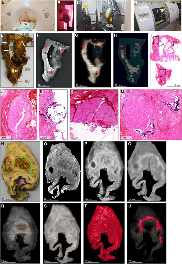
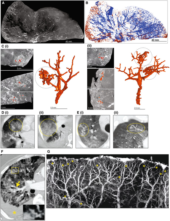
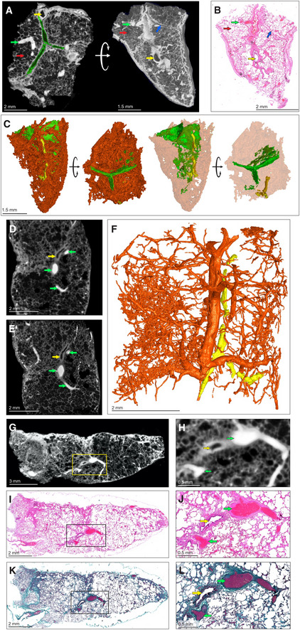
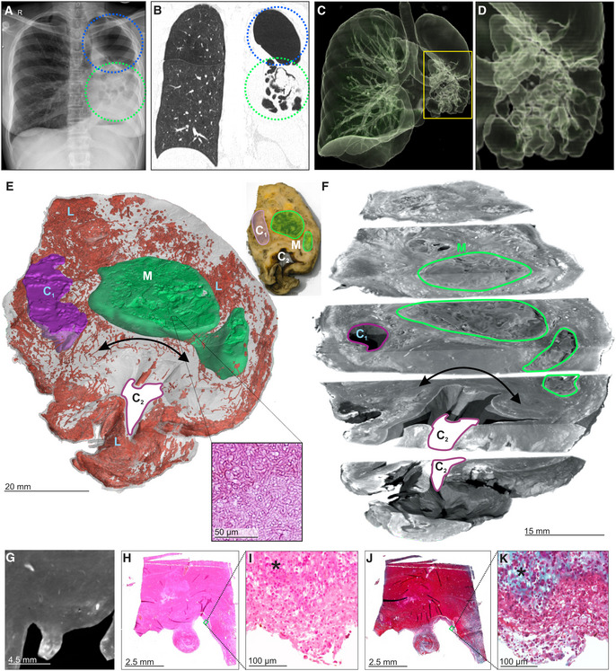
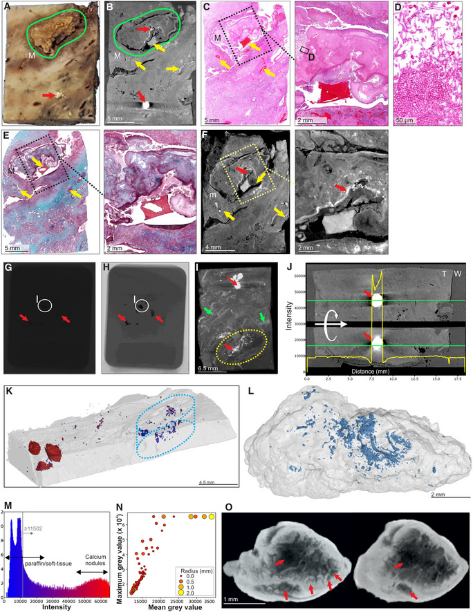
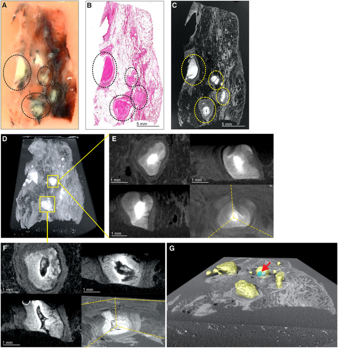
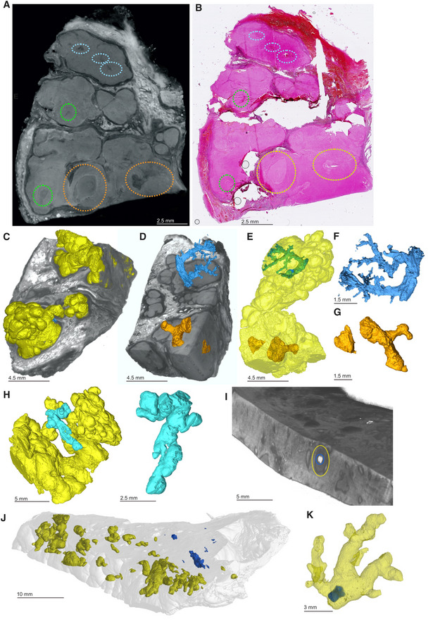
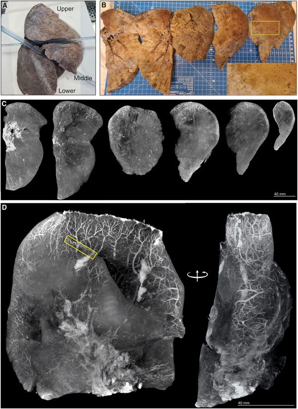
Our current understanding of the spectrum of TB and COVID-19 lesions in the human lung is limited by a reliance on low-resolution imaging platforms that cannot provide accurate 3D representations of lesion types within the context of the whole lung. To characterize TB and COVID-19 lesions in 3D, we applied micro/nanocomputed tomography to surgically resected, postmortem, and paraffin-embedded human lung tissue. We define a spectrum of TB pathologies, including cavitary lesions, calcium deposits outside and inside necrotic granulomas and mycetomas, and vascular rearrangement. We identified an unusual spatial arrangement of vasculature within an entire COVID-19 lobe, and 3D segmentation of blood vessels revealed microangiopathy associated with hemorrhage. Notably, segmentation of pathological anomalies reveals hidden pathological structures that might otherwise be disregarded, demonstrating a powerful method to visualize pathologies in 3D in TB lung tissue and whole COVID-19 lobes. These findings provide unexpected new insight into the spatial organization of the spectrum of TB and COVID-19 lesions within the framework of the entire lung.
我们目前对人类肺部中结核和 COVID-19 病变的认识受到了限制,因为我们依赖于低分辨率的成像平台,这些平台无法在整个肺部的背景下提供病变类型的准确 3D 表示。为了对结核和 COVID-19 病变进行 3D 分析,我们应用微/纳米计算机断层扫描技术对手术切除、尸检和石蜡包埋的人类肺组织进行了分析。我们定义了一系列结核病理学特征,包括空洞性病变、坏死性肉芽肿和真菌病内外的钙沉积,以及血管重排。我们发现了 COVID-19 整个肺叶中一种不寻常的血管空间排列方式,并且对血管的 3D 分割揭示了与出血相关的微血管病变。值得注意的是,病理异常的分割揭示了隐藏的病理结构,如果不进行分割,这些结构可能会被忽视,这证明了一种在结核肺组织和整个 COVID-19 肺叶中可视化 3D 病理的强大方法。这些发现为我们提供了在整个肺部框架内对结核和 COVID-19 病变谱的空间组织的意外新见解。