• 文献检索
  • 文档翻译
  • 深度研究
  • 学术资讯
  • Suppr Zotero 插件Zotero 插件
  • 邀请有礼
  • 套餐&价格
  • 历史记录
应用&插件
Suppr Zotero 插件Zotero 插件浏览器插件Mac 客户端Windows 客户端微信小程序
定价
高级版会员购买积分包购买API积分包
服务
文献检索文档翻译深度研究API 文档MCP 服务
关于我们
关于 Suppr公司介绍联系我们用户协议隐私条款
关注我们

Suppr 超能文献

核心技术专利:CN118964589B侵权必究
粤ICP备2023148730 号-1Suppr @ 2026

文献检索

告别复杂PubMed语法,用中文像聊天一样搜索,搜遍4000万医学文献。AI智能推荐,让科研检索更轻松。

立即免费搜索

文件翻译

保留排版,准确专业,支持PDF/Word/PPT等文件格式,支持 12+语言互译。

免费翻译文档

深度研究

AI帮你快速写综述,25分钟生成高质量综述,智能提取关键信息,辅助科研写作。

立即免费体验

遗传性额颞叶痴呆症无症状携带者的纵向脑萎缩率。

Longitudinal Brain Atrophy Rates in Presymptomatic Carriers of Genetic Frontotemporal Dementia.

机构信息

From the Department of Neurology and Alzheimer Center Erasmus MC (Jackie M. Poos, L.D.M.G., E.L.E., J.L.P., Janne M. Papma, H.S., Esther van den Berg, J.S., L.C.J.), Erasmus MC University Medical Center; Quantib B.V. (R.K.), Rotterdam; Departments of Radiology and Nuclear Medicine (Esther Bron, R.S., M.W.V.) and Epidemiology (M.W.V.), Erasmus MC University Medical Center Rotterdam; Department of Neurology (Y.A.L.P.), Alzheimer Center, Location VU University Medical Center Amsterdam Neuroscience, Amsterdam University Medical Center; Department of Radiology (S.A.R.B.R.), Leiden University Medical Center; Institute of Psychology (S.A.R.B.R.) and Leiden Institute for Brain and Cognition (S.A.R.B.R.), Leiden University, The Netherlands; and Dementia Research Centre (L.C.J.), Department of Neurodegenerative Disease, UCL Institute of Neurology, London, United Kingdom.

出版信息

Neurology. 2022 Dec 12;99(24):e2661-e2671. doi: 10.1212/WNL.0000000000201292.

DOI:10.1212/WNL.0000000000201292
PMID:36288997
原文链接:https://pmc.ncbi.nlm.nih.gov/articles/PMC9757869/
Abstract

BACKGROUND AND OBJECTIVES

It is important to identify at what age brain atrophy rates in genetic frontotemporal dementia (FTD) start to accelerate and deviate from normal aging effects to find the optimal starting point for treatment. We investigated longitudinal brain atrophy rates in the presymptomatic stage of genetic FTD using normative brain volumetry software.

METHODS

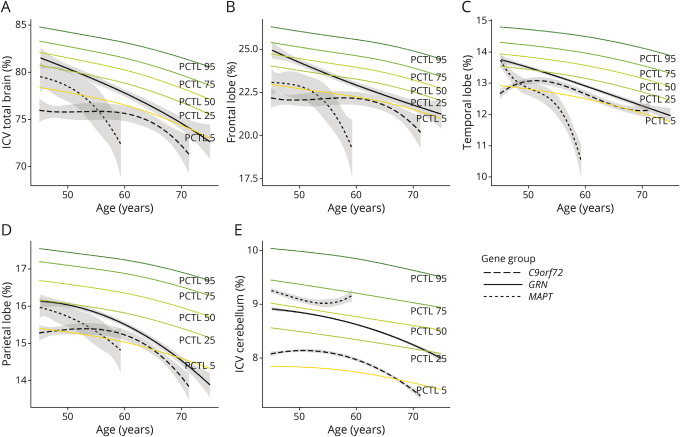
Presymptomatic , , and pathogenic variant carriers underwent longitudinal volumetric T1-weighted magnetic resonance imaging of the brain as part of a prospective cohort study. Images were automatically analyzed with Quantib® ND, which consisted of volume measurements (CSF and sum of gray and white matter) of lobes, cerebellum, and hippocampus. All volumes were compared with reference centile curves based on a large population-derived sample of nondemented individuals (n = 4,951). Mixed-effects models were fitted to analyze atrophy rates of the different gene groups as a function of age.

RESULTS

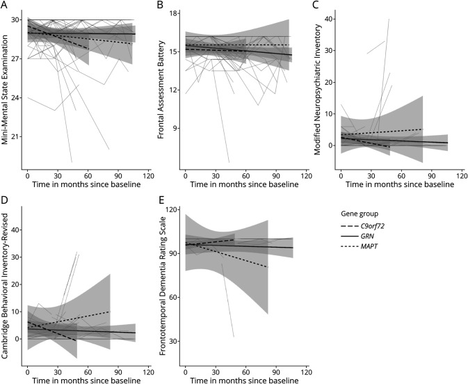
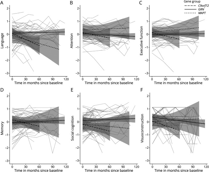
Thirty-four , 8 , and 14 pathogenic variant carriers were included (mean age = 52.1, standard deviation = 7.2; 66% female). The mean follow-up duration of the study was 64 ± 33 months (median = 52; range 13-108). pathogenic variant carriers showed a faster decline than the reference centile curves for all brain areas, though relative volumes remained between the 5th and 75th percentiles between the ages of 45 and 70 years. In pathogenic variant carriers, frontal lobe volume was already at the 5th percentile at age 45 years and showed a further decline between the ages 50 and 60 years. Temporal lobe volume started in the 50th percentile at age 45 years but showed fastest decline over time compared with other brain structures. Frontal, temporal, parietal, and cerebellar volume already started below the 5th percentile compared with the reference centile curves at age 45 years for pathogenic variant carriers, but there was minimal decline over time until the age of 60 years.

DISCUSSION

We provide evidence for longitudinal brain atrophy in the presymptomatic stage of genetic FTD. The affected brain areas and the age after which atrophy rates start to accelerate and diverge from normal aging slopes differed between gene groups. These results highlight the value of normative volumetry software for disease tracking and staging biomarkers in genetic FTD. These techniques could help in identifying the optimal time window for starting treatment and monitoring treatment response.

摘要

背景与目的

确定遗传性额颞叶痴呆(FTD)患者的脑萎缩速度从何时开始加速并偏离正常衰老效应,从而找到治疗的最佳起始点非常重要。我们使用标准脑容量测定软件研究了遗传性 FTD 患者的无症状期的纵向脑萎缩速度。

方法

作为前瞻性队列研究的一部分,无症状的、携带致病性变异的患者接受了脑纵向容积 T1 加权磁共振成像检查。使用 Quantib® ND 自动分析图像,该软件包括基于无痴呆个体(n = 4951)的大型人群样本的脑叶、小脑和海马体的容积测量值(CSF 和灰质及白质总和)。所有体积均与参考百分位曲线进行比较。采用混合效应模型分析不同基因组的萎缩率随年龄的变化。

结果

共纳入 34 名、8 名和 14 名携带致病性变异的患者(平均年龄 = 52.1,标准差 = 7.2;66%为女性)。研究的平均随访时间为 64 ± 33 个月(中位数 = 52;范围 13-108)。所有脑区的 携带致病性变异的患者的下降速度均快于参考百分位曲线,但在 45 至 70 岁之间,相对体积仍保持在第 5 至 75 百分位之间。在 携带致病性变异的患者中,额叶体积在 45 岁时已处于第 5 百分位,并在 50 至 60 岁之间进一步下降。颞叶体积在 45 岁时处于第 50 百分位,但与其他脑结构相比,随时间的推移下降最快。携带致病性变异的患者的额、颞、顶和小脑体积在 45 岁时已经低于参考百分位曲线的第 5 百分位,但直到 60 岁时,体积才出现最小程度的下降。

讨论

我们提供了遗传性 FTD 无症状期纵向脑萎缩的证据。受影响的脑区以及萎缩速度开始加速并偏离正常衰老斜率的年龄因基因组而异。这些结果突出了标准容量测定软件在遗传性 FTD 疾病跟踪和分期生物标志物方面的价值。这些技术可以帮助确定开始治疗的最佳时间窗口,并监测治疗反应。

https://cdn.ncbi.nlm.nih.gov/pmc/blobs/3e56/9757869/54ca8489306f/WNL-2022-201191f3.jpg
https://cdn.ncbi.nlm.nih.gov/pmc/blobs/3e56/9757869/ab29a9a13c81/WNL-2022-201191f1.jpg
https://cdn.ncbi.nlm.nih.gov/pmc/blobs/3e56/9757869/6a38f55cb8ea/WNL-2022-201191f2.jpg
https://cdn.ncbi.nlm.nih.gov/pmc/blobs/3e56/9757869/54ca8489306f/WNL-2022-201191f3.jpg
https://cdn.ncbi.nlm.nih.gov/pmc/blobs/3e56/9757869/ab29a9a13c81/WNL-2022-201191f1.jpg
https://cdn.ncbi.nlm.nih.gov/pmc/blobs/3e56/9757869/6a38f55cb8ea/WNL-2022-201191f2.jpg
https://cdn.ncbi.nlm.nih.gov/pmc/blobs/3e56/9757869/54ca8489306f/WNL-2022-201191f3.jpg

相似文献

1
Longitudinal Brain Atrophy Rates in Presymptomatic Carriers of Genetic Frontotemporal Dementia.遗传性额颞叶痴呆症无症状携带者的纵向脑萎缩率。
Neurology. 2022 Dec 12;99(24):e2661-e2671. doi: 10.1212/WNL.0000000000201292.
2
Rates of Brain Atrophy Across Disease Stages in Familial Frontotemporal Dementia Associated With MAPT, GRN, and C9orf72 Pathogenic Variants.携带 MAPT、GRN 和 C9orf72 致病性变异的家族性额颞叶痴呆在疾病各阶段的脑萎缩率。
JAMA Netw Open. 2020 Oct 1;3(10):e2022847. doi: 10.1001/jamanetworkopen.2020.22847.
3
Frequency and Longitudinal Course of Behavioral and Neuropsychiatric Symptoms in Participants With Genetic Frontotemporal Dementia.遗传性额颞叶痴呆患者行为和神经精神症状的频率及纵向病程
Neurology. 2024 Oct 22;103(8):e209569. doi: 10.1212/WNL.0000000000209569. Epub 2024 Sep 16.
4
Patterns of regional cerebellar atrophy in genetic frontotemporal dementia.遗传性额颞叶痴呆的小脑区域萎缩模式
Neuroimage Clin. 2016 Feb 21;11:287-290. doi: 10.1016/j.nicl.2016.02.008. eCollection 2016.
5
Brain atrophy over time in genetic and sporadic frontotemporal dementia: a study of 198 serial magnetic resonance images.遗传型和散发型额颞叶痴呆患者脑萎缩随时间变化的研究:198例连续磁共振成像分析
Eur J Neurol. 2015 May;22(5):745-52. doi: 10.1111/ene.12675. Epub 2015 Feb 12.
6
Distinct Neuroanatomical Correlates of Neuropsychiatric Symptoms in the Three Main Forms of Genetic Frontotemporal Dementia in the GENFI Cohort.在 GENFI 队列中,三种主要遗传形式额颞叶痴呆的神经精神症状有不同的神经解剖学相关性。
J Alzheimers Dis. 2018;65(1):147-163. doi: 10.3233/JAD-180053.
7
Patterns of gray matter atrophy in genetic frontotemporal dementia: results from the GENFI study.遗传性额颞叶痴呆的灰质萎缩模式:GENFI 研究结果。
Neurobiol Aging. 2018 Feb;62:191-196. doi: 10.1016/j.neurobiolaging.2017.10.008. Epub 2017 Oct 19.
8
Gray and white matter changes in presymptomatic genetic frontotemporal dementia: a longitudinal MRI study.遗传前驱性额颞叶痴呆的灰白质变化:一项纵向 MRI 研究。
Neurobiol Aging. 2019 Apr;76:115-124. doi: 10.1016/j.neurobiolaging.2018.12.017. Epub 2019 Jan 7.
9
Prodromal language impairment in genetic frontotemporal dementia within the GENFI cohort.遗传型额颞叶痴呆患者在 GENFI 队列中的前驱性语言障碍。
J Neurol Sci. 2023 Aug 15;451:120711. doi: 10.1016/j.jns.2023.120711. Epub 2023 Jun 10.
10
A modified Camel and Cactus Test detects presymptomatic semantic impairment in genetic frontotemporal dementia within the GENFI cohort.改良骆驼和仙人掌测试在 GENFI 队列中检测到遗传额颞叶痴呆的无症状语义障碍。
Appl Neuropsychol Adult. 2022 Jan-Feb;29(1):112-119. doi: 10.1080/23279095.2020.1716357. Epub 2020 Feb 5.

引用本文的文献

1
AI-based staging, causal hypothesis and progression of subjects at risk of Alzheimer's disease: a multicenter study.基于人工智能的阿尔茨海默病高危受试者的分期、因果假设及病情进展:一项多中心研究
Front Neurol. 2025 May 19;16:1568086. doi: 10.3389/fneur.2025.1568086. eCollection 2025.
2
Atlas of Proteomic signatures of brain structure and its links to brain disorders.脑结构蛋白质组学特征图谱及其与脑部疾病的关联
Nat Commun. 2025 Jun 2;16(1):5092. doi: 10.1038/s41467-025-60185-7.
3
Sex differences in the executive and behavioral reserve of autosomal dominant frontotemporal dementia.
常染色体显性遗传性额颞叶痴呆患者执行功能及行为储备方面的性别差异
Alzheimers Dement. 2025 Apr;21(4):e70070. doi: 10.1002/alz.70070.
4
Biomarker-guided decision making in clinical drug development for neurodegenerative disorders.神经退行性疾病临床药物研发中的生物标志物引导决策
Nat Rev Drug Discov. 2025 Apr 4. doi: 10.1038/s41573-025-01165-w.
5
Neuropsychological Profiles in Genetic Frontotemporal Dementia: A Meta-Analysis and Systematic Review.遗传性额颞叶痴呆的神经心理学特征:一项荟萃分析与系统评价
Aging Dis. 2024 Jun 24;16(3):1378-1396. doi: 10.14336/AD.2024.0183.
6
Genetic evidence for the causal relationships between migraine, dementia, and longitudinal brain atrophy.遗传证据表明偏头痛、痴呆症与纵向脑萎缩之间存在因果关系。
J Headache Pain. 2024 Jun 5;25(1):93. doi: 10.1186/s10194-024-01801-7.
7
A mutation in the PRKAR1B gene drives pathological mechanisms of neurodegeneration across species.PRKAR1B 基因突变驱动跨物种的神经退行性病变理机制。
Brain. 2024 Nov 4;147(11):3890-3905. doi: 10.1093/brain/awae154.
8
Clinical and neuroimaging characterization of the first frontotemporal dementia family carrying the MAPT p.K298E mutation.携带 MAPT p.K298E 突变的首个额颞叶痴呆家系的临床和神经影像学特征。
Neurogenetics. 2024 Jul;25(3):215-223. doi: 10.1007/s10048-024-00756-w. Epub 2024 Apr 9.
9
Cortical thickness modeling and variability in Alzheimer's disease and frontotemporal dementia.阿尔茨海默病和额颞叶痴呆的皮质厚度建模和变异性。
J Neurol. 2024 Mar;271(3):1428-1438. doi: 10.1007/s00415-023-12087-1. Epub 2023 Nov 27.
10
Presymptomatic and early pathological features of MAPT-associated frontotemporal lobar degeneration.MAPT 相关额颞叶痴呆的前驱期和早期病理学特征。
Acta Neuropathol Commun. 2023 Aug 2;11(1):126. doi: 10.1186/s40478-023-01588-9.