Ploeg Meike C, Munts Chantal, Seddiqi Tayeba, Ten Brink Tim J L, Breemhaar Jonathan, Moroni Lorenzo, Prinzen Frits W, van Nieuwenhoven Frans A
Department of Physiology, Cardiovascular Research Institute Maastricht (CARIM), Maastricht University, 6200 MD Maastricht, The Netherlands.
Institute for Technology-Inspired Regenerative Medicine (MERLN), Maastricht University, 6200 MD Maastricht, The Netherlands.
Bioengineering (Basel). 2022 Oct 14;9(10):551. doi: 10.3390/bioengineering9100551.
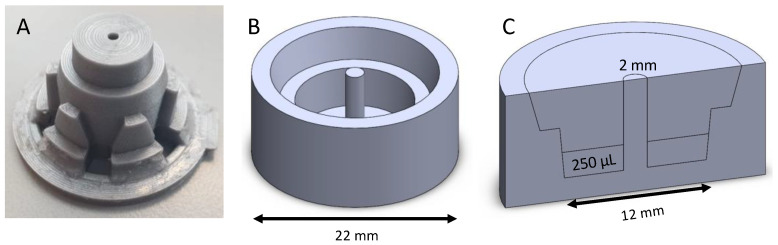
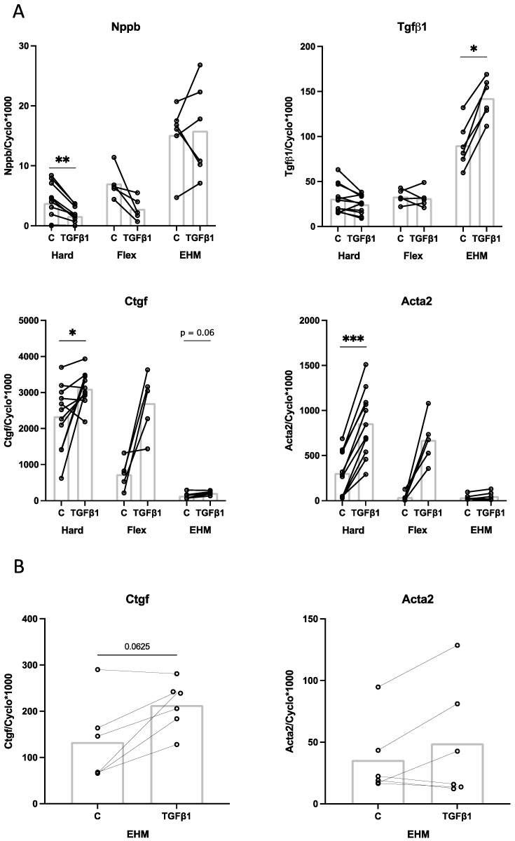
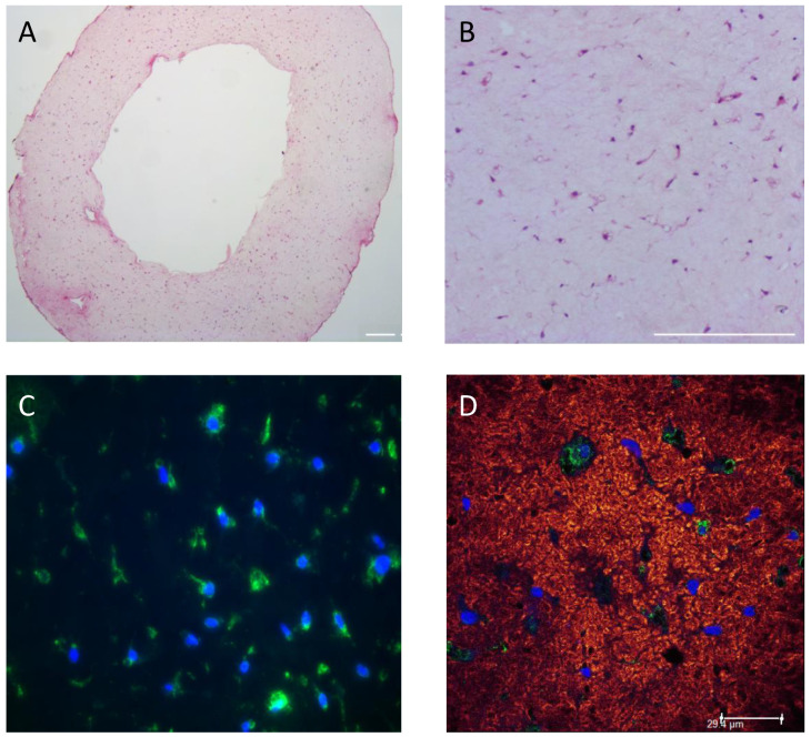
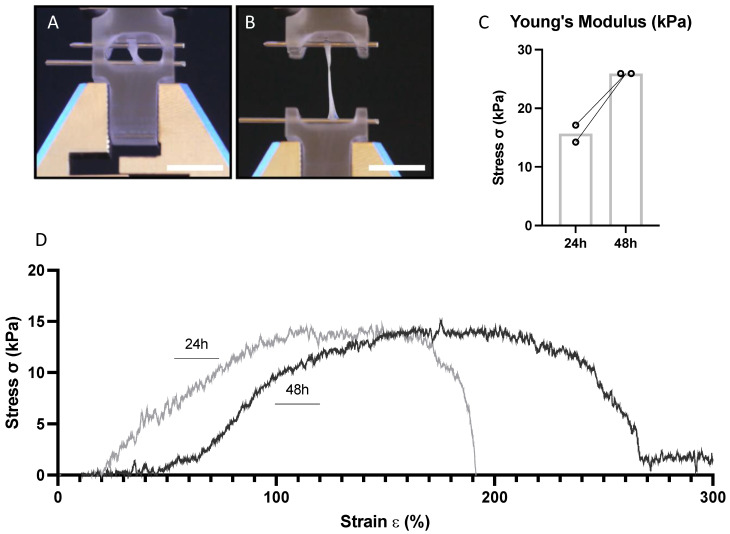
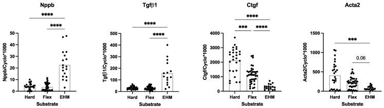
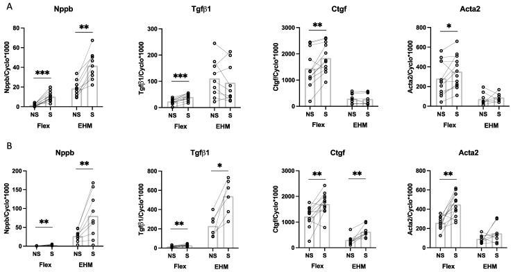
Isolation and culturing of cardiac fibroblasts (CF) induces rapid differentiation toward a myofibroblast phenotype, which is partly mediated by the high substrate stiffness of the culture plates. In the present study, a 3D model of Engineered Heart Matrix (EHM) of physiological stiffness (Youngs modulus ~15 kPa) was developed using primary adult rat CF and a natural hydrogel collagen type 1 matrix. CF were equally distributed, viable and quiescent for at least 13 days in EHM and the baseline gene expression of myofibroblast-markers alfa-smooth muscle actin (Acta2), and connective tissue growth factor (Ctgf) was significantly lower, compared to CF cultured in 2D monolayers. CF baseline gene expression of transforming growth factor-beta1 (Tgfβ1) and brain natriuretic peptide (Nppb) was higher in EHM-fibers compared to the monolayers. EHM stimulation by 10% cyclic stretch (1 Hz) increased the gene expression of Nppb (3.0-fold), Ctgf (2.1-fold) and Tgfβ1 (2.3-fold) after 24 h. Stimulation of EHM with TGFβ1 (1 ng/mL, 24 h) induced Tgfβ1 (1.6-fold) and Ctgf (1.6-fold). In conclusion, culturing CF in EHM of physiological stiffness reduced myofibroblast marker gene expression, while the CF response to stretch or TGFβ1 was maintained, indicating that our novel EHM structure provides a good physiological model to study CF function and myofibroblast differentiation.
心脏成纤维细胞(CF)的分离和培养会诱导其迅速向肌成纤维细胞表型分化,这部分是由培养板的高底物硬度介导的。在本研究中,使用原代成年大鼠CF和天然水凝胶I型胶原基质构建了具有生理硬度(杨氏模量~15 kPa)的工程心脏基质(EHM)三维模型。CF在EHM中均匀分布、存活且至少13天保持静止,与二维单层培养的CF相比,肌成纤维细胞标志物α-平滑肌肌动蛋白(Acta2)和结缔组织生长因子(Ctgf)的基线基因表达显著降低。与单层培养相比,EHM纤维中转化生长因子-β1(Tgfβ1)和脑钠肽(Nppb)的CF基线基因表达更高。10%循环拉伸(1 Hz)刺激EHM 24小时后,Nppb(3.0倍)、Ctgf(2.1倍)和Tgfβ1(2.3倍)的基因表达增加。用TGFβ1(1 ng/mL,24小时)刺激EHM可诱导Tgfβ1(1.6倍)和Ctgf(1.6倍)。总之,在具有生理硬度的EHM中培养CF可降低肌成纤维细胞标志物基因表达,同时维持CF对拉伸或TGFβ1的反应,这表明我们的新型EHM结构为研究CF功能和肌成纤维细胞分化提供了一个良好的生理模型。