• 文献检索
  • 文档翻译
  • 深度研究
  • 学术资讯
  • Suppr Zotero 插件Zotero 插件
  • 邀请有礼
  • 套餐&价格
  • 历史记录
应用&插件
Suppr Zotero 插件Zotero 插件浏览器插件Mac 客户端Windows 客户端微信小程序
定价
高级版会员购买积分包购买API积分包
服务
文献检索文档翻译深度研究API 文档MCP 服务
关于我们
关于 Suppr公司介绍联系我们用户协议隐私条款
关注我们

Suppr 超能文献

核心技术专利:CN118964589B侵权必究
粤ICP备2023148730 号-1Suppr @ 2026

文献检索

告别复杂PubMed语法,用中文像聊天一样搜索,搜遍4000万医学文献。AI智能推荐,让科研检索更轻松。

立即免费搜索

文件翻译

保留排版,准确专业,支持PDF/Word/PPT等文件格式,支持 12+语言互译。

免费翻译文档

深度研究

AI帮你快速写综述,25分钟生成高质量综述,智能提取关键信息,辅助科研写作。

立即免费体验

基于 CBCT 的自适应放疗的测试时优化分割。

Segmentation by test-time optimization for CBCT-based adaptive radiation therapy.

机构信息

Medical Artificial Intelligence and Automation (MAIA) Laboratory, Department of Radiation Oncology, University of Texas Southwestern Medical Center, Dallas, Texas, USA.

Department of Radiation Oncology, Yonsei Cancer Center, Yonsei University College of Medicine, Seoul, South Korea.

出版信息

Med Phys. 2023 Apr;50(4):1947-1961. doi: 10.1002/mp.15960. Epub 2022 Nov 12.

DOI:10.1002/mp.15960
PMID:36310403
原文链接:https://pmc.ncbi.nlm.nih.gov/articles/PMC10121749/
Abstract

PURPOSE

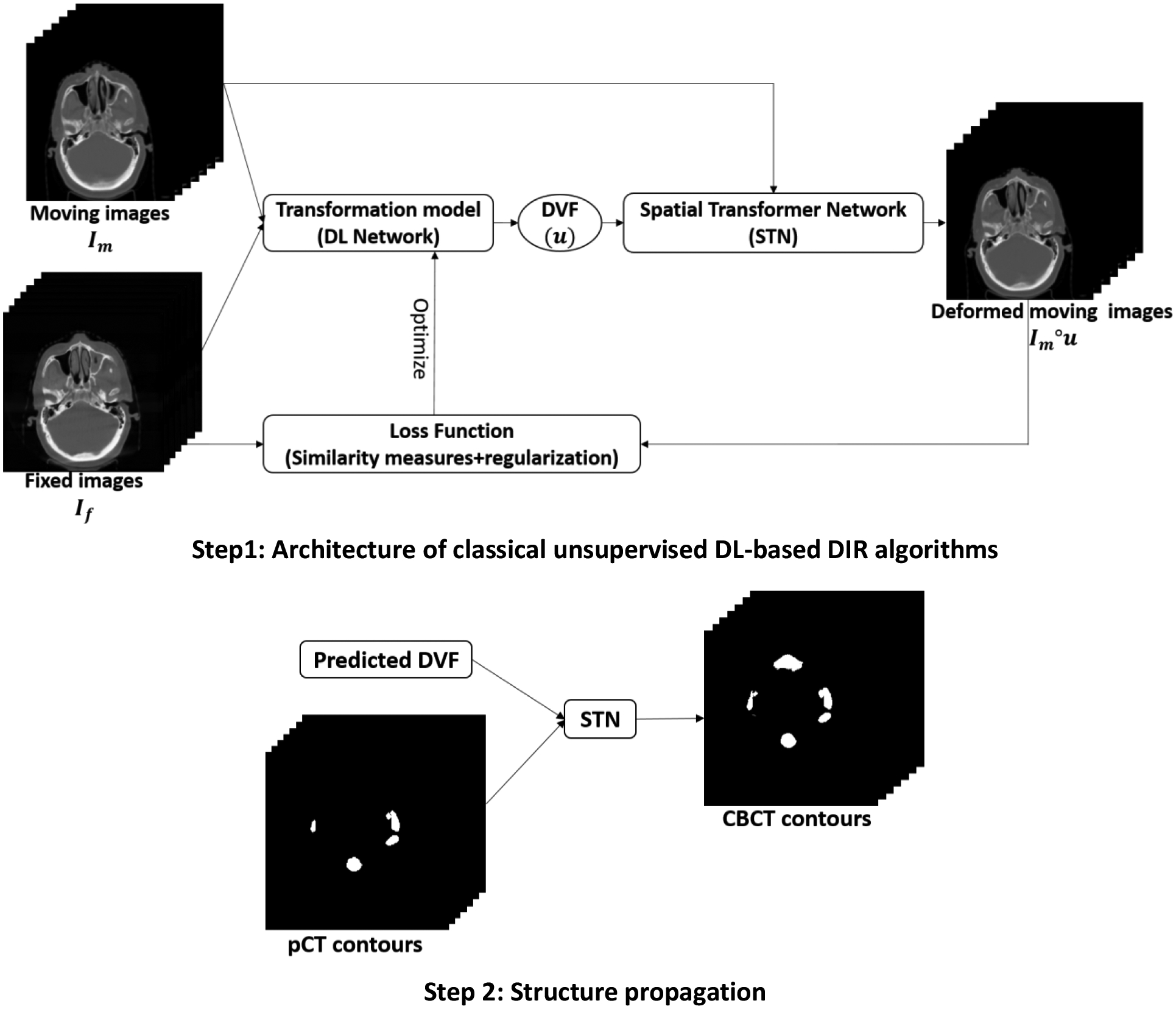
Online adaptive radiotherapy (ART) requires accurate and efficient auto-segmentation of target volumes and organs-at-risk (OARs) in mostly cone-beam computed tomography (CBCT) images, which often have severe artifacts and lack soft-tissue contrast, making direct segmentation very challenging. Propagating expert-drawn contours from the pretreatment planning CT through traditional or deep learning (DL)-based deformable image registration (DIR) can achieve improved results in many situations. Typical DL-based DIR models are population based, that is, trained with a dataset for a population of patients, and so they may be affected by the generalizability problem.

METHODS

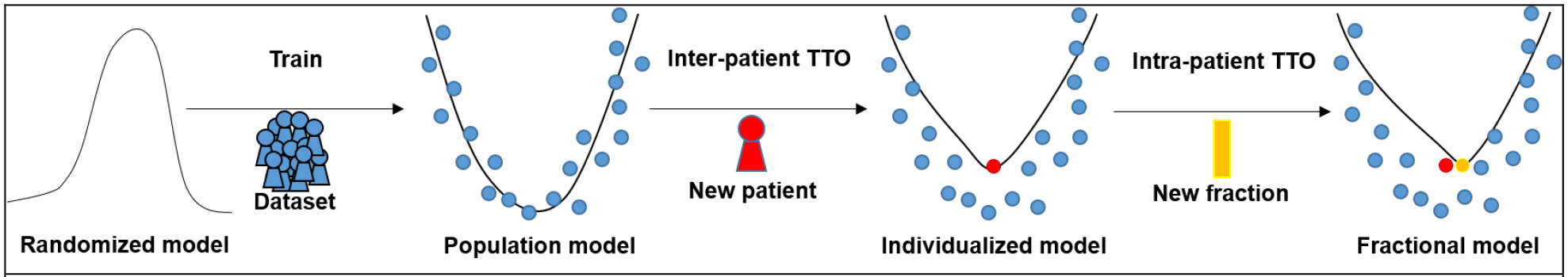
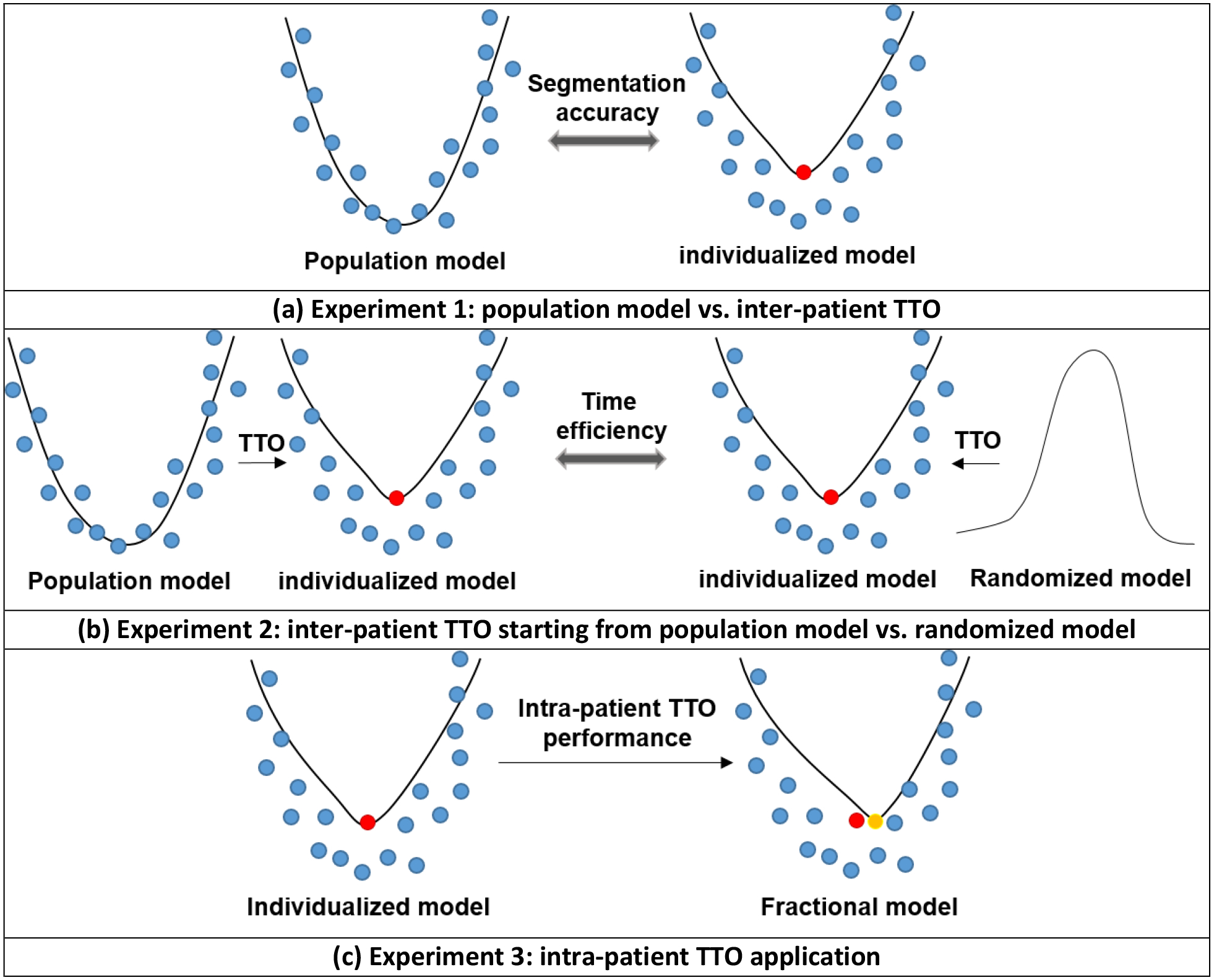
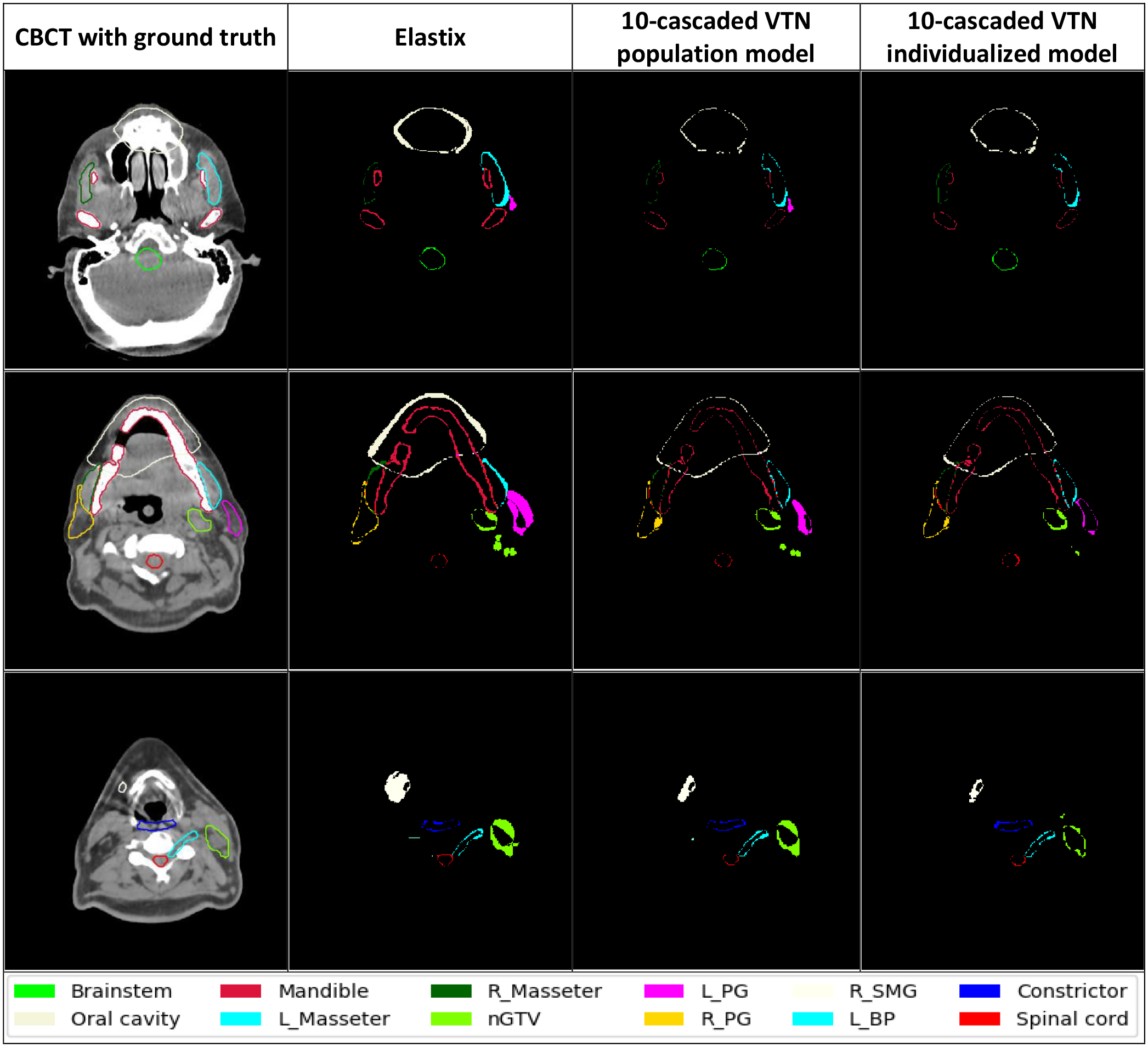
In this paper, we propose a method called test-time optimization (TTO) to refine a pretrained DL-based DIR population model, first for each individual test patient, and then progressively for each fraction of online ART treatment. Our proposed method is less susceptible to the generalizability problem and thus can improve overall performance of different DL-based DIR models by improving model accuracy, especially for outliers. Our experiments used data from 239 patients with head-and-neck squamous cell carcinoma to test the proposed method. First, we trained a population model with 200 patients and then applied TTO to the remaining 39 test patients by refining the trained population model to obtain 39 individualized models. We compared each of the individualized models with the population model in terms of segmentation accuracy.

RESULTS

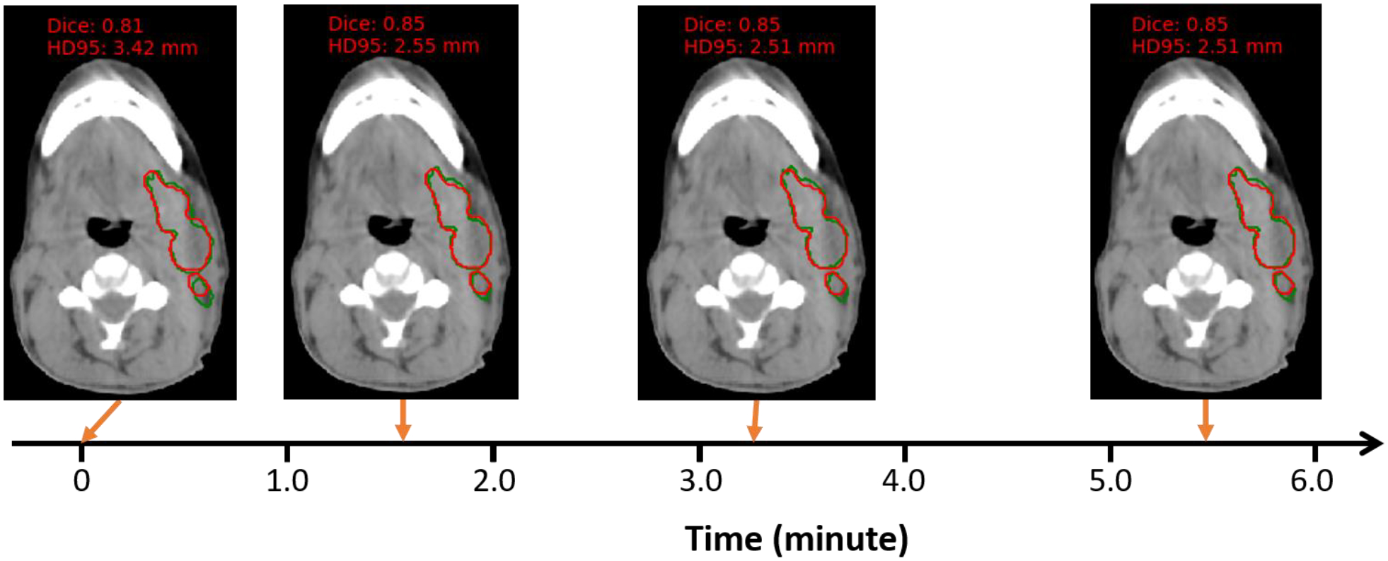
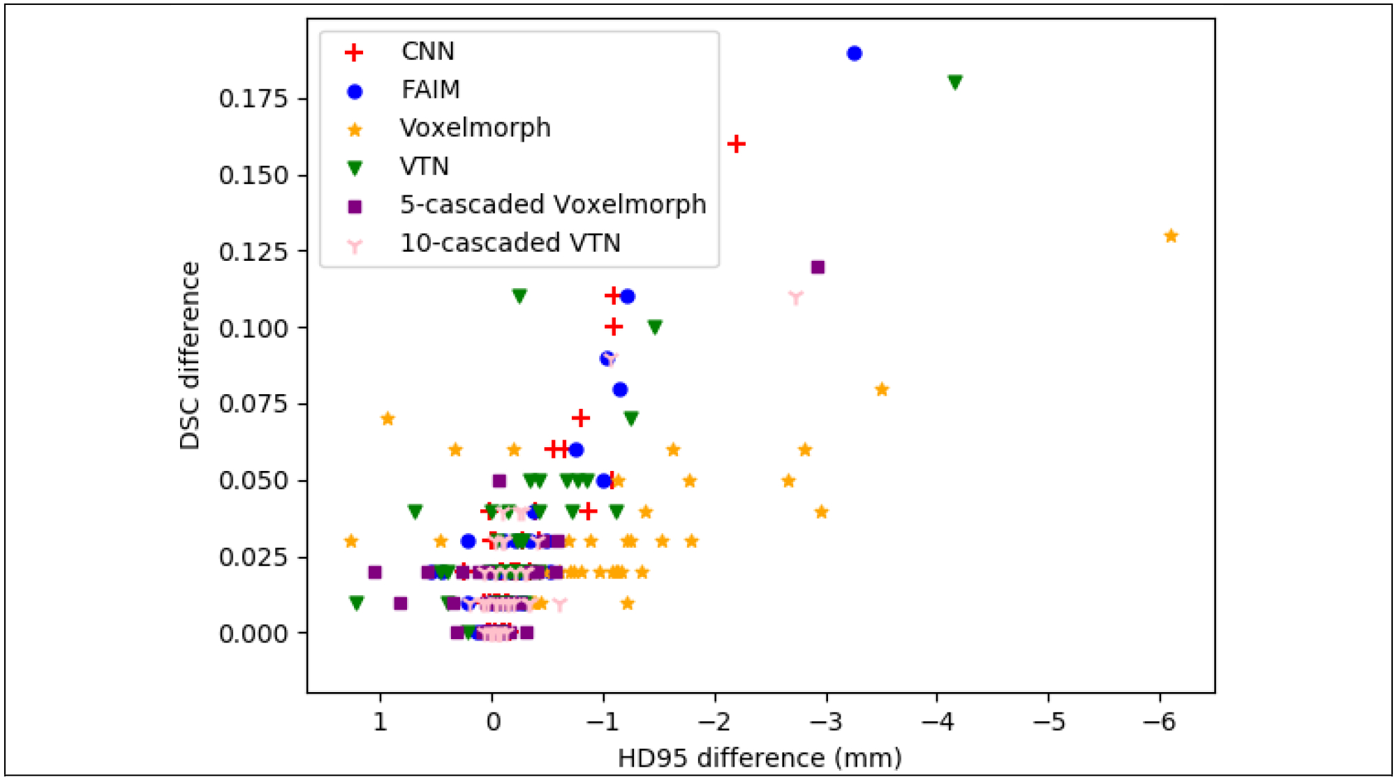
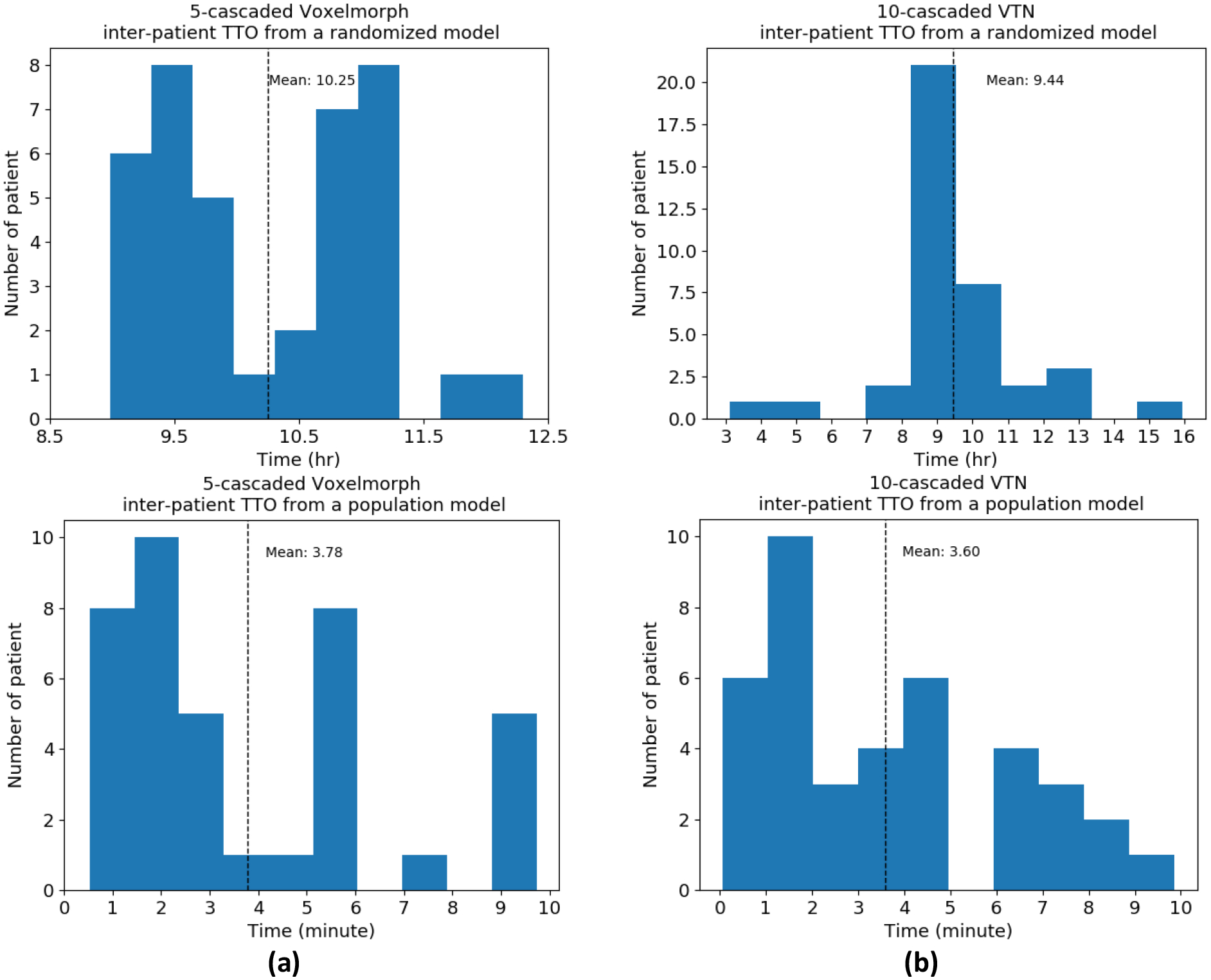
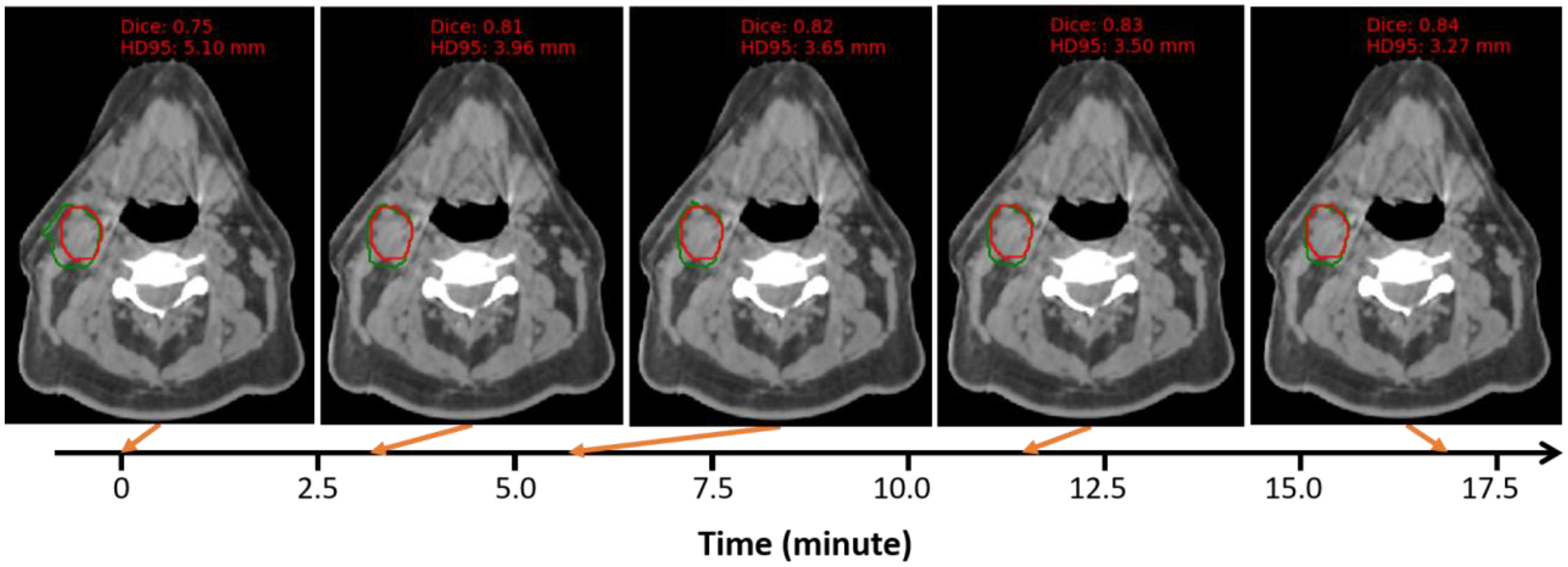
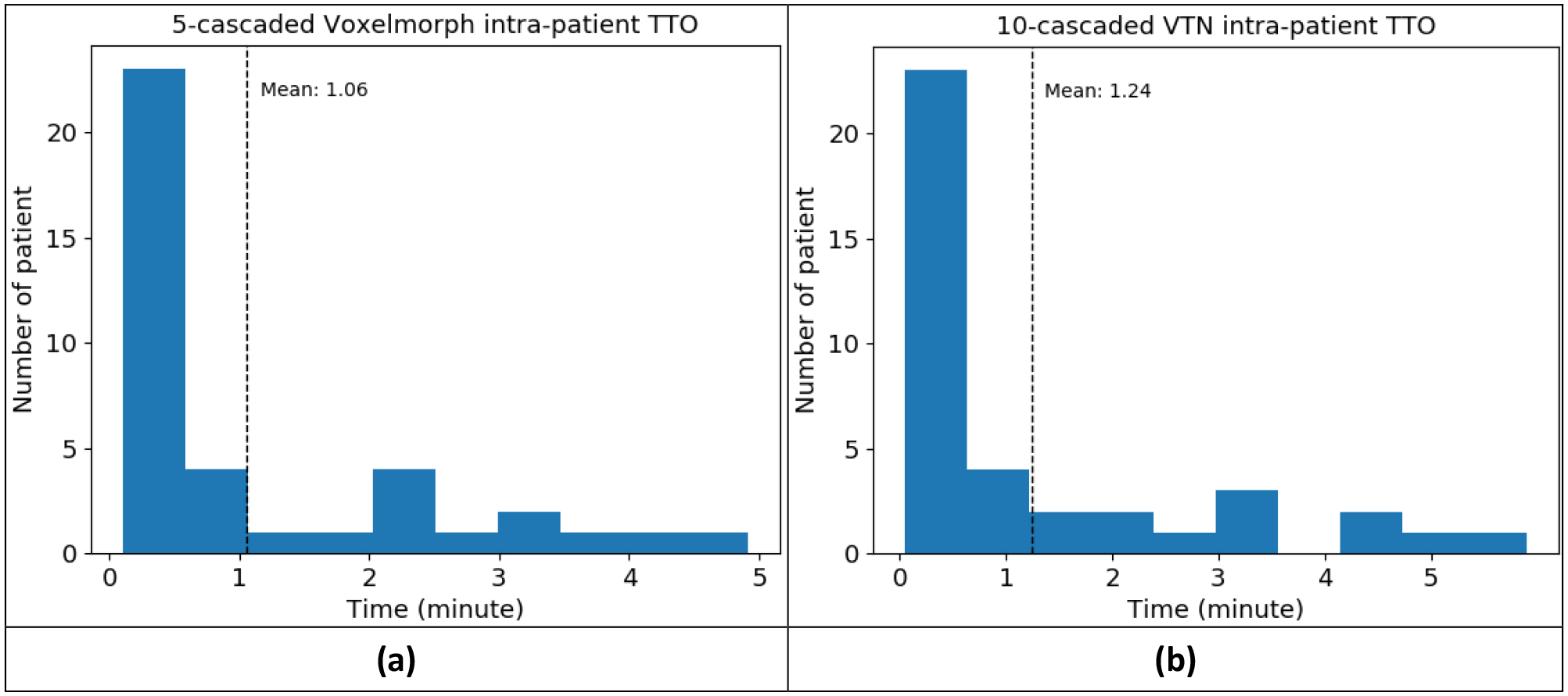
The average improvement of the Dice similarity coefficient (DSC) and 95% Hausdorff distance (HD95) of segmentation can be up to 0.04 (5%) and 0.98 mm (25%), respectively, with the individualized models compared to the population model over 17 selected OARs and a target of 39 patients. Although the average improvement may seem mild, we found that the improvement for outlier patients with structures of large anatomical changes is significant. The number of patients with at least 0.05 DSC improvement or 2 mm HD95 improvement by TTO averaged over the 17 selected structures for the state-of-the-art architecture VoxelMorph is 10 out of 39 test patients. By deriving the individualized model using TTO from the pretrained population model, TTO models can be ready in about 1 min. We also generated the adapted fractional models for each of the 39 test patients by progressively refining the individualized models using TTO to CBCT images acquired at later fractions of online ART treatment. When adapting the individualized model to a later fraction of the same patient, the model can be ready in less than a minute with slightly improved accuracy.

CONCLUSIONS

The proposed TTO method is well suited for online ART and can boost segmentation accuracy for DL-based DIR models, especially for outlier patients where the pretrained models fail.

摘要

目的

在线自适应放疗(ART)需要在大多数锥形束 CT(CBCT)图像中准确高效地对靶区和危及器官(OAR)进行自动分割,这些图像通常存在严重的伪影,且软组织对比度差,使得直接分割极具挑战性。通过传统或基于深度学习(DL)的变形图像配准(DIR)将专家绘制的轮廓从预处理计划 CT 传播到可以在许多情况下获得改进的结果。典型的基于 DL 的 DIR 模型是基于人群的,也就是说,使用针对人群患者的数据集进行训练,因此它们可能受到泛化问题的影响。

方法

在本文中,我们提出了一种称为测试时优化(TTO)的方法,用于首先为每个单独的测试患者,然后为在线 ART 治疗的每个部分逐渐改进预先训练的基于 DL 的 DIR 人群模型。我们提出的方法不易受到泛化问题的影响,因此可以通过提高模型准确性(特别是对异常值)来提高不同基于 DL 的 DIR 模型的整体性能。我们的实验使用了 239 名头颈部鳞状细胞癌患者的数据来测试所提出的方法。首先,我们使用 200 名患者训练了一个人群模型,然后通过对其余 39 名测试患者应用 TTO 来细化训练好的人群模型,以获得 39 个个体化模型。我们比较了每个个体化模型与人群模型在分割准确性方面的差异。

结果

与人群模型相比,17 个选定 OAR 和 39 名患者的目标的分割的 Dice 相似系数(DSC)和 95%Hausdorff 距离(HD95)的平均改善可达 0.04(5%)和 0.98mm(25%)。虽然平均提高幅度可能看起来很小,但我们发现,对于结构变化较大的异常值患者,改进效果显著。通过 TTO 从预先训练的人群模型中得出个体化模型,对于 17 个选定结构,使用 VoxelMorph 等最先进的架构,39 名测试患者中至少有 0.05 DSC 改善或 2mm HD95 改善的患者人数平均为 10 名。通过使用 TTO 从预先训练的人群模型中生成个体化模型,大约 1 分钟内即可准备好 TTO 模型。我们还通过使用 TTO 逐步细化个体化模型,为在线 ART 治疗的每个后续部分生成了每个 39 名测试患者的适应分数模型。当将个体化模型适配到同一患者的后续部分时,模型可以在不到一分钟的时间内准备就绪,并且准确性略有提高。

结论

所提出的 TTO 方法非常适合在线 ART,可以提高基于 DL 的 DIR 模型的分割准确性,特别是对于预训练模型失败的异常值患者。

https://cdn.ncbi.nlm.nih.gov/pmc/blobs/be35/10121749/6b2ac91bacaa/nihms-1863124-f0009.jpg
https://cdn.ncbi.nlm.nih.gov/pmc/blobs/be35/10121749/5b00408328c5/nihms-1863124-f0001.jpg
https://cdn.ncbi.nlm.nih.gov/pmc/blobs/be35/10121749/c232582e942b/nihms-1863124-f0002.jpg
https://cdn.ncbi.nlm.nih.gov/pmc/blobs/be35/10121749/0ff53c21b67d/nihms-1863124-f0003.jpg
https://cdn.ncbi.nlm.nih.gov/pmc/blobs/be35/10121749/d54b5bc4ccd0/nihms-1863124-f0004.jpg
https://cdn.ncbi.nlm.nih.gov/pmc/blobs/be35/10121749/4f6176b830b5/nihms-1863124-f0005.jpg
https://cdn.ncbi.nlm.nih.gov/pmc/blobs/be35/10121749/a2c351c30ac5/nihms-1863124-f0006.jpg
https://cdn.ncbi.nlm.nih.gov/pmc/blobs/be35/10121749/361a02152184/nihms-1863124-f0007.jpg
https://cdn.ncbi.nlm.nih.gov/pmc/blobs/be35/10121749/423c94e7ff40/nihms-1863124-f0008.jpg
https://cdn.ncbi.nlm.nih.gov/pmc/blobs/be35/10121749/6b2ac91bacaa/nihms-1863124-f0009.jpg

相似文献

1
Segmentation by test-time optimization for CBCT-based adaptive radiation therapy.基于 CBCT 的自适应放疗的测试时优化分割。
Med Phys. 2023 Apr;50(4):1947-1961. doi: 10.1002/mp.15960. Epub 2022 Nov 12.
2
Registration-guided deep learning image segmentation for cone beam CT-based online adaptive radiotherapy.基于锥形束 CT 的在线自适应放疗中基于配准引导的深度学习图像分割。
Med Phys. 2022 Aug;49(8):5304-5316. doi: 10.1002/mp.15677. Epub 2022 May 4.
3
Deep learning based direct segmentation assisted by deformable image registration for cone-beam CT based auto-segmentation for adaptive radiotherapy.基于深度学习的直接分割辅助的可变形图像配准在自适应放疗中基于锥形束 CT 的自动分割。
Phys Med Biol. 2023 Feb 10;68(4). doi: 10.1088/1361-6560/acb4d7.
4
Comprehensive evaluation of ten deformable image registration algorithms for contour propagation between CT and cone-beam CT images in adaptive head & neck radiotherapy.自适应头颈部放疗中用于CT与锥形束CT图像间轮廓传播的十种可变形图像配准算法的综合评估
PLoS One. 2017 Apr 17;12(4):e0175906. doi: 10.1371/journal.pone.0175906. eCollection 2017.
5
Progressively refined deep joint registration segmentation (ProRSeg) of gastrointestinal organs at risk: Application to MRI and cone-beam CT.渐进式精细化深度联合注册分割(ProRSeg)胃肠道危险器官:MRI 和锥形束 CT 的应用。
Med Phys. 2023 Aug;50(8):4758-4774. doi: 10.1002/mp.16527. Epub 2023 Jun 2.
6
Quantifying the accuracy of deformable image registration for cone-beam computed tomography with a physical phantom.使用物理体模定量评估锥形束 CT 中的形变图像配准精度。
J Appl Clin Med Phys. 2019 Oct;20(10):92-100. doi: 10.1002/acm2.12717. Epub 2019 Sep 21.
7
Synthetic CT-aided multiorgan segmentation for CBCT-guided adaptive pancreatic radiotherapy.基于合成 CT 的多器官自动勾画在锥形束 CT 引导下自适应胰腺放疗中的应用。
Med Phys. 2021 Nov;48(11):7063-7073. doi: 10.1002/mp.15264. Epub 2021 Oct 13.
8
Accuracy of software-assisted contour propagation from planning CT to cone beam CT in head and neck radiotherapy.头颈部放疗中从计划CT到锥形束CT的软件辅助轮廓传播的准确性。
Acta Oncol. 2016 Nov;55(11):1324-1330. doi: 10.1080/0284186X.2016.1185149. Epub 2016 Aug 24.
9
Deep-learning-based image registration and automatic segmentation of organs-at-risk in cone-beam CT scans from high-dose radiation treatment of pancreatic cancer.基于深度学习的图像配准和自动分割在胰腺癌高剂量放射治疗中的锥形束 CT 扫描中的危险器官
Med Phys. 2021 Jun;48(6):3084-3095. doi: 10.1002/mp.14906. Epub 2021 May 14.
10
Transfer learning for auto-segmentation of 17 organs-at-risk in the head and neck: Bridging the gap between institutional and public datasets.基于迁移学习的头颈部 17 个危及器官自动分割:弥合机构数据集和公共数据集之间的差距。
Med Phys. 2024 Jul;51(7):4767-4777. doi: 10.1002/mp.16997. Epub 2024 Feb 20.

引用本文的文献

1
Nasopharyngeal cancer adaptive radiotherapy with CBCT-derived synthetic CT: deep learning-based auto-segmentation precision and dose calculation consistency on a C-Arm linac.基于CBCT的合成CT的鼻咽癌自适应放疗:在C型臂直线加速器上基于深度学习的自动分割精度和剂量计算一致性
Radiat Oncol. 2025 Aug 28;20(1):135. doi: 10.1186/s13014-025-02715-7.
2
A systematic review of the role of artificial intelligence in automating computed tomography-based adaptive radiotherapy for head and neck cancer.人工智能在头颈部癌基于计算机断层扫描的自适应放射治疗自动化中作用的系统评价。
Phys Imaging Radiat Oncol. 2025 Feb 14;33:100731. doi: 10.1016/j.phro.2025.100731. eCollection 2025 Jan.
3

本文引用的文献

1
Clinical implementation of artificial intelligence-driven cone-beam computed tomography-guided online adaptive radiotherapy in the pelvic region.人工智能驱动的锥形束计算机断层扫描引导的盆腔区域在线自适应放射治疗的临床应用。
Phys Imaging Radiat Oncol. 2020 Dec 18;17:1-7. doi: 10.1016/j.phro.2020.12.004. eCollection 2021 Jan.
2
Adaptive Radiation Therapy (ART) Strategies and Technical Considerations: A State of the ART Review From NRG Oncology.自适应放射治疗(ART)策略与技术考量:来自NRG肿瘤学的ART综述现状
Int J Radiat Oncol Biol Phys. 2021 Mar 15;109(4):1054-1075. doi: 10.1016/j.ijrobp.2020.10.021. Epub 2020 Oct 24.
3
Progressive auto-segmentation for cone-beam computed tomography-based online adaptive radiotherapy.
基于锥束计算机断层扫描的在线自适应放疗的渐进式自动分割
Phys Imaging Radiat Oncol. 2024 Jul 14;31:100610. doi: 10.1016/j.phro.2024.100610. eCollection 2024 Jul.
4
Enhancing Radiotherapy Workflow for Head and Neck Cancer with Artificial Intelligence: A Systematic Review.利用人工智能优化头颈癌放疗流程:一项系统综述
J Pers Med. 2023 Jun 2;13(6):946. doi: 10.3390/jpm13060946.
Head-and-neck organs-at-risk auto-delineation using dual pyramid networks for CBCT-guided adaptive radiotherapy.
基于双金字塔网络的头颈部危及器官自动勾画技术在锥形束 CT 引导自适应放疗中的应用
Phys Med Biol. 2021 Feb 11;66(4):045021. doi: 10.1088/1361-6560/abd953.
4
Generating anthropomorphic phantoms using fully unsupervised deformable image registration with convolutional neural networks.使用全监督的可变形图像配准卷积神经网络生成拟人化体模。
Med Phys. 2020 Dec;47(12):6366-6380. doi: 10.1002/mp.14545. Epub 2020 Nov 9.
5
One-Shot Learning for Deformable Medical Image Registration and Periodic Motion Tracking.单次学习在可变形医学图像配准和周期性运动跟踪中的应用。
IEEE Trans Med Imaging. 2020 Jul;39(7):2506-2517. doi: 10.1109/TMI.2020.2972616. Epub 2020 Feb 10.
6
Unsupervised 3D End-to-End Medical Image Registration With Volume Tweening Network.无监督的 3D 端到端医学图像配准方法,采用体素插值网络。
IEEE J Biomed Health Inform. 2020 May;24(5):1394-1404. doi: 10.1109/JBHI.2019.2951024. Epub 2019 Nov 1.
7
Unsupervised learning of probabilistic diffeomorphic registration for images and surfaces.图像和曲面的概率微分同胚配准的无监督学习
Med Image Anal. 2019 Oct;57:226-236. doi: 10.1016/j.media.2019.07.006. Epub 2019 Jul 12.
8
Universal Approximation Using Radial-Basis-Function Networks.使用径向基函数网络的通用逼近
Neural Comput. 1991 Summer;3(2):246-257. doi: 10.1162/neco.1991.3.2.246.
9
Generating synthesized computed tomography (CT) from cone-beam computed tomography (CBCT) using CycleGAN for adaptive radiation therapy.使用 CycleGAN 从锥形束 CT(CBCT)生成合成计算机断层扫描(CT),用于自适应放射治疗。
Phys Med Biol. 2019 Jun 10;64(12):125002. doi: 10.1088/1361-6560/ab22f9.
10
Advances in Auto-Segmentation.自动分割技术的进展
Semin Radiat Oncol. 2019 Jul;29(3):185-197. doi: 10.1016/j.semradonc.2019.02.001.