Department of Bioengineering, Imperial College London, London SW7 2AZ, UK.
Laboratory for Molecular Cell Biology, University College London, London WC1E 6BT, UK.
Cell Rep Methods. 2022 Sep 30;2(10):100311. doi: 10.1016/j.crmeth.2022.100311. eCollection 2022 Oct 24.
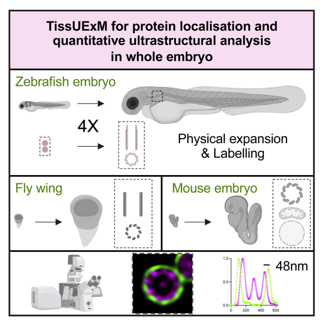
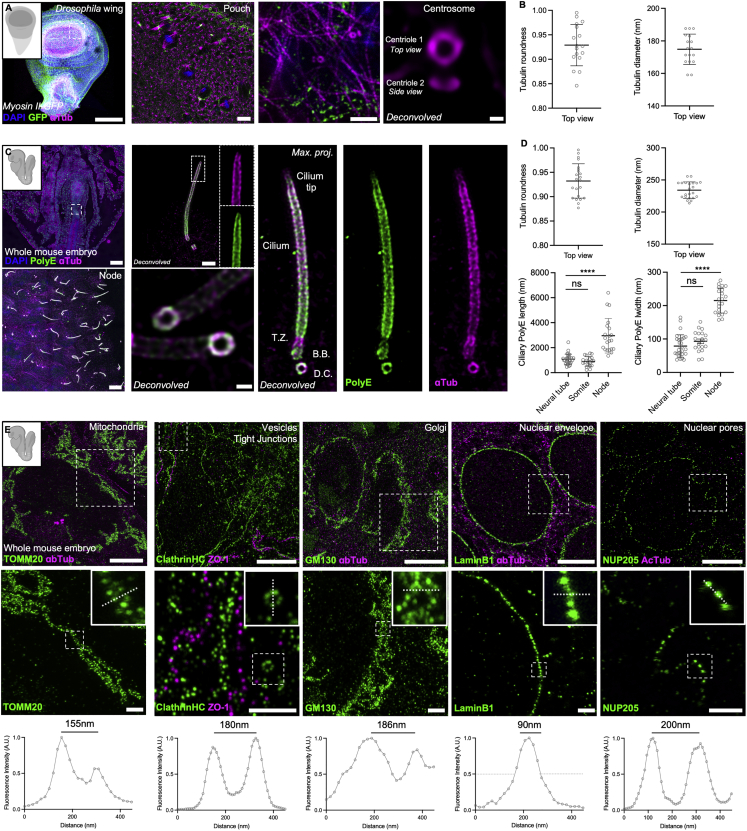
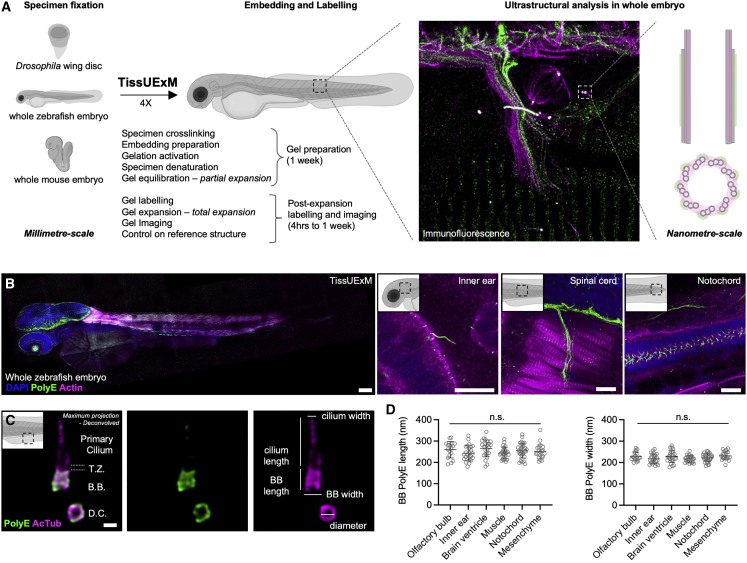
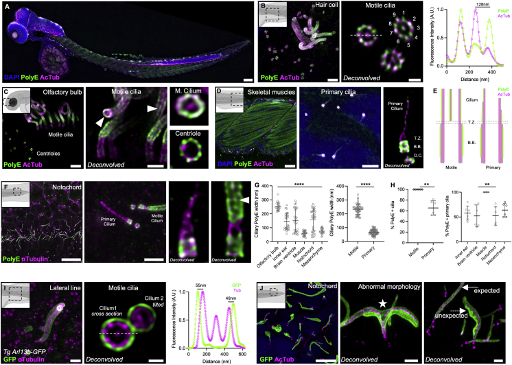
Super-resolution microscopy reveals the molecular organization of biological structures down to the nanoscale. While it allows the study of protein complexes in single cells, small organisms, or thin tissue sections, there is currently no versatile approach for ultrastructural analysis compatible with whole vertebrate embryos. Here, we present tissue ultrastructure expansion microscopy (TissUExM), a method to expand millimeter-scale and mechanically heterogeneous whole embryonic tissues, including wing discs, whole zebrafish, and mouse embryos. TissUExM is designed for the observation of endogenous proteins. It permits quantitative characterization of protein complexes in various organelles at super-resolution in a range of ∼3 mm-sized tissues using conventional microscopes. We demonstrate its strength by investigating tissue-specific ciliary architecture heterogeneity and ultrastructural defects observed upon ciliary protein overexpression. Overall, TissUExM is ideal for performing ultrastructural studies and molecular mapping in whole embryos.
超分辨率显微镜能够揭示生物结构的分子组织,达到纳米级分辨率。虽然它可以研究单细胞、小型生物或薄组织切片中的蛋白质复合物,但目前还没有一种通用的方法可以用于与整个脊椎动物胚胎兼容的超微结构分析。在这里,我们提出了组织超微结构扩展显微镜(TissUExM),这是一种扩展毫米级和机械异质的整个胚胎组织的方法,包括翅盘、整个斑马鱼和小鼠胚胎。TissUExM 是为观察内源性蛋白质而设计的。它允许使用传统显微镜在各种细胞器中以超分辨率对范围内的约 3mm 大小的组织中的蛋白质复合物进行定量表征。我们通过研究组织特异性纤毛结构异质性和纤毛蛋白过表达观察到的超微结构缺陷来证明其优势。总的来说,TissUExM 非常适合在整个胚胎中进行超微结构研究和分子作图。