Département de Médecine, Université de Montréal, Montréal, QC, H3C 3J7, Canada.
Division of Immunology-Oncology, Centre de Recherche Hopital Maisonneuve-Rosemont, Montréal, QC, H1T 2M4, Canada.
Sci Rep. 2022 Nov 2;12(1):18509. doi: 10.1038/s41598-022-23016-z.
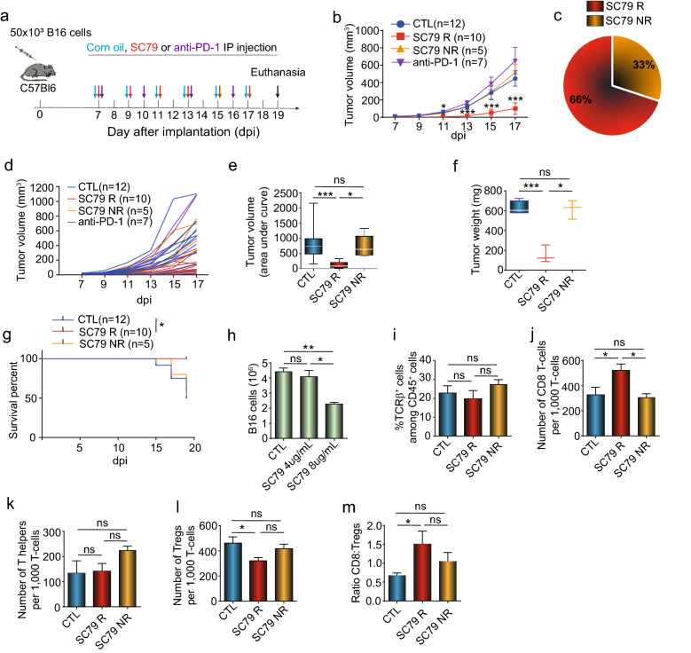
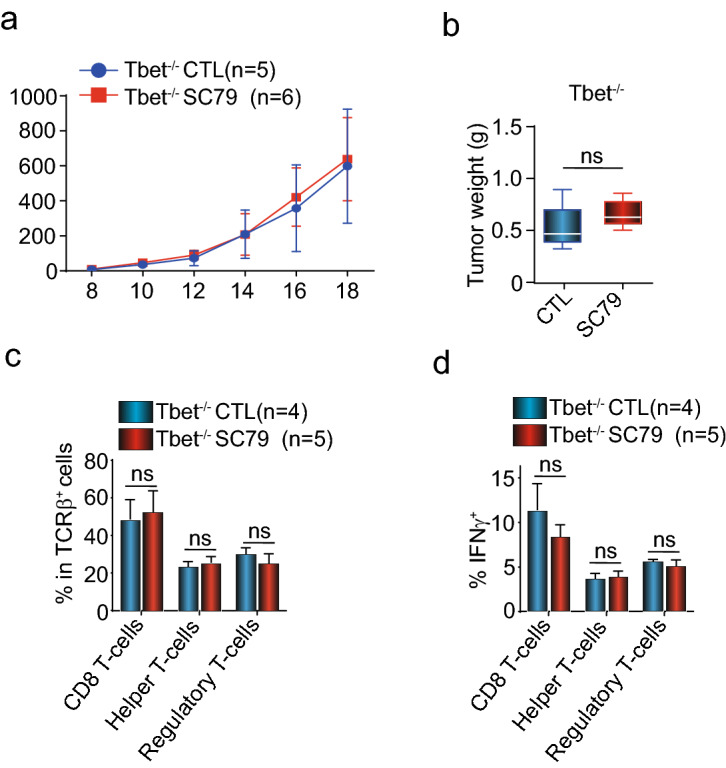
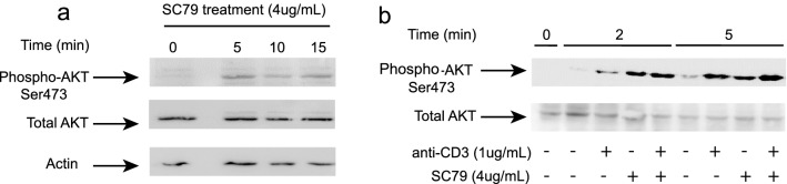
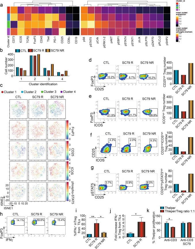
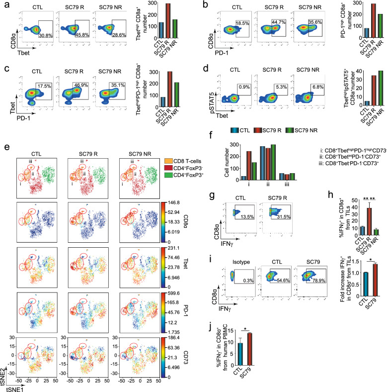
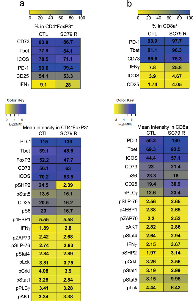
PD-1 immune checkpoint blockade against inhibitory receptors such as receptor programmed cell death-1 (PD-1), has revolutionized cancer treatment. Effective immune reactivity against tumour antigens requires the infiltration and activation of tumour-infiltrating T-cells (TILs). In this context, ligation of the antigen-receptor complex (TCR) in combination with the co-receptor CD28 activates the intracellular mediator AKT (or PKB, protein kinase B) and its downstream targets. PD-1 inhibits the activation of AKT/PKB. Given this, we assessed whether the direct activation of AKT might be effective in activating the immune system to limit the growth of tumors that are resistant to PD-1 checkpoint blockade. We found that the small molecule activator of AKT (SC79) limited growth of a B16 tumor and an EMT-6 syngeneic breast tumor model that are poorly responsive to PD-1 immunotherapy. In the case of B16 tumors, direct AKT activation induced (i) a reduction of suppressor regulatory (Treg) TILs and (ii) an increase in effector CD8+ TILs. SC79 in vivo therapy caused a major increase in the numbers of CD4+ and CD8+ TILs to express interferon-γ (IFN-γ). This effect on IFN-γ expression distinguished responsive from non-responsive anti-tumor responses and could be recapitulated ex vivo with human T-cells. In CD4+FoxP3+Treg TILs, AKT induced IFN-γ expression was accompanied by a loss of suppressor activity, the conversation to CD4 helper Th1-like TILs and a marked reduction in phospho-SHP2. In CD8+ TILs, we observed an increase in the phospho-activation of PLC-γ. Further, the genetic deletion of the transcription factor T-bet (Tbx21) blocked the increased IFN-γ expression on all subsets while ablating the therapeutic benefits of SC79 on tumor growth. Our study shows that AKT activation therapy acts to induce IFN-γ on CD4 and CD8 TILs that is accompanied by the intra-tumoral conversation of suppressive Tregs into CD4Th1-like T-cells and augmented CD8 responses.
PD-1 免疫检查点阻断针对抑制性受体,如受体程序性细胞死亡-1(PD-1),彻底改变了癌症治疗。有效针对肿瘤抗原的免疫反应需要肿瘤浸润 T 细胞(TILs)的浸润和激活。在这种情况下,抗原受体复合物(TCR)的连接与共受体 CD28 结合,激活细胞内介质 AKT(或 PKB,蛋白激酶 B)及其下游靶标。PD-1 抑制 AKT/PKB 的激活。鉴于此,我们评估了直接激活 AKT 是否可以有效激活免疫系统,以限制对 PD-1 检查点阻断反应不佳的肿瘤的生长。我们发现,AKT 的小分子激活剂(SC79)限制了 B16 肿瘤和 EMT-6 同源乳腺癌肿瘤模型的生长,这些肿瘤对 PD-1 免疫治疗反应不佳。在 B16 肿瘤的情况下,直接 AKT 激活诱导(i)抑制性调节性(Treg)TILs 的减少和(ii)效应性 CD8+TILs 的增加。SC79 体内治疗导致 CD4+和 CD8+TILs 表达干扰素-γ(IFN-γ)的数量大幅增加。这种对 IFN-γ 表达的影响将对肿瘤的反应性与非反应性区分开来,并可以在体外用人 T 细胞重现。在 CD4+FoxP3+Treg TILs 中,AKT 诱导的 IFN-γ 表达伴随着抑制活性的丧失,转化为 CD4 辅助 Th1 样 TILs,以及磷酸化 SHP2 的显著减少。在 CD8+TILs 中,我们观察到 PLC-γ 的磷酸化激活增加。此外,转录因子 T-bet(Tbx21)的基因缺失阻断了所有亚群上 IFN-γ 表达的增加,同时消除了 SC79 对肿瘤生长的治疗益处。我们的研究表明,AKT 激活治疗可诱导 CD4 和 CD8 TILs 上的 IFN-γ 表达,同时伴有肿瘤内抑制性 Tregs 转化为 CD4Th1 样 T 细胞和增强的 CD8 反应。