Chen Xiuyuan, Xu Hao, Qi Qingyi, Sun Chao, Jin Jian, Zhao Heng, Wang Xun, Weng Wenhan, Wang Shaodong, Sui Xizhao, Wang Zhenfan, Dai Chenyang, Peng Muyun, Wang Dawei, Hao Zenghao, Huang Yafen, Wang Xiang, Duan Liang, Zhu Yuming, Hong Nan, Yang Fan
Department of Thoracic Surgery, Peking University People's Hospital, Beijing, China.
Thoracic Oncology Institute, Peking University People's Hospital, Beijing, China.
Front Oncol. 2022 Oct 13;12:1021084. doi: 10.3389/fonc.2022.1021084. eCollection 2022.
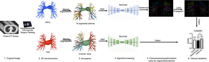
The recognition of anatomical variants is essential in preoperative planning for lung cancer surgery. Although three-dimensional (3-D) reconstruction provided an intuitive demonstration of the anatomical structure, the recognition process remains fully manual. To render a semiautomated approach for surgery planning, we developed an artificial intelligence (AI)-based chest CT semantic segmentation algorithm that recognizes pulmonary vessels on lobular or segmental levels. Hereby, we present a retrospective validation of the algorithm comparing surgeons' performance.
The semantic segmentation algorithm to be validated was trained on non-contrast CT scans from a single center. A retrospective pilot study was performed. An independent validation dataset was constituted by an arbitrary selection from patients who underwent lobectomy or segmentectomy in three institutions during Apr. 2020 to Jun. 2021. The golden standard of anatomical variants of each enrolled case was obtained via expert surgeons' judgments based on chest CT, 3-D reconstruction, and surgical observation. The performance of the algorithm is compared against the performance of two junior thoracic surgery attendings based on chest CT.
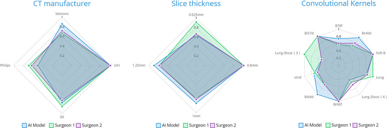
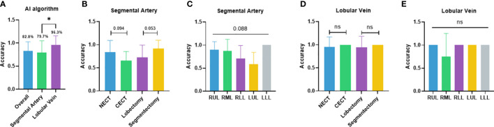
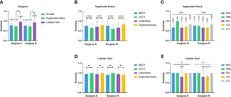
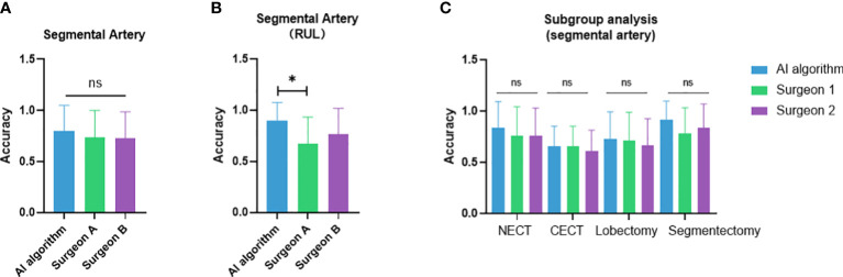
A total of 27 cases were included in this study. The overall case-wise accuracy of the AI model was 82.8% in pulmonary vessels compared to 78.8% and 77.0% for the two surgeons, respectively. Segmental artery accuracy was 79.7%, 73.6%, and 72.7%; lobular vein accuracy was 96.3%, 96.3%, and 92.6% by the AI model and two surgeons, respectively. No statistical significance was found. In subgroup analysis, the anatomic structure-wise analysis of the AI algorithm showed a significant difference in accuracies between different lobes (p = 0.012). Higher AI accuracy in the right-upper lobe (RUL) and left-lower lobe (LLL) arteries was shown. A trend of better performance in non-contrast CT was also detected. Most recognition errors by the algorithm were the misclassification of LA and LA. Radiological parameters did not exhibit a significant impact on the performance of both AI and surgeons.
The semantic segmentation algorithm achieves the recognition of the segmental pulmonary artery and the lobular pulmonary vein. The performance of the model approximates that of junior thoracic surgery attendings. Our work provides a novel semiautomated surgery planning approach that is potentially beneficial to lung cancer patients.
识别解剖变异对于肺癌手术的术前规划至关重要。尽管三维(3-D)重建能直观展示解剖结构,但识别过程仍完全依赖人工。为实现手术规划的半自动化方法,我们开发了一种基于人工智能(AI)的胸部CT语义分割算法,用于在小叶或节段水平识别肺血管。在此,我们对该算法进行回顾性验证,并比较外科医生的表现。
待验证的语义分割算法在来自单一中心的非增强CT扫描上进行训练。开展了一项回顾性试点研究。独立验证数据集由从2020年4月至2021年6月期间在三个机构接受肺叶切除术或节段切除术的患者中任意选取的病例组成。通过专家外科医生基于胸部CT、3-D重建和手术观察对每个纳入病例的解剖变异进行判断,从而获得金标准。将该算法的表现与两名胸外科初级主治医师基于胸部CT的表现进行比较。
本研究共纳入27例病例。人工智能模型在肺血管方面的总体逐例准确率为82.8%,而两名外科医生的准确率分别为78.8%和77.0%。节段动脉准确率分别为79.7%、73.6%和72.7%;小叶静脉准确率方面,人工智能模型和两名外科医生分别为96.3%、96.3%和92.6%。未发现统计学意义。在亚组分析中,人工智能算法的解剖结构逐结构分析显示不同肺叶之间的准确率存在显著差异(p = 0.012)。结果显示在右上叶(RUL)和左下叶(LLL)动脉中人工智能的准确率更高。还检测到在非增强CT中表现更佳的趋势。该算法的大多数识别错误是左心房(LA)和左心房的错误分类。放射学参数对人工智能和外科医生的表现均未显示出显著影响。
语义分割算法实现了对节段性肺动脉和小叶性肺静脉的识别。该模型的表现接近胸外科初级主治医师的表现。我们的工作提供了一种新颖的半自动化手术规划方法,可能对肺癌患者有益。