• 文献检索
  • 文档翻译
  • 深度研究
  • 学术资讯
  • Suppr Zotero 插件Zotero 插件
  • 邀请有礼
  • 套餐&价格
  • 历史记录
应用&插件
Suppr Zotero 插件Zotero 插件浏览器插件Mac 客户端Windows 客户端微信小程序
定价
高级版会员购买积分包购买API积分包
服务
文献检索文档翻译深度研究API 文档MCP 服务
关于我们
关于 Suppr公司介绍联系我们用户协议隐私条款
关注我们

Suppr 超能文献

核心技术专利:CN118964589B侵权必究
粤ICP备2023148730 号-1Suppr @ 2026

文献检索

告别复杂PubMed语法,用中文像聊天一样搜索,搜遍4000万医学文献。AI智能推荐,让科研检索更轻松。

立即免费搜索

文件翻译

保留排版,准确专业,支持PDF/Word/PPT等文件格式,支持 12+语言互译。

免费翻译文档

深度研究

AI帮你快速写综述,25分钟生成高质量综述,智能提取关键信息,辅助科研写作。

立即免费体验

基于人工智能算法的超声图像特征与内镜联合检测在早期上消化道癌诊断中的应用。

Utilization of Ultrasonic Image Characteristics Combined with Endoscopic Detection on the Basis of Artificial Intelligence Algorithm in Diagnosis of Early Upper Gastrointestinal Cancer.

机构信息

Digestive Endoscope Room, Cangzhou Central Hospital, Cangzhou 061001, Hebei, China.

Department of Gastroenterology, Cangzhou Central Hospital, Cangzhou 061001, Hebei, China.

出版信息

J Healthc Eng. 2021 Nov 29;2021:2773022. doi: 10.1155/2021/2773022. eCollection 2021.

DOI:10.1155/2021/2773022
PMID:34880973
原文链接:https://pmc.ncbi.nlm.nih.gov/articles/PMC8648460/
Abstract

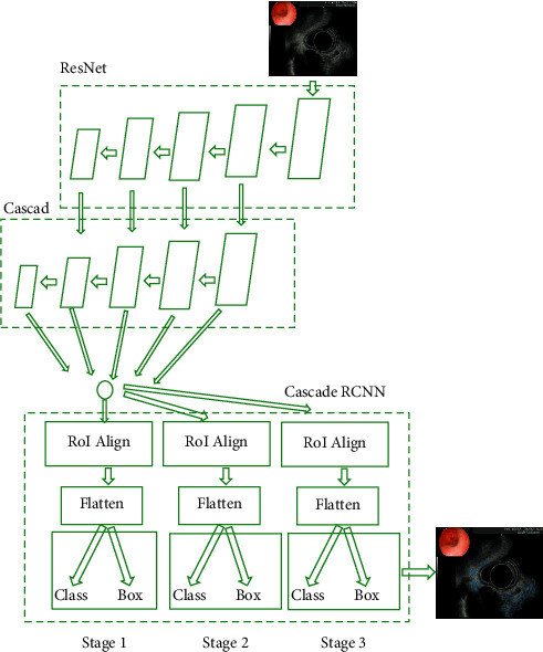
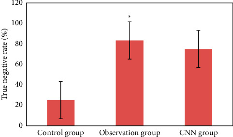
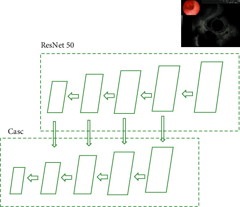
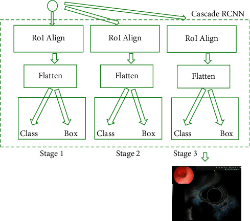
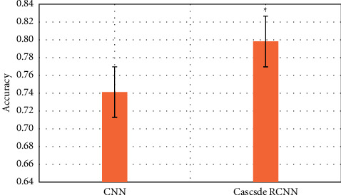
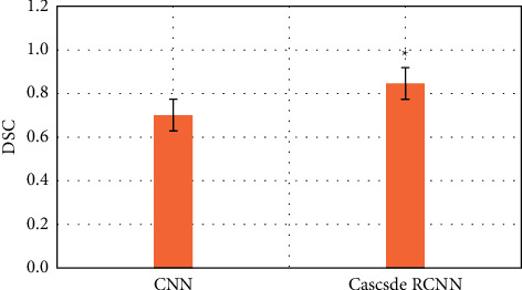
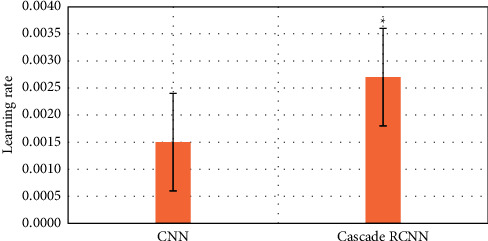
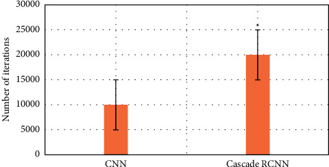
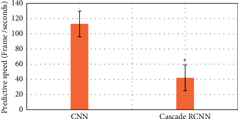
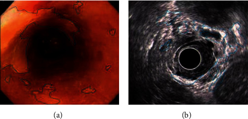
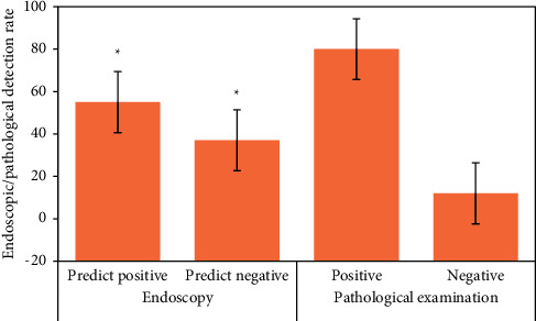
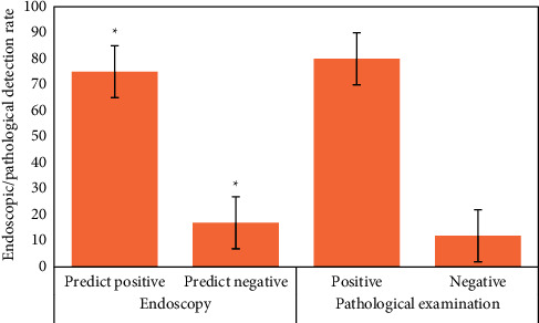
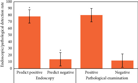
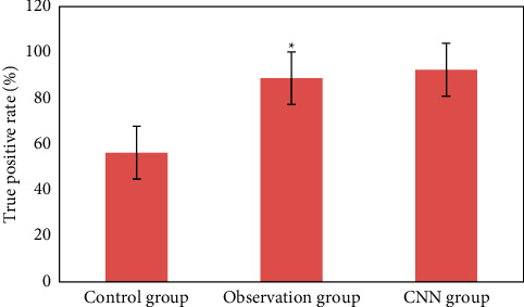
The aim of this study was to evaluate the diagnostic value of artificial intelligence algorithm combined with ultrasound endoscopy in early esophageal cancer and precancerous lesions by comparing the examination of conventional endoscopy and artificial intelligence algorithm combined with ultrasound endoscopy, and by comparing the real-time diagnosis of endoscopy and the ultrasonic image characteristics of artificial intelligence algorithm combined with endoscopic detection and pathological results. 120 cases were selected. According to the inclusion and exclusion criteria, 80 patients who met the criteria were selected and randomly divided into two groups: endoscopic examination combined with ultrasound imaging based on intelligent algorithm processing (cascade region-convolutional neural network (Cascade RCNN) model algorithm group) and simple use of endoscopy group (control group). This study shows that the ultrasonic image of artificial intelligence algorithm is effective, and the detection performance is better than that of endoscopic detection. The results are close to the gold standard of doctor recognition, and the detection time is greatly shortened, and the recognition time is shortened by 71 frames per second. Compared with the traditional convolutional neural network (CNN) algorithm, the accuracy and recall of image analysis and segmentation using feature pyramid network are increased. The detection rates of CNN model, Cascade RCNN model, and endoscopic detection alone in early esophageal cancer and precancerous lesions are 56.3% (45/80), 88.8% (71/80), and 44.1% (35/80), respectively. The detection rate of Cascade RCNN model and CNN model was higher than that of endoscopy alone, and the difference was statistically significant ( < 0.05). The sensitivity, specificity, positive predictive value, and negative predictive value of Cascade RCNN model were higher than those of CNN model, which was close to the gold standard for physician identification. This provided a reference basis for endoscopic ultrasound identification of early upper gastrointestinal cancer or other gastrointestinal cancers.

摘要

本研究旨在通过比较常规内镜检查和人工智能算法联合超声内镜检查、比较内镜实时诊断与人工智能算法联合内镜检测和病理结果的超声图像特征,评估人工智能算法联合超声内镜在早期食管癌及癌前病变中的诊断价值。选取 120 例患者,根据纳入和排除标准,选择符合标准的 80 例患者,并随机分为两组:基于智能算法处理的内镜联合超声成像检查(级联区域卷积神经网络(Cascade RCNN)模型算法组)和单纯使用内镜组(对照组)。本研究表明,人工智能算法的超声图像是有效的,检测性能优于内镜检测。结果接近医生识别的金标准,检测时间大大缩短,识别时间缩短了 71 帧/秒。与传统卷积神经网络(CNN)算法相比,使用特征金字塔网络的图像分析和分割的准确性和召回率都有所提高。CNN 模型、Cascade RCNN 模型和内镜单独检测早期食管癌及癌前病变的检出率分别为 56.3%(45/80)、88.8%(71/80)和 44.1%(35/80)。Cascade RCNN 模型和 CNN 模型的检出率均高于内镜单独检测,差异有统计学意义( < 0.05)。Cascade RCNN 模型的灵敏度、特异度、阳性预测值和阴性预测值均高于 CNN 模型,与医生识别的金标准接近。这为内镜超声对上消化道癌或其他胃肠道癌的早期诊断提供了参考依据。

https://cdn.ncbi.nlm.nih.gov/pmc/blobs/c92d/8648460/fa81a344c8cc/JHE2021-2773022.017.jpg
https://cdn.ncbi.nlm.nih.gov/pmc/blobs/c92d/8648460/a9b9c7c7bb05/JHE2021-2773022.001.jpg
https://cdn.ncbi.nlm.nih.gov/pmc/blobs/c92d/8648460/1e4921fbd5da/JHE2021-2773022.002.jpg
https://cdn.ncbi.nlm.nih.gov/pmc/blobs/c92d/8648460/41a39034034a/JHE2021-2773022.003.jpg
https://cdn.ncbi.nlm.nih.gov/pmc/blobs/c92d/8648460/ee0781402e05/JHE2021-2773022.004.jpg
https://cdn.ncbi.nlm.nih.gov/pmc/blobs/c92d/8648460/6a2ec30678b3/JHE2021-2773022.005.jpg
https://cdn.ncbi.nlm.nih.gov/pmc/blobs/c92d/8648460/0999082f3259/JHE2021-2773022.006.jpg
https://cdn.ncbi.nlm.nih.gov/pmc/blobs/c92d/8648460/e6651a57dd8e/JHE2021-2773022.007.jpg
https://cdn.ncbi.nlm.nih.gov/pmc/blobs/c92d/8648460/55ede720c631/JHE2021-2773022.008.jpg
https://cdn.ncbi.nlm.nih.gov/pmc/blobs/c92d/8648460/37a5f94e2f7b/JHE2021-2773022.009.jpg
https://cdn.ncbi.nlm.nih.gov/pmc/blobs/c92d/8648460/0e1b36edc41c/JHE2021-2773022.010.jpg
https://cdn.ncbi.nlm.nih.gov/pmc/blobs/c92d/8648460/13bd94fc82c0/JHE2021-2773022.011.jpg
https://cdn.ncbi.nlm.nih.gov/pmc/blobs/c92d/8648460/9c16210155bf/JHE2021-2773022.012.jpg
https://cdn.ncbi.nlm.nih.gov/pmc/blobs/c92d/8648460/e2e1222eb481/JHE2021-2773022.013.jpg
https://cdn.ncbi.nlm.nih.gov/pmc/blobs/c92d/8648460/aea233ed6692/JHE2021-2773022.014.jpg
https://cdn.ncbi.nlm.nih.gov/pmc/blobs/c92d/8648460/f5e0f1192986/JHE2021-2773022.015.jpg
https://cdn.ncbi.nlm.nih.gov/pmc/blobs/c92d/8648460/9da42b90f965/JHE2021-2773022.016.jpg
https://cdn.ncbi.nlm.nih.gov/pmc/blobs/c92d/8648460/fa81a344c8cc/JHE2021-2773022.017.jpg

相似文献

1
Utilization of Ultrasonic Image Characteristics Combined with Endoscopic Detection on the Basis of Artificial Intelligence Algorithm in Diagnosis of Early Upper Gastrointestinal Cancer.基于人工智能算法的超声图像特征与内镜联合检测在早期上消化道癌诊断中的应用。
J Healthc Eng. 2021 Nov 29;2021:2773022. doi: 10.1155/2021/2773022. eCollection 2021.
2
Multimodal MRI Analysis of Cervical Cancer on the Basis of Artificial Intelligence Algorithm.基于人工智能算法的宫颈癌多模态 MRI 分析。
Contrast Media Mol Imaging. 2021 Nov 8;2021:1673490. doi: 10.1155/2021/1673490. eCollection 2021.
3
Real-time artificial intelligence for detection of upper gastrointestinal cancer by endoscopy: a multicentre, case-control, diagnostic study.实时人工智能用于内窥镜检查上消化道癌的检测:一项多中心、病例对照、诊断研究。
Lancet Oncol. 2019 Dec;20(12):1645-1654. doi: 10.1016/S1470-2045(19)30637-0. Epub 2019 Oct 4.
4
Artificial intelligence for cancer detection of the upper gastrointestinal tract.人工智能在上消化道癌症检测中的应用。
Dig Endosc. 2021 Jan;33(2):254-262. doi: 10.1111/den.13897. Epub 2020 Dec 28.
5
Utilizing artificial intelligence in endoscopy: a clinician's guide.利用人工智能进行内镜检查:临床医生指南。
Expert Rev Gastroenterol Hepatol. 2020 Aug;14(8):689-706. doi: 10.1080/17474124.2020.1779058. Epub 2020 Jun 17.
6
Application of artificial intelligence using a convolutional neural network for diagnosis of early gastric cancer based on magnifying endoscopy with narrow-band imaging.应用卷积神经网络的人工智能技术在窄带成像放大内镜下对早期胃癌的诊断
J Gastroenterol Hepatol. 2021 Feb;36(2):482-489. doi: 10.1111/jgh.15190. Epub 2020 Jul 28.
7
Convolutional neural network-based artificial intelligence for the diagnosis of early esophageal cancer based on endoscopic images: A meta-analysis.基于卷积神经网络的内镜图像早期食管癌人工智能诊断:一项荟萃分析。
Saudi J Gastroenterol. 2022 Sep-Oct;28(5):332-340. doi: 10.4103/sjg.sjg_178_22.
8
Automatic detection of early gastric cancer in endoscopy based on Mask region-based convolutional neural networks (Mask R-CNN)(with video).基于掩码区域卷积神经网络(Mask R-CNN)的内镜检查中早期胃癌自动检测(附视频)
Front Oncol. 2022 Oct 20;12:927868. doi: 10.3389/fonc.2022.927868. eCollection 2022.
9
Recent Progress of Image-Enhanced Endoscopy for Upper Gastrointestinal Neoplasia and Associated Lesions.上消化道肿瘤及相关病变的图像增强内镜检查的最新进展
Dig Dis. 2024;42(2):186-198. doi: 10.1159/000535055. Epub 2023 Nov 10.
10
Application of artificial intelligence using a convolutional neural network for detecting gastric cancer in endoscopic images.应用卷积神经网络的人工智能技术用于内镜图像中胃癌的检测。
Gastric Cancer. 2018 Jul;21(4):653-660. doi: 10.1007/s10120-018-0793-2. Epub 2018 Jan 15.

引用本文的文献

1
Concordance with CONSORT-AI guidelines in reporting of randomised controlled trials investigating artificial intelligence in oncology: a systematic review.肿瘤学中人工智能随机对照试验报告与CONSORT-AI指南的一致性:一项系统评价
BMJ Oncol. 2025 Aug 24;4(1):e000733. doi: 10.1136/bmjonc-2025-000733. eCollection 2025.
2
The application of artificial intelligence in EUS.人工智能在超声内镜检查中的应用。
Endosc Ultrasound. 2024 Mar-Apr;13(2):65-75. doi: 10.1097/eus.0000000000000053. Epub 2024 Apr 10.
3
Concordance of randomised controlled trials for artificial intelligence interventions with the CONSORT-AI reporting guidelines.

本文引用的文献

1
Evolving role of artificial intelligence in gastrointestinal endoscopy.人工智能在胃肠内镜中的不断发展的作用。
World J Gastroenterol. 2020 Dec 14;26(46):7287-7298. doi: 10.3748/wjg.v26.i46.7287.
2
Artificial intelligence for cancer detection of the upper gastrointestinal tract.人工智能在上消化道癌症检测中的应用。
Dig Endosc. 2021 Jan;33(2):254-262. doi: 10.1111/den.13897. Epub 2020 Dec 28.
3
Accuracy of endoscopic size measurements of early gastric signet ring cell carcinoma.早期胃印戒细胞癌内镜下大小测量的准确性。
人工智能干预随机对照试验与 CONSORT-AI 报告指南的一致性。
Nat Commun. 2024 Feb 22;15(1):1619. doi: 10.1038/s41467-024-45355-3.
4
Applications and Prospects of Artificial Intelligence-Assisted Endoscopic Ultrasound in Digestive System Diseases.人工智能辅助内镜超声在消化系统疾病中的应用及前景
Diagnostics (Basel). 2023 Aug 30;13(17):2815. doi: 10.3390/diagnostics13172815.
5
Machine learning applications for early detection of esophageal cancer: a systematic review.机器学习在食管癌早期检测中的应用:系统评价。
BMC Med Inform Decis Mak. 2023 Jul 17;23(1):124. doi: 10.1186/s12911-023-02235-y.
6
Evaluation of deep learning methods for early gastric cancer detection using gastroscopic images.利用胃镜图像评估深度学习方法在早期胃癌检测中的应用。
Technol Health Care. 2023;31(S1):313-322. doi: 10.3233/THC-236027.
7
A deep learning-based approach toward differentiating scalp psoriasis and seborrheic dermatitis from dermoscopic images.一种基于深度学习的从皮肤镜图像中区分头皮银屑病和脂溢性皮炎的方法。
Front Med (Lausanne). 2022 Nov 3;9:965423. doi: 10.3389/fmed.2022.965423. eCollection 2022.
8
AI-based chest CT semantic segmentation algorithm enables semi-automated lung cancer surgery planning by recognizing anatomical variants of pulmonary vessels.基于人工智能的胸部CT语义分割算法通过识别肺血管的解剖变异实现肺癌手术的半自动规划。
Front Oncol. 2022 Oct 13;12:1021084. doi: 10.3389/fonc.2022.1021084. eCollection 2022.
9
Global research trends of artificial intelligence applied in esophageal carcinoma: A bibliometric analysis (2000-2022) CiteSpace and VOSviewer.人工智能应用于食管癌的全球研究趋势:一项文献计量分析(2000 - 2022年) CiteSpace和VOSviewer
Front Oncol. 2022 Aug 25;12:972357. doi: 10.3389/fonc.2022.972357. eCollection 2022.
10
Application Value of Emergency Bedside Echocardiography in Early Warning of Acute and Severe Shock and Clinical Classification.床旁超声心动图在急性和严重休克早期预警及临床分类中的应用价值
Comput Math Methods Med. 2022 Jul 19;2022:1634866. doi: 10.1155/2022/1634866. eCollection 2022.
Surg Endosc. 2021 May;35(5):2324-2331. doi: 10.1007/s00464-020-07646-w. Epub 2020 May 19.
4
Esophagus and Gastrointestinal Junction Tumors.食管胃结合部肿瘤。
Surg Clin North Am. 2020 Jun;100(3):507-521. doi: 10.1016/j.suc.2020.02.003. Epub 2020 Apr 11.
5
Recent advances in diagnostic upper endoscopy.诊断性上消化道内镜的最新进展。
World J Gastroenterol. 2020 Jan 28;26(4):433-447. doi: 10.3748/wjg.v26.i4.433.
6
Artificial intelligence in gastrointestinal endoscopy: general overview.人工智能在胃肠内镜中的应用:概述。
Chin Med J (Engl). 2020 Feb 5;133(3):326-334. doi: 10.1097/CM9.0000000000000623.
7
Cascade R-CNN: High Quality Object Detection and Instance Segmentation.级联 R-CNN:高质量目标检测和实例分割。
IEEE Trans Pattern Anal Mach Intell. 2021 May;43(5):1483-1498. doi: 10.1109/TPAMI.2019.2956516. Epub 2021 Apr 1.
8
[Cost-effectiveness and cost-benefit analysis of upper gastrointestinal cancer screening in Yangzhong City, Jiangsu Province, from 2009 to 2015].[2009年至2015年江苏省扬中市上消化道癌筛查的成本效益与成本效果分析]
Zhonghua Yu Fang Yi Xue Za Zhi. 2019 Nov 6;53(11):1115-1118. doi: 10.3760/cma.j.issn.0253-9624.2019.11.008.
9
Application of artificial intelligence in gastrointestinal endoscopy.人工智能在胃肠内镜中的应用。
J Dig Dis. 2019 Dec;20(12):623-630. doi: 10.1111/1751-2980.12827. Epub 2019 Nov 20.
10
Usefulness of Full-spectrum Endoscopy for the Upper Gastrointestinal Tract.全谱内镜在上消化道的应用
J Clin Gastroenterol. 2020 Apr;54(4):344-349. doi: 10.1097/MCG.0000000000001226.