Graduate School, Tianjin Medical University, No. 22 Qixiangtai Road, Heping District, Tianjin, 300210, China.
Yuncheng Central Hospital, No. 3690 Hedong East Road, Yuncheng, 044000, Shanxi, China.
Sci Rep. 2022 Nov 4;12(1):18675. doi: 10.1038/s41598-022-23541-x.
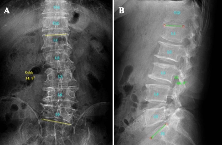
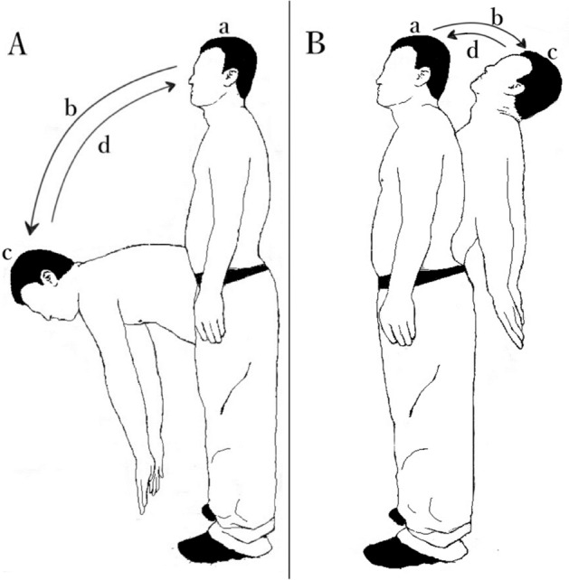
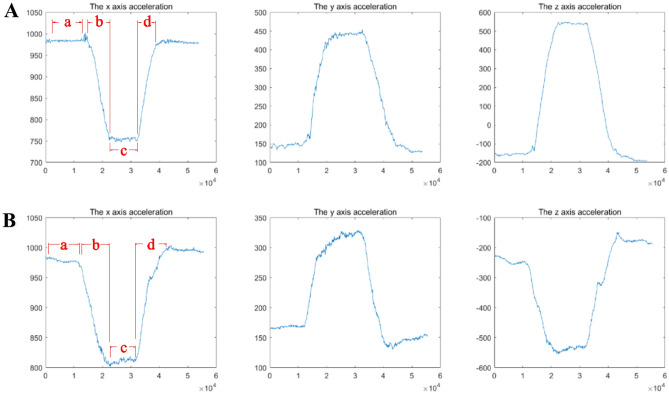
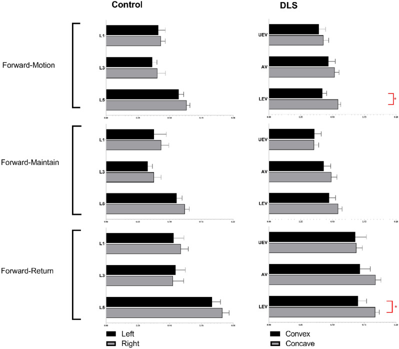
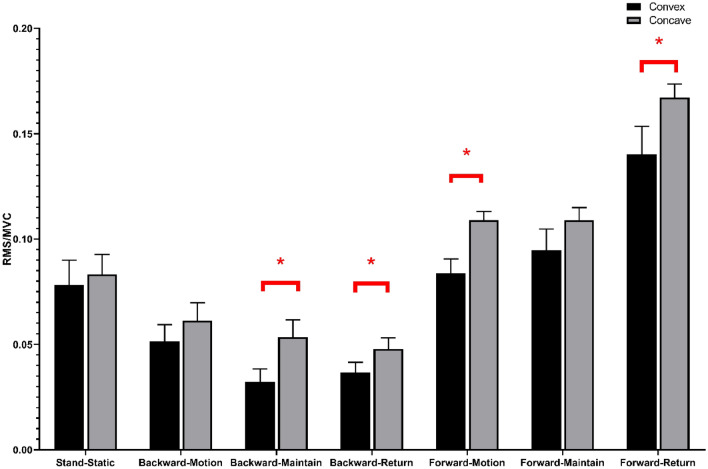
The asymmetry of paravertebral muscle (PVM) degeneration in degenerative lumbar scoliosis (DLS) patients has been extensively studied by imaging and histological examination and has not yet been verified by surface electromyography (sEMG) techniques. To study the relationship between the surface electromyography (sEMG) and degenerative characteristics of paravertebral muscles (PVMs) in patients with degenerative lumbar scoliosis (DLS). In twenty DLS patients and fifteen healthy subjects, sEMG activity of the PVMs at the level of the upper end vertebra (UEV), apical vertebra (AV) and lower end vertebra (LEV) was measured during static standing and dynamic standing forward flexion and backward extension tasks. Action segmentation was achieved according to inertial measurement unit (IMU) data. The sEMG characteristics of the PVMs on the convex and concave sides were compared, and the relationship of these data with the Cobb angle and lumbar lordotic angle (LL) was analyzed. In the DLS group, there was no difference in sEMG activity between the convex and concave sides at the UEV or AV level, but in the motion and return phases of the standing forward flexion task (P = 0.000, P = 0.015) and the maintenance and return phases of the standing backward extension task (P = 0.001, P = 0.01), there was a significant difference in sEMG activity between the convex and concave sides at the LEV level. Asymmetrical sEMG activity at the LEV level was negatively correlated with the Cobb angle (F = 93.791, P < 0.001) and LL angle (F = 65.564, P < 0.001). In the DLS group, asymmetrical sEMG activity of the PVMs appeared at the LEV level, with the concave side being more active than the convex side. This sEMG characteristics were consistent with their imaging and histological degenerative features and correlated with bone structural parameters.
在退行性腰椎侧凸(DLS)患者中,椎旁肌(PVM)退变的不对称性已通过影像学和组织学检查进行了广泛研究,但尚未通过表面肌电图(sEMG)技术得到验证。为了研究退行性腰椎侧凸(DLS)患者的表面肌电图(sEMG)与椎旁肌(PVM)退变特征之间的关系。在 20 例 DLS 患者和 15 例健康受试者中,在静态站立和动态站立前屈和后伸任务中测量了上终椎(UEV)、顶椎(AV)和下终椎(LEV)水平的 PVMs 的 sEMG 活动。根据惯性测量单元(IMU)数据实现动作分割。比较了凸侧和凹侧 PVMs 的 sEMG 特征,并分析了这些数据与 Cobb 角和腰椎前凸角(LL)的关系。在 DLS 组中,UEV 或 AV 水平的凸侧和凹侧的 sEMG 活动没有差异,但在站立前屈任务的运动和返回阶段(P = 0.000,P = 0.015)和站立后伸任务的维持和返回阶段(P = 0.001,P = 0.01),LEV 水平的凸侧和凹侧的 sEMG 活动存在显著差异。LEV 水平不对称的 sEMG 活性与 Cobb 角(F = 93.791,P < 0.001)和 LL 角(F = 65.564,P < 0.001)呈负相关。在 DLS 组中,PVMs 的 LEV 水平出现了不对称的 sEMG 活动,凹侧比凸侧更活跃。这些 sEMG 特征与影像学和组织学退变特征一致,并与骨结构参数相关。