German Center for Neurodegenerative Diseases (DZNE), Bonn, Germany; Department of Neurology, University Hospital Bonn, Germany.
German Center for Neurodegenerative Diseases (DZNE), Bonn, Germany.
Neuroimage. 2022 Dec 1;264:119703. doi: 10.1016/j.neuroimage.2022.119703. Epub 2022 Oct 27.
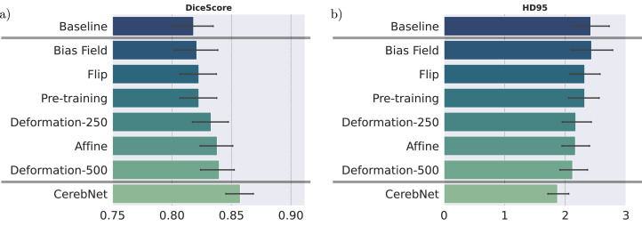
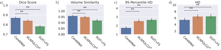
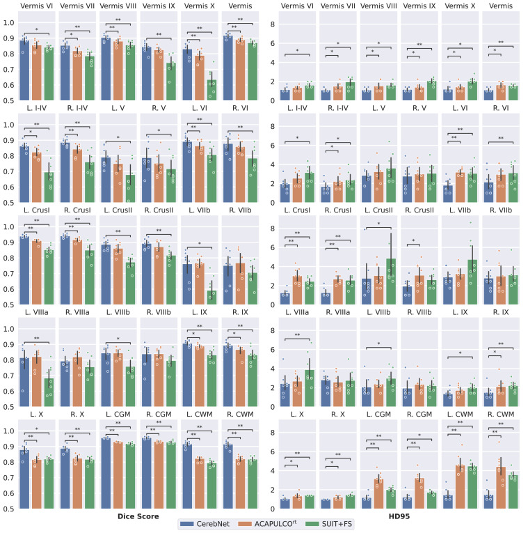
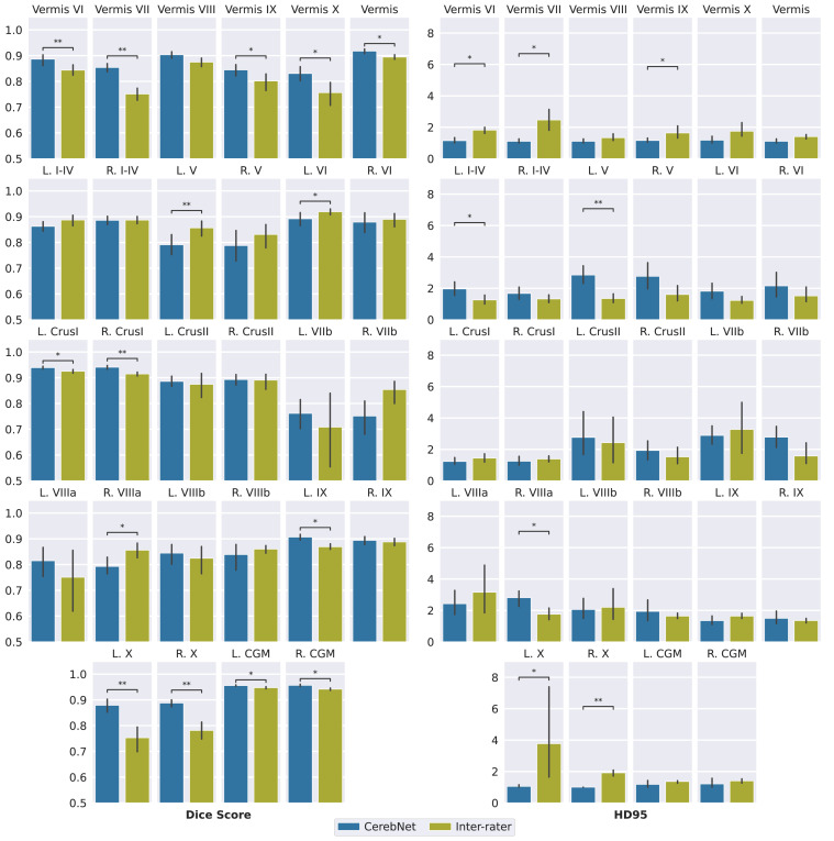
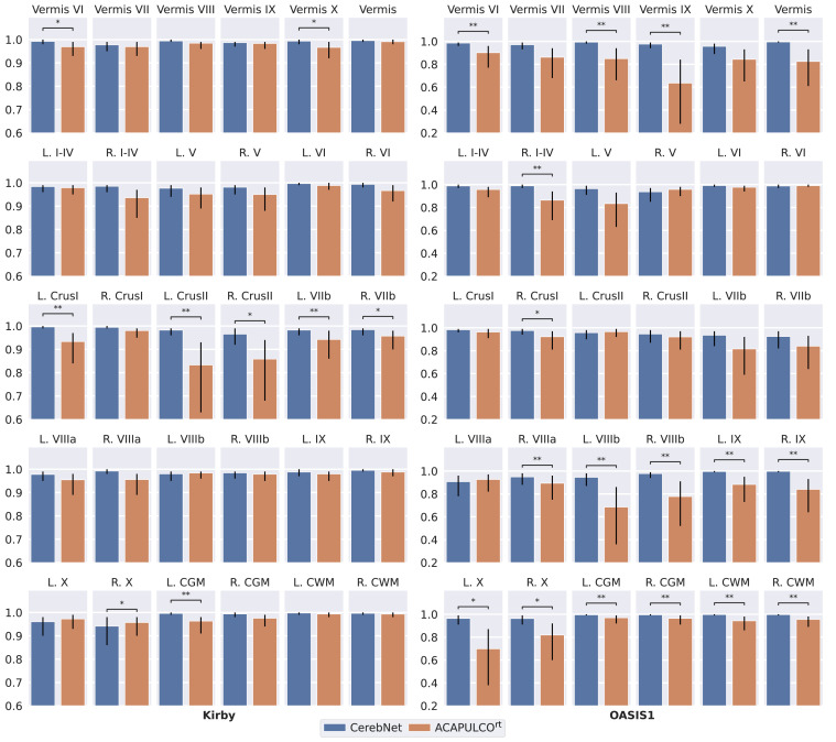
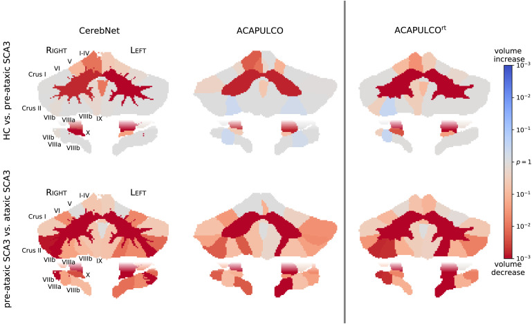
Quantifying the volume of the cerebellum and its lobes is of profound interest in various neurodegenerative and acquired diseases. Especially for the most common spinocerebellar ataxias (SCA), for which the first antisense oligonculeotide-base gene silencing trial has recently started, there is an urgent need for quantitative, sensitive imaging markers at pre-symptomatic stages for stratification and treatment assessment. This work introduces CerebNet, a fully automated, extensively validated, deep learning method for the lobular segmentation of the cerebellum, including the separation of gray and white matter. For training, validation, and testing, T1-weighted images from 30 participants were manually annotated into cerebellar lobules and vermal sub-segments, as well as cerebellar white matter. CerebNet combines FastSurferCNN, a UNet-based 2.5D segmentation network, with extensive data augmentation, e.g. realistic non-linear deformations to increase the anatomical variety, eliminating additional preprocessing steps, such as spatial normalization or bias field correction. CerebNet demonstrates a high accuracy (on average 0.87 Dice and 1.742mm Robust Hausdorff Distance across all structures) outperforming state-of-the-art approaches. Furthermore, it shows high test-retest reliability (average ICC >0.97 on OASIS and Kirby) as well as high sensitivity to disease effects, including the pre-ataxic stage of spinocerebellar ataxia type 3 (SCA3). CerebNet is compatible with FreeSurfer and FastSurfer and can analyze a 3D volume within seconds on a consumer GPU in an end-to-end fashion, thus providing an efficient and validated solution for assessing cerebellum sub-structure volumes. We make CerebNet available as source-code (https://github.com/Deep-MI/FastSurfer).
量化小脑及其叶的体积在各种神经退行性和获得性疾病中具有重要意义。特别是对于最常见的脊髓小脑共济失调(SCA),最近已经开始了第一个反义寡核苷酸基基因沉默试验,因此迫切需要在无症状阶段进行定量、敏感的成像标志物,以进行分层和治疗评估。这项工作介绍了 CerebNet,这是一种全自动、经过广泛验证的深度学习方法,用于小脑叶的分割,包括灰质和白质的分离。为了训练、验证和测试,我们对 30 名参与者的 T1 加权图像进行了手动注释,将小脑叶和蚓部亚段以及小脑白质进行了分割。CerebNet 将基于 UNet 的 2.5D 分割网络 FastSurferCNN 与广泛的数据增强相结合,例如逼真的非线性变形以增加解剖学多样性,消除了额外的预处理步骤,例如空间归一化或偏置场校正。CerebNet 表现出很高的准确性(平均在所有结构上的 Dice 为 0.87,稳健 Hausdorff 距离为 1.742mm),优于最先进的方法。此外,它还表现出很高的测试-重测可靠性(在 OASIS 和 Kirby 上平均 ICC>0.97),并且对疾病效应也很敏感,包括脊髓小脑共济失调 3 型(SCA3)的前共济失调阶段。CerebNet 与 FreeSurfer 和 FastSurfer 兼容,可以在消费级 GPU 上以端到端的方式在几秒钟内分析一个 3D 体积,从而为评估小脑亚结构体积提供了一种高效且经过验证的解决方案。我们将 CerebNet 作为源代码提供(https://github.com/Deep-MI/FastSurfer)。