• 文献检索
  • 文档翻译
  • 深度研究
  • 学术资讯
  • Suppr Zotero 插件Zotero 插件
  • 邀请有礼
  • 套餐&价格
  • 历史记录
应用&插件
Suppr Zotero 插件Zotero 插件浏览器插件Mac 客户端Windows 客户端微信小程序
定价
高级版会员购买积分包购买API积分包
服务
文献检索文档翻译深度研究API 文档MCP 服务
关于我们
关于 Suppr公司介绍联系我们用户协议隐私条款
关注我们

Suppr 超能文献

核心技术专利:CN118964589B侵权必究
粤ICP备2023148730 号-1Suppr @ 2026

文献检索

告别复杂PubMed语法,用中文像聊天一样搜索,搜遍4000万医学文献。AI智能推荐,让科研检索更轻松。

立即免费搜索

文件翻译

保留排版,准确专业,支持PDF/Word/PPT等文件格式,支持 12+语言互译。

免费翻译文档

深度研究

AI帮你快速写综述,25分钟生成高质量综述,智能提取关键信息,辅助科研写作。

立即免费体验

纳米级骨的层次结构与特性

Hierarchical Structure and Properties of the Bone at Nano Level.

作者信息

Hamandi Farah, Goswami Tarun

机构信息

Department of Biomedical, Industrial and Human Factors Engineering, Wright State University, Dayton, OH 45435, USA.

Department of Orthopedic Surgery, Sports Medicine and Rehabilitation, Wright State University, Dayton, OH 45435, USA.

出版信息

Bioengineering (Basel). 2022 Nov 10;9(11):677. doi: 10.3390/bioengineering9110677.

DOI:10.3390/bioengineering9110677
PMID:36354587
原文链接:https://pmc.ncbi.nlm.nih.gov/articles/PMC9687701/
Abstract

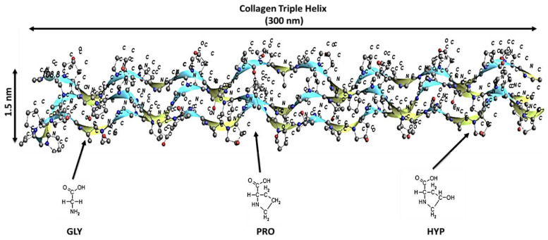
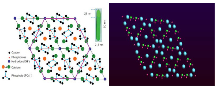
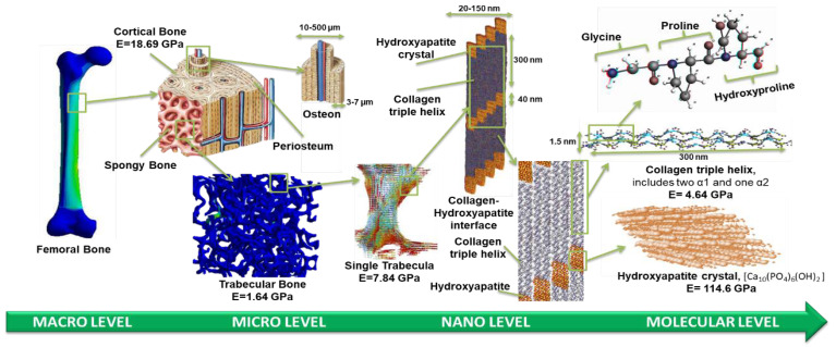
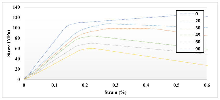
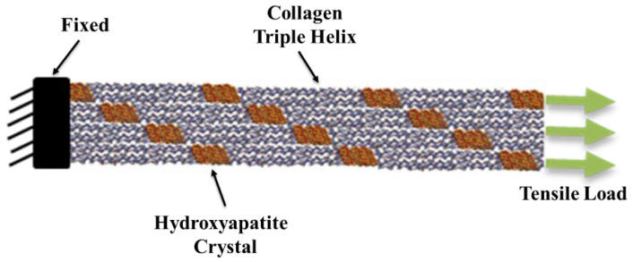
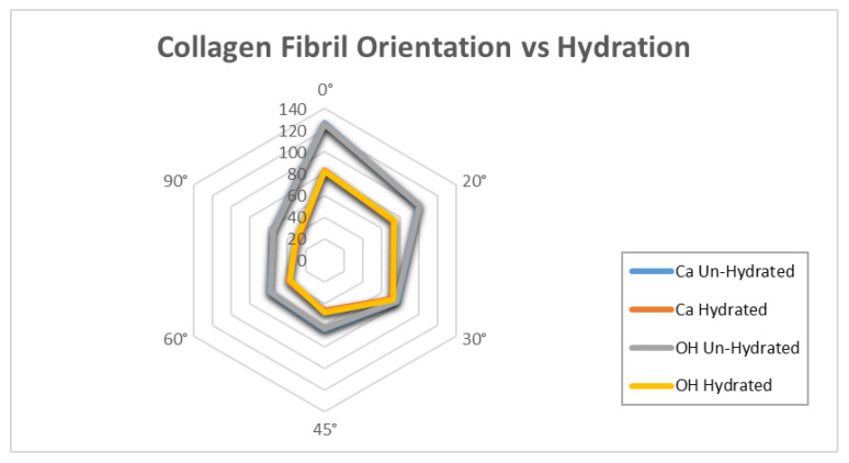
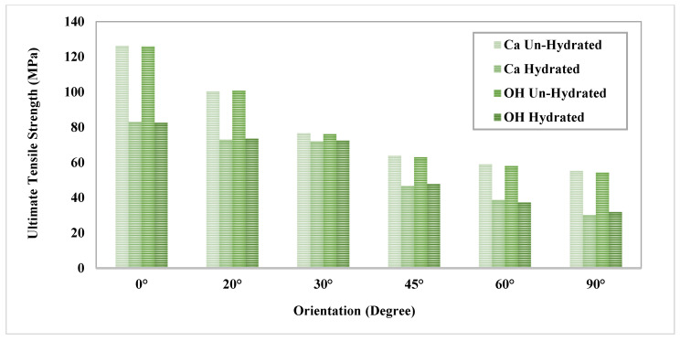
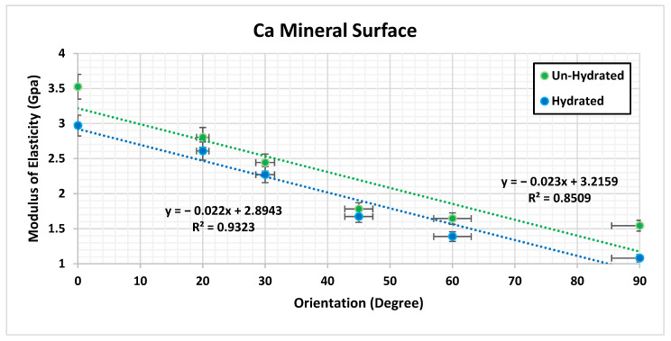
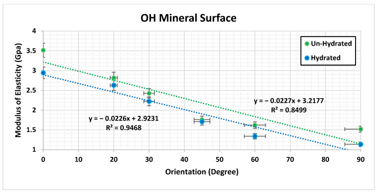
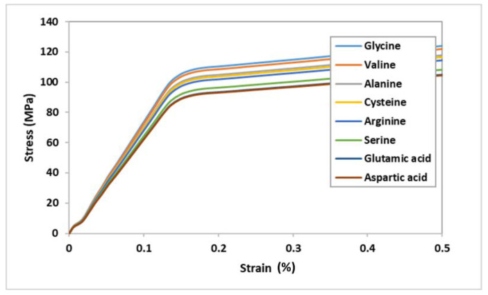
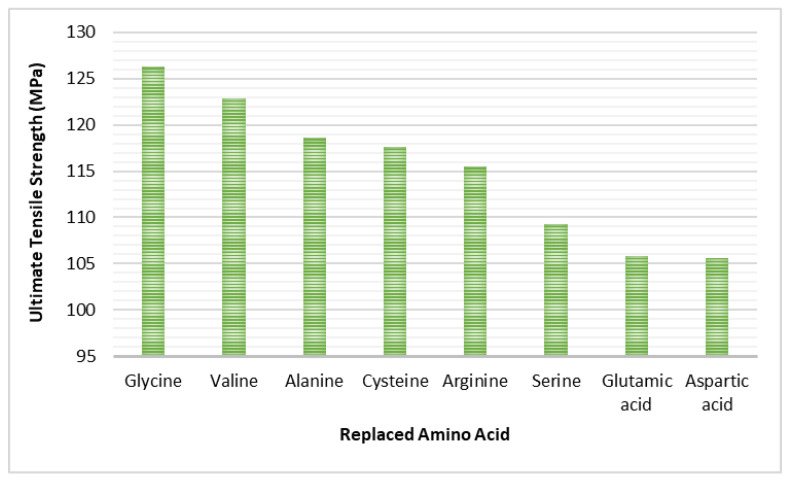
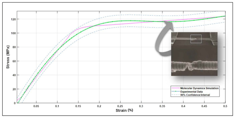
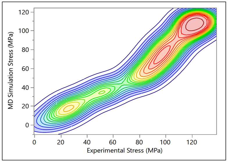
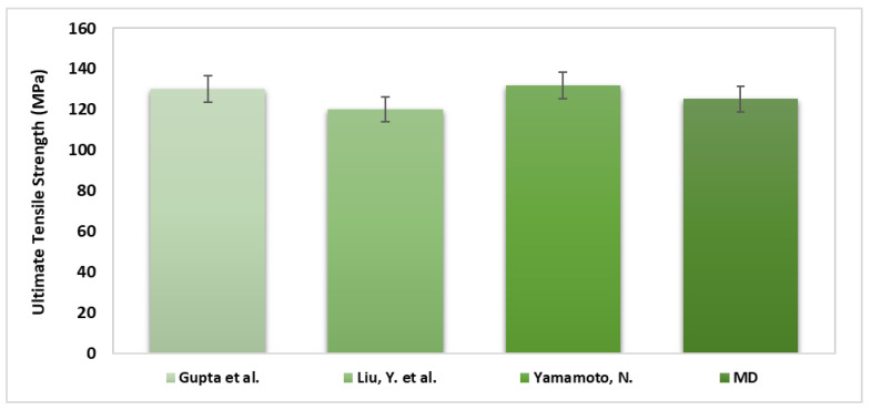
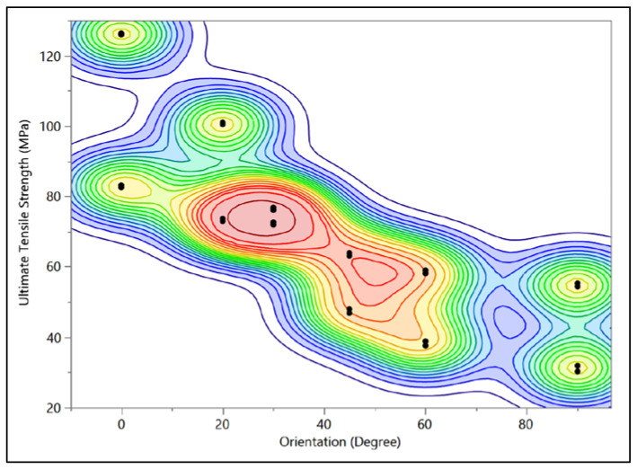
Bone is a highly hierarchical complex structure that consists of organic and mineral components represented by collagen molecules (CM) and hydroxyapatite crystals (HAC), respectively. The nanostructure of bone can significantly affect its mechanical properties. There is a lack of understanding how collagen fibrils (CF) in different orientations may affect the mechanical properties of the bone. The objective of this study is to investigate the effect of interaction, orientation, and hydration on atomic models of the bone composed of collagen helix (CH) and HAC, using molecular dynamics simulations and therefrom bone-related disease origins. The results demonstrate that the mechanical properties of the bone are affected significantly by the orientation of the CF attributed to contact areas at 0° and 90° models. The molecular dynamics simulation illustrated that there is significant difference (p < 0.005) in the ultimate tensile strength and toughness with respect to the orientation of the hydrated and un-hydrated CF. Additionally, the results indicated that having the force in a longitudinal direction (0°) provides more strength compared with the CF in the perpendicular direction (90°). Furthermore, the results show that substituting glycine (GLY) with any other amino acid affects the mechanical properties and strength of the CH, collagen−hydroxyapatite interface, and eventually affects the HAC. Generally, hydration dramatically influences bone tissue elastic properties, and any change in the orientation or any abnormality in the atomic structure of either the CM or the HAC would be the main reason of the fragility in the bone, affecting bone pathology.

摘要

骨骼是一种高度分层的复杂结构,由分别以胶原分子(CM)和羟基磷灰石晶体(HAC)为代表的有机和矿物质成分组成。骨骼的纳米结构会显著影响其力学性能。目前尚不清楚不同取向的胶原纤维(CF)如何影响骨骼的力学性能。本研究的目的是使用分子动力学模拟研究相互作用、取向和水合作用对由胶原螺旋(CH)和HAC组成的骨骼原子模型的影响,并由此探究与骨骼相关的疾病起源。结果表明,由于0°和90°模型中的接触面积,CF的取向对骨骼的力学性能有显著影响。分子动力学模拟表明,水合和未水合CF的取向在极限拉伸强度和韧性方面存在显著差异(p < 0.005)。此外,结果表明,与垂直方向(90°)的CF相比,纵向(0°)受力时能提供更大的强度。此外,结果表明,用任何其他氨基酸取代甘氨酸(GLY)会影响CH、胶原 - 羟基磷灰石界面的力学性能和强度,并最终影响HAC。一般来说,水合作用会显著影响骨组织的弹性性能,CM或HAC的取向变化或原子结构异常将是骨骼脆性的主要原因,影响骨骼病理学。

https://cdn.ncbi.nlm.nih.gov/pmc/blobs/bd4d/9687701/c26cfda76d59/bioengineering-09-00677-g019.jpg
https://cdn.ncbi.nlm.nih.gov/pmc/blobs/bd4d/9687701/bf973a39a7e4/bioengineering-09-00677-g001.jpg
https://cdn.ncbi.nlm.nih.gov/pmc/blobs/bd4d/9687701/493a88b4d024/bioengineering-09-00677-g002.jpg
https://cdn.ncbi.nlm.nih.gov/pmc/blobs/bd4d/9687701/af26252ac35e/bioengineering-09-00677-g003.jpg
https://cdn.ncbi.nlm.nih.gov/pmc/blobs/bd4d/9687701/5acc93c635e4/bioengineering-09-00677-g004.jpg
https://cdn.ncbi.nlm.nih.gov/pmc/blobs/bd4d/9687701/8c4f4821063d/bioengineering-09-00677-g005.jpg
https://cdn.ncbi.nlm.nih.gov/pmc/blobs/bd4d/9687701/b6196d80e54b/bioengineering-09-00677-g006.jpg
https://cdn.ncbi.nlm.nih.gov/pmc/blobs/bd4d/9687701/75b534627dbd/bioengineering-09-00677-g007.jpg
https://cdn.ncbi.nlm.nih.gov/pmc/blobs/bd4d/9687701/6b6a7e3c5193/bioengineering-09-00677-g008a.jpg
https://cdn.ncbi.nlm.nih.gov/pmc/blobs/bd4d/9687701/1bd31630fd94/bioengineering-09-00677-g009.jpg
https://cdn.ncbi.nlm.nih.gov/pmc/blobs/bd4d/9687701/728d5f9614ef/bioengineering-09-00677-g010.jpg
https://cdn.ncbi.nlm.nih.gov/pmc/blobs/bd4d/9687701/197dac8c9662/bioengineering-09-00677-g011.jpg
https://cdn.ncbi.nlm.nih.gov/pmc/blobs/bd4d/9687701/73870ada190e/bioengineering-09-00677-g012.jpg
https://cdn.ncbi.nlm.nih.gov/pmc/blobs/bd4d/9687701/f88c93f282f9/bioengineering-09-00677-g013.jpg
https://cdn.ncbi.nlm.nih.gov/pmc/blobs/bd4d/9687701/c7f97abe9353/bioengineering-09-00677-g014.jpg
https://cdn.ncbi.nlm.nih.gov/pmc/blobs/bd4d/9687701/7a7a0dbc8349/bioengineering-09-00677-g015.jpg
https://cdn.ncbi.nlm.nih.gov/pmc/blobs/bd4d/9687701/3c721fa3fb3a/bioengineering-09-00677-g016.jpg
https://cdn.ncbi.nlm.nih.gov/pmc/blobs/bd4d/9687701/9a9ebe87640c/bioengineering-09-00677-g017.jpg
https://cdn.ncbi.nlm.nih.gov/pmc/blobs/bd4d/9687701/adab1801c177/bioengineering-09-00677-g018.jpg
https://cdn.ncbi.nlm.nih.gov/pmc/blobs/bd4d/9687701/c26cfda76d59/bioengineering-09-00677-g019.jpg

相似文献

1
Hierarchical Structure and Properties of the Bone at Nano Level.纳米级骨的层次结构与特性
Bioengineering (Basel). 2022 Nov 10;9(11):677. doi: 10.3390/bioengineering9110677.
2
A multiscale modelling of bone ultrastructure elastic proprieties using finite elements simulation and neural network method.一种使用有限元模拟和神经网络方法对骨超微结构弹性特性进行的多尺度建模。
Comput Methods Programs Biomed. 2016 Oct;134:69-78. doi: 10.1016/j.cmpb.2016.07.005. Epub 2016 Jul 5.
3
Large Deformation Mechanisms, Plasticity, and Failure of an Individual Collagen Fibril With Different Mineral Content.不同矿物质含量的单个胶原纤维的大变形机制、塑性与破坏
J Bone Miner Res. 2016 Feb;31(2):380-90. doi: 10.1002/jbmr.2705.
4
Disuse Impairs the Mechanical Competence of Bone by Regulating the Characterizations of Mineralized Collagen Fibrils in Cortical Bone.废用通过调节皮质骨中矿化胶原纤维的特性损害骨的力学性能。
Front Physiol. 2019 Jun 21;10:775. doi: 10.3389/fphys.2019.00775. eCollection 2019.
5
Thickness of hydroxyapatite nanocrystal controls mechanical properties of the collagen-hydroxyapatite interface.羟基磷灰石纳米晶的厚度控制着胶原-羟基磷灰石界面的力学性能。
Langmuir. 2012 Jan 31;28(4):1982-92. doi: 10.1021/la204052a. Epub 2012 Jan 18.
6
A finite element study evaluating the influence of mineralization distribution and content on the tensile mechanical response of mineralized collagen fibril networks.一种评估矿化分布和含量对矿化胶原纤维网络拉伸力学响应影响的有限元研究。
J Mech Behav Biomed Mater. 2019 Dec;100:103361. doi: 10.1016/j.jmbbm.2019.07.019. Epub 2019 Jul 20.
7
Mineral and cross-linking in collagen fibrils: The mechanical behavior of bone tissue at the nano-scale.胶原纤维中的矿物质与交联:骨组织在纳米尺度下的力学行为。
ArXiv. 2024 Mar 18:arXiv:2403.11753v1.
8
Deformation regimes of collagen fibrils in cortical bone revealed by in situ morphology and elastic modulus observations under mechanical loading.原位形貌和力学加载下弹性模量观察揭示皮质骨中胶原原纤维的变形机制。
J Mech Behav Biomed Mater. 2018 Mar;79:115-121. doi: 10.1016/j.jmbbm.2017.12.015. Epub 2017 Dec 16.
9
Hierarchical structure and nanomechanics of collagen microfibrils from the atomistic scale up.从原子尺度到胶原微纤维的层次结构和纳米力学。
Nano Lett. 2011 Feb 9;11(2):757-66. doi: 10.1021/nl103943u. Epub 2011 Jan 5.
10
Effects of hydration and mineralization on the deformation mechanisms of collagen fibrils in bone at the nanoscale.水合作用和矿化作用对纳米尺度下骨胶原原纤维变形机制的影响。
Biomech Model Mechanobiol. 2019 Feb;18(1):57-68. doi: 10.1007/s10237-018-1067-y. Epub 2018 Aug 7.

引用本文的文献

1
Analysis of Biomechanical Characteristics of Bone Tissues Using a Bayesian Neural Network: A Narrative Review.使用贝叶斯神经网络分析骨组织的生物力学特性:一篇综述。
J Funct Biomater. 2025 May 8;16(5):168. doi: 10.3390/jfb16050168.
2
Bridging the Gap in Understanding Bone Metastasis: A Multifaceted Perspective.弥合对骨转移认识的差距:多维度视角
Int J Mol Sci. 2024 Feb 29;25(5):2846. doi: 10.3390/ijms25052846.
3
Harnessing the Potential of PLGA Nanoparticles for Enhanced Bone Regeneration.利用聚乳酸-羟基乙酸共聚物纳米颗粒促进骨再生的潜力

本文引用的文献

1
Exploring the Mechanical Properties and Performance of Type-I Collagen at Various Length Scales: A Progress Report.探索不同长度尺度下I型胶原蛋白的力学性能和性能:进展报告。
Materials (Basel). 2022 Apr 8;15(8):2753. doi: 10.3390/ma15082753.
2
Collagen Fibril Orientation in Tissue Specimens From Atherosclerotic Plaque Explored Using Small Angle X-Ray Scattering.采用小角 X 射线散射技术研究动脉粥样硬化斑块组织标本中的胶原纤维取向。
J Biomech Eng. 2022 Feb 1;144(2). doi: 10.1115/1.4052432.
3
Multiscale characterization of pathological bone tissue.
Pharmaceutics. 2024 Feb 15;16(2):273. doi: 10.3390/pharmaceutics16020273.
病理性骨组织的多尺度特征分析。
Microsc Res Tech. 2022 Feb;85(2):469-486. doi: 10.1002/jemt.23920. Epub 2021 Sep 7.
4
On the Crystallization of Hydroxyapatite under Hydrothermal Conditions: Role of Sebacic Acid as an Additive.水热条件下羟基磷灰石的结晶:癸二酸作为添加剂的作用
ACS Omega. 2020 Oct 16;5(42):27204-27210. doi: 10.1021/acsomega.0c03297. eCollection 2020 Oct 27.
5
Validation study of small-angle X-ray scattering tensor tomography.小角 X 射线散射张量层析成像的验证研究。
J Synchrotron Radiat. 2020 May 1;27(Pt 3):779-787. doi: 10.1107/S1600577520003860. Epub 2020 Apr 22.
6
50 years of scanning electron microscopy of bone-a comprehensive overview of the important discoveries made and insights gained into bone material properties in health, disease, and taphonomy.50年的骨扫描电子显微镜研究——对在健康、疾病和埋藏学方面所取得的重要发现以及对骨材料特性的深入见解的全面概述。
Bone Res. 2019 May 22;7:15. doi: 10.1038/s41413-019-0053-z. eCollection 2019.
7
Macrodamage Accumulation Model for a Human Femur.人体股骨的宏观损伤累积模型。
Appl Bionics Biomech. 2017;2017:4539178. doi: 10.1155/2017/4539178. Epub 2017 Aug 29.
8
Orientation anisotropy of quantitative MRI relaxation parameters in ordered tissue.有序组织中定量 MRI 弛豫参数的各向异性
Sci Rep. 2017 Aug 29;7(1):9606. doi: 10.1038/s41598-017-10053-2.
9
Ultrastructure Organization of Human Trabeculae Assessed by 3D sSAXS and Relation to Bone Microarchitecture.通过三维小角X射线散射评估的人小梁超微结构组织及其与骨微结构的关系
PLoS One. 2016 Aug 22;11(8):e0159838. doi: 10.1371/journal.pone.0159838. eCollection 2016.
10
Techniques to assess bone ultrastructure organization: orientation and arrangement of mineralized collagen fibrils.评估骨超微结构组织的技术:矿化胶原纤维的取向和排列
J R Soc Interface. 2016 Jun;13(119). doi: 10.1098/rsif.2016.0088.