• 文献检索
  • 文档翻译
  • 深度研究
  • 学术资讯
  • Suppr Zotero 插件Zotero 插件
  • 邀请有礼
  • 套餐&价格
  • 历史记录
应用&插件
Suppr Zotero 插件Zotero 插件浏览器插件Mac 客户端Windows 客户端微信小程序
定价
高级版会员购买积分包购买API积分包
服务
文献检索文档翻译深度研究API 文档MCP 服务
关于我们
关于 Suppr公司介绍联系我们用户协议隐私条款
关注我们

Suppr 超能文献

核心技术专利:CN118964589B侵权必究
粤ICP备2023148730 号-1Suppr @ 2026

文献检索

告别复杂PubMed语法,用中文像聊天一样搜索,搜遍4000万医学文献。AI智能推荐,让科研检索更轻松。

立即免费搜索

文件翻译

保留排版,准确专业,支持PDF/Word/PPT等文件格式,支持 12+语言互译。

免费翻译文档

深度研究

AI帮你快速写综述,25分钟生成高质量综述,智能提取关键信息,辅助科研写作。

立即免费体验

大鼠下颌后移后的组织学和生化分析

Histological and Biochemical Analysis after Posterior Mandibular Displacement in Rats.

作者信息

Lyros Ioannis, Perrea Despoina, Tosios Konstantinos, Nikitakis Nikolaos, Tsolakis Ioannis A, Ferdianakis Efstratios, Fora Eleni, Lykogeorgos Theodoros, Maroulakos Michael P, Vardas Emmanouil, Georgaki Maria, Papadopoulou Erofili, Tsolakis Apostolos I

机构信息

Department of Orthodontics, School of Dentistry, National and Kapodistrian University of Athens, 11527 Athens, Greece.

Laboratory of Experimental Surgery and Surgical Research "N.S. Christeas", Medical School, National and Kapodistrian University of Athens, 11527 Athens, Greece.

出版信息

Vet Sci. 2022 Nov 10;9(11):625. doi: 10.3390/vetsci9110625.

DOI:10.3390/vetsci9110625
PMID:36356102
原文链接:https://pmc.ncbi.nlm.nih.gov/articles/PMC9697094/
Abstract

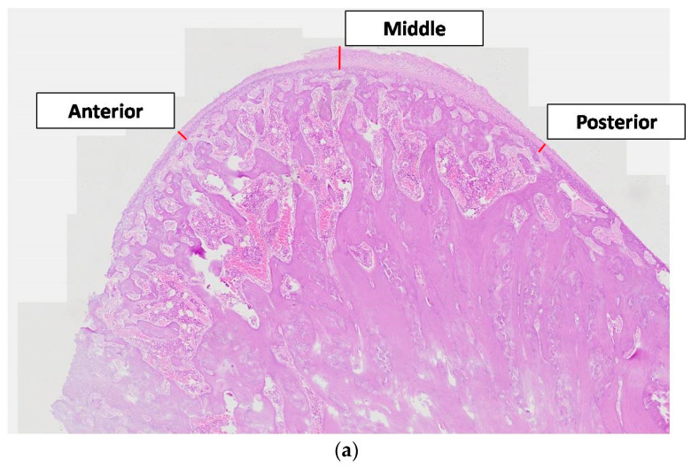
The present study aimed to investigate any biochemical and histological changes of the rat condyle and mandible in animals that had sustained mandibular growth restriction. Seventy-two male Wistar rats were divided into two equal groups, experimental and control. Each group consisted of three equal subgroups. The animals were sacrificed 30, 60, and 90 days after the start of the experiment. Blood samples were collected from the eye, and the osteoprotegerin (OPG), Receptor Activator of Nuclear Factor Kappa B Ligand (RANKL), and Macrophage Colony-Stimulating factor (MCSF)concentrations were measured by using enzyme-linked immunosorbent assay (ELISA) kits. A histological analysis was performed on the mandibular condyles. The blood serum values of OPG, RANKL, and MCSF did not exhibit any statistically significant difference between groups or subgroups. However, significant histological changes became evident after a histomorphometric condylar examination was performed. The Bone Surface/Total Surface ratio appeared reduced in the anterior and posterior regions of the condyle. In addition, the Posterior Condylar Cartilage Thickness was measured and determined to be significantly diminished. The present intervention that employed orthodontic/orthopedic devices did not prove to have any significant effect on the circulating proteins under study. Posterior displacement of the mandible may culminate only in local histological alterations in condylar cartilage thickness and its osseous microarchitecture.

摘要

本研究旨在调查下颌生长受限动物的大鼠髁突和下颌骨的任何生化和组织学变化。72只雄性Wistar大鼠被分为两组,即实验组和对照组,每组又分为三个相等的亚组。在实验开始后的30天、60天和90天处死动物。从眼眶采集血样,使用酶联免疫吸附测定(ELISA)试剂盒测量骨保护素(OPG)、核因子κB受体活化因子配体(RANKL)和巨噬细胞集落刺激因子(MCSF)的浓度。对下颌髁突进行组织学分析。OPG、RANKL和MCSF的血清值在组间或亚组间未显示出任何统计学上的显著差异。然而,在进行髁突组织形态计量学检查后,明显的组织学变化变得明显。髁突前后区域的骨表面积/总表面积比值似乎降低。此外,测量了髁突后软骨厚度,发现其显著减小。采用正畸/矫形装置的当前干预措施并未证明对所研究的循环蛋白有任何显著影响。下颌后移可能仅导致髁突软骨厚度及其骨微结构的局部组织学改变。

https://cdn.ncbi.nlm.nih.gov/pmc/blobs/338a/9697094/76ab0db47863/vetsci-09-00625-g006a.jpg
https://cdn.ncbi.nlm.nih.gov/pmc/blobs/338a/9697094/6058dec27ff7/vetsci-09-00625-g001.jpg
https://cdn.ncbi.nlm.nih.gov/pmc/blobs/338a/9697094/27af70df28ab/vetsci-09-00625-g002.jpg
https://cdn.ncbi.nlm.nih.gov/pmc/blobs/338a/9697094/a1eb0931170c/vetsci-09-00625-g003.jpg
https://cdn.ncbi.nlm.nih.gov/pmc/blobs/338a/9697094/4dd9174e6bae/vetsci-09-00625-g004a.jpg
https://cdn.ncbi.nlm.nih.gov/pmc/blobs/338a/9697094/c72e30a39396/vetsci-09-00625-g005a.jpg
https://cdn.ncbi.nlm.nih.gov/pmc/blobs/338a/9697094/76ab0db47863/vetsci-09-00625-g006a.jpg

相似文献

1
Histological and Biochemical Analysis after Posterior Mandibular Displacement in Rats.大鼠下颌后移后的组织学和生化分析
Vet Sci. 2022 Nov 10;9(11):625. doi: 10.3390/vetsci9110625.
2
[Cone-beam CT analysis of vertical control of mandible and changes of temporomandibular joint in adult patients with skeletal class Ⅱ malocclusion with high angle].[成人骨性Ⅱ类高角错牙合患者下颌垂直向控制及颞下颌关节变化的锥形束CT分析]
Zhonghua Kou Qiang Yi Xue Za Zhi. 2022 Nov 9;57(11):1147-1155. doi: 10.3760/cma.j.cn112144-20220301-00086.
3
[Effect of gradually induced disordered occlusion on the expression of osteoprotegerin and receptor activator of nuclear factor-kappaB ligand in mandibular condylar cartilage of rats].[逐渐诱导的紊乱咬合对大鼠下颌髁突软骨中骨保护素及核因子-κB受体活化因子配体表达的影响]
Hua Xi Kou Qiang Yi Xue Za Zhi. 2012 Apr;30(2):119-22, 127.
4
[The effect of lowered serum levels of estrogen on the expression of ERα, ERβ, OPG and RANKL in rat mandibular condylar cartilage].[血清雌激素水平降低对大鼠下颌髁突软骨中ERα、ERβ、OPG和RANKL表达的影响]
Shanghai Kou Qiang Yi Xue. 2015 Aug;24(4):437-41.
5
Morphometric evaluation of condylar cartilage of growing rats in response to mandibular retractive forces.生长大鼠髁突软骨对下颌后缩力反应的形态计量学评估
Dental Press J Orthod. 2013 Jul-Aug;18(4):113-9. doi: 10.1590/s2176-94512013000400016.
6
Three-Dimensional Analysis of Posterior Mandibular Displacement in Rats.大鼠下颌骨后移位的三维分析
Vet Sci. 2022 Mar 20;9(3):144. doi: 10.3390/vetsci9030144.
7
Histological and histomorphometric investigation of the condylar cartilage of juvenile pigs after anterior mandibular displacement.幼年猪下颌前移位后髁突软骨的组织学和组织形态计量学研究
Ann Anat. 2007;189(3):269-75. doi: 10.1016/j.aanat.2006.09.006.
8
Expression of lubricin in rat posterior mandibular condylar cartilage following functional mandibular forward repositioning.功能性下颌前伸复位后大鼠下颌后髁突软骨中润滑素的表达
J Orofac Orthop. 2019 May;80(3):128-135. doi: 10.1007/s00056-019-00173-x. Epub 2019 Apr 5.
9
Systemic administration of strontium or NBD peptide ameliorates early stage cartilage degradation of mouse mandibular condyles.锶或NBD肽的全身给药可改善小鼠下颌髁突早期软骨降解。
Osteoarthritis Cartilage. 2016 Jan;24(1):178-187. doi: 10.1016/j.joca.2015.07.022. Epub 2015 Aug 6.
10
Biomechanically stimulated chondrocytes promote osteoclastic bone resorption in the mandibular condyle.生物力学刺激的软骨细胞促进下颌髁突的破骨细胞骨吸收。
Arch Oral Biol. 2019 Feb;98:248-257. doi: 10.1016/j.archoralbio.2018.12.002. Epub 2018 Dec 3.

引用本文的文献

1
Growth Prediction in Orthodontics: ASystematic Review of Past Methods up to Artificial Intelligence.正畸学中的生长预测:对直至人工智能的既往方法的系统评价。
Children (Basel). 2025 Aug 3;12(8):1023. doi: 10.3390/children12081023.
2
Effects of orthodontic tooth movement on periodontal tissues after ligature-induced periodontitis through the mechanism of RANKL-induced osteoclastogenesis: an animal study.通过RANKL诱导破骨细胞生成机制,结扎诱导性牙周炎后正畸牙齿移动对牙周组织的影响:一项动物研究。
BMC Oral Health. 2025 Jul 30;25(1):1278. doi: 10.1186/s12903-025-06627-6.
3
Immunohistochemical Evaluation of Bone Remodeling Following Compressive Force on Mandibular Condyle.

本文引用的文献

1
Anterior Mandibular Displacement in Growing Rats-A Systematic Review.生长中大鼠的下颌前移位——一项系统评价
Animals (Basel). 2022 Aug 12;12(16):2059. doi: 10.3390/ani12162059.
2
Effect of Diet Consistency on Rat Mandibular Growth: A Geometric Morphometric and Linear Cephalometric Study.饮食稠度对大鼠下颌生长的影响:一项几何形态测量学和线性头影测量学研究。
Biology (Basel). 2022 Jun 11;11(6):901. doi: 10.3390/biology11060901.
3
Three-Dimensional Analysis of Posterior Mandibular Displacement in Rats.大鼠下颌骨后移位的三维分析
下颌髁突压缩力作用后骨重塑的免疫组织化学评估
Biology (Basel). 2025 Feb 23;14(3):228. doi: 10.3390/biology14030228.
Vet Sci. 2022 Mar 20;9(3):144. doi: 10.3390/vetsci9030144.
4
Impact of Diet Consistency on the Mandibular Morphology: A Systematic Review of Studies on Rat Models.饮食一致性对下颌形态的影响:大鼠模型研究的系统评价。
Int J Environ Res Public Health. 2022 Feb 25;19(5):2706. doi: 10.3390/ijerph19052706.
5
Breast implant illness: scientific evidence of its existence.乳房植入病:其存在的科学证据。
Expert Rev Clin Immunol. 2022 Jan;18(1):15-29. doi: 10.1080/1744666X.2022.2010546. Epub 2022 Jan 5.
6
Regulation of Osteoclastogenesis and Bone Resorption by miRNAs.微小RNA对破骨细胞生成和骨吸收的调控
Front Cell Dev Biol. 2021 Jun 18;9:651161. doi: 10.3389/fcell.2021.651161. eCollection 2021.
7
Posterior Mandibular Displacement-A Systematic Review Based on Animal Studies.下颌后移位——基于动物研究的系统评价
Animals (Basel). 2021 Mar 15;11(3):823. doi: 10.3390/ani11030823.
8
Transcriptional Regulation of Osteoclastogenesis: The Emerging Role of KLF2.破骨细胞生成的转录调控:KLF2 的新作用。
Front Immunol. 2020 May 13;11:937. doi: 10.3389/fimmu.2020.00937. eCollection 2020.
9
Mandibular Condyle Remodeling after Functional Orthopedic Treatment: A Clinical Report of Four Cases.功能性正畸治疗后下颌髁突重塑:4例临床报告
J Contemp Dent Pract. 2019 Dec 1;20(12):1461-1465.
10
Selection of Models: Evolution and the Choice of Species for Translational Research.模型的选择:进化与转化研究的物种选择
Brain Behav Evol. 2019;93(2-3):82-91. doi: 10.1159/000500317. Epub 2019 Aug 15.