Fernández-Albarral José A, Salobrar-García Elena, Matamoros José A, Fernández-Mendívil Cristina, Del Sastre Eric, Chen Lejing, de Hoz Rosa, López-Cuenca Inés, Sánchez-Puebla Lidia, Ramírez José M, Salazar Juan J, Lopez Manuela G, Ramírez Ana I
Instituto de Investigaciones Oftalmológicas Ramón Castroviejo, Grupo UCM 920105, IdISSC, Universidad Complutense de Madrid, 28040 Madrid, Spain.
Facultad de Óptica y Optometría, Departamento de Inmunología, Oftalmología y ORL, Universidad Complutense de Madrid, 28037 Madrid, Spain.
Antioxidants (Basel). 2022 Oct 30;11(11):2151. doi: 10.3390/antiox11112151.
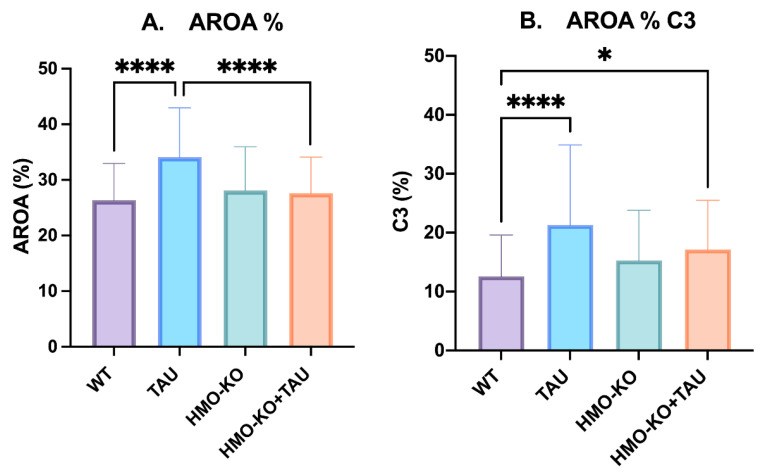
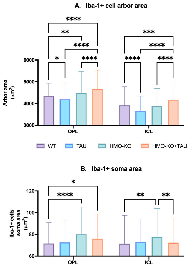
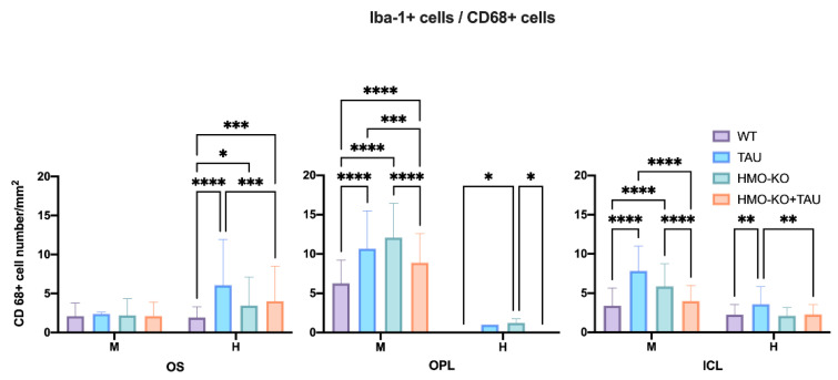
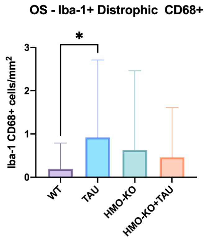
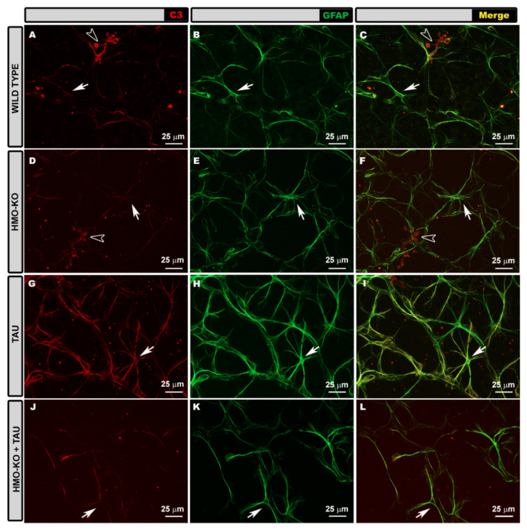
Tauopathies such as Alzheimer's disease are characterized by the accumulation of neurotoxic aggregates of tau protein. With aging and, especially, in Alzheimer's patients, the inducible enzyme heme oxygenase 1 (HO-1) progressively increases in microglia, causing iron accumulation, neuroinflammation, and neurodegeneration. The retina is an organ that can be readily accessed and can reflect changes that occur in the brain. In this context, we evaluated how the lack of microglial HO-1, using mice that do not express HO-1 in microglia (HMO-KO), impacts retinal macro and microgliosis of aged subjects (18 months old mice) subjected to tauopathy by intrahippocampal delivery of AAV-hTau (TAU). Our results show that although tauopathy, measured as anti-TAUY9 and anti-AT8 positive immunostaining, was not observed in the retina of WT-TAU or HMO-KO+TAU mice, a morphometric study of retinal microglia and macroglia showed significant retinal changes in the TAU group compared to the WT group, such as: (i) increased number of activated microglia, (ii) retraction of microglial processes, (iii) increased number of CD68+ microglia, and (iv) increased retinal area occupied by GFAP (AROA) and C3 (AROC3). This retinal inflammatory profile was reduced in HMO-KO+TAU mice. Conclusion: Reduction of microglial HO-1 could be beneficial to prevent tauopathy-induced neuroinflammation.
诸如阿尔茨海默病之类的tau蛋白病的特征是tau蛋白神经毒性聚集体的积累。随着年龄增长,尤其是在阿尔茨海默病患者中,诱导型酶血红素加氧酶1(HO-1)在小胶质细胞中逐渐增加,导致铁积累、神经炎症和神经退行性变。视网膜是一个易于观察的器官,能够反映大脑中发生的变化。在此背景下,我们利用在小胶质细胞中不表达HO-1的小鼠(HMO-KO),评估了小胶质细胞HO-1的缺失如何影响通过海马内注射AAV-hTau(TAU)诱导tau蛋白病的老年受试者(18月龄小鼠)的视网膜大胶质细胞增生和小胶质细胞增生。我们的结果表明,尽管在WT-TAU或HMO-KO+TAU小鼠的视网膜中未观察到以抗TAUY9和抗AT8阳性免疫染色衡量的tau蛋白病,但与WT组相比,对视网膜小胶质细胞和大胶质细胞的形态计量学研究显示TAU组的视网膜有显著变化,例如:(i)活化小胶质细胞数量增加,(ii)小胶质细胞突起回缩,(iii)CD68+小胶质细胞数量增加,以及(iv)GFAP(AROA)和C3(AROC3)占据的视网膜面积增加。HMO-KO+TAU小鼠的这种视网膜炎症特征有所减轻。结论:小胶质细胞HO-1的减少可能有利于预防tau蛋白病诱导的神经炎症。