Kiamehr Pegah, Shahidi Minoo, Samii Amir, Zaker Farhad
Department of Hematology and Blood Banking, School of Allied Medical Sciences, Iran University of Medical Sciences, Tehran, Iran .
Int J Mol Cell Med. 2022;11(1):16-30. doi: 10.22088/IJMCM.BUMS.11.1.16. Epub 2022 Oct 3.
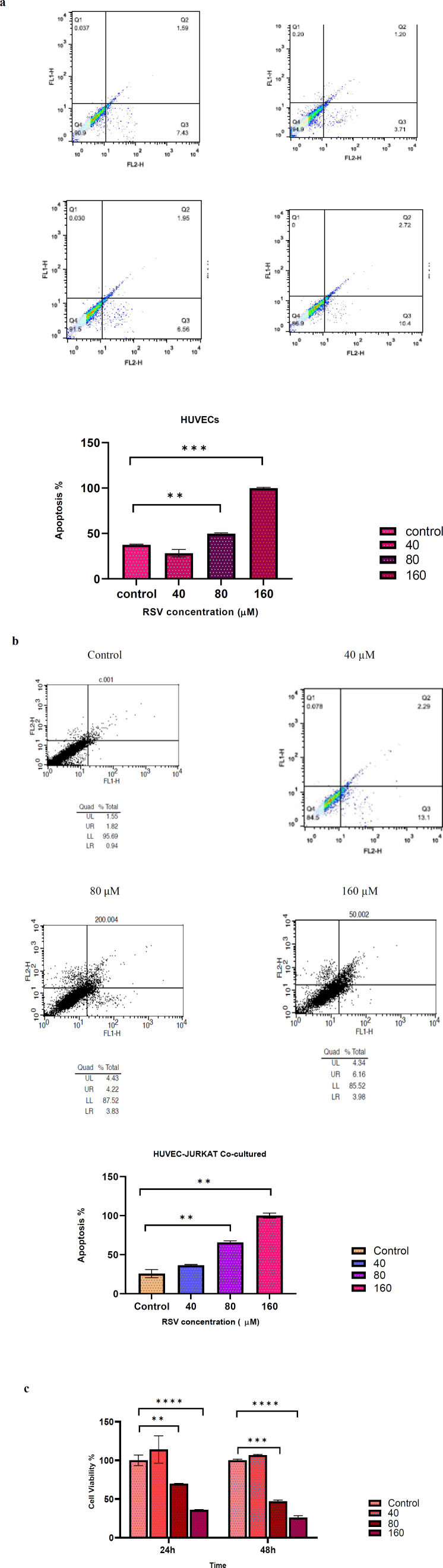
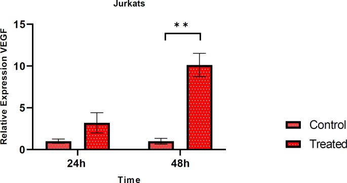
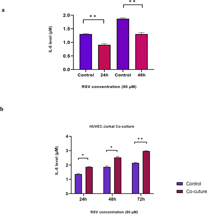
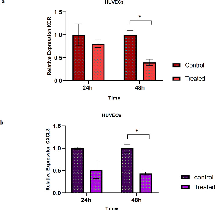
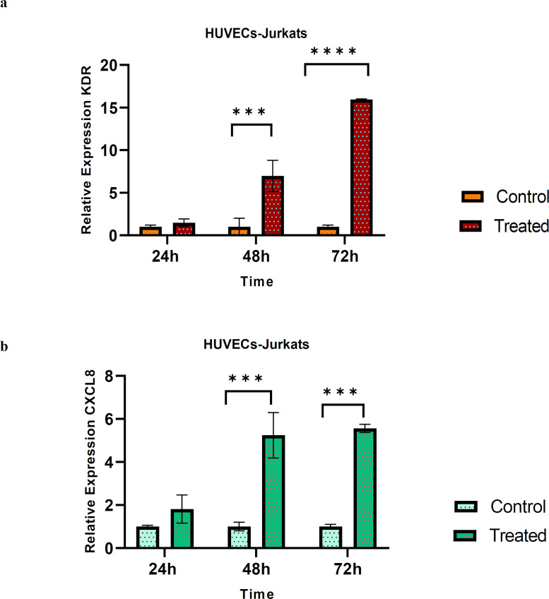
Angiogenesis is an essential process in the growth, development, and transition of tumors from dormancy to proliferating state. Resveratrol (RSV), as a natural polyphenolic compound, is claimed to be effective in regulating angiogenesis. This study aimed to evaluate the impact of RSV onthe angiogenesis process in HUVECs (human umbilical vein endothelial cells) alone and co-cultured with Jurkat cells. The effects of RSV on HUVECs and Jurkat cell viability and apoptosis were measured by MTT and Annexin-V/PI methods. HUVECs were co-cultured with pre-treated Jurkat cells and incubated for 24 h, 48 h and 72 h. The angiogenesis process in HUVECs and Jurkat cells alone and in co-culture models was investigated by analyzing the expression of VEGF, VEGFR-2, and Interleukin-8 (IL-8) employing qPCR and ELISA. RSV at low concentration (40 µM) had no significant effects on apoptosis rate of HUVECs, but higher concentrations (80-160 µM) increased apoptosis in co-culture method and HUVECs alone. RSV significantly reduced VEGFR2 and IL-8 gene expression also, IL-8 protein concentration in HUVECs, but the effects of this drug in the HUVECs-Jurkats co-culture were different. Expression of VEGF in Jurkat cells increased following treatment with RSV. RSV had direct anti-angiogenic effects on HUVECs. Unexpectedly its indirect effects were not significant on HUVECs-Jurkats co-culture. Results of our study showed, RSV may be effective in anti-angiogenesis therapy, but in some situations, it may induce angiogenesis. So, appropriate concentrations should achieve to minimize the unpredicted effects of RSV.
血管生成是肿瘤生长、发育以及从休眠状态转变为增殖状态过程中的一个重要过程。白藜芦醇(RSV)作为一种天然多酚化合物,据称在调节血管生成方面具有功效。本研究旨在评估RSV对单独培养的人脐静脉内皮细胞(HUVECs)以及与Jurkat细胞共培养时血管生成过程的影响。通过MTT法和Annexin-V/PI法测定RSV对HUVECs和Jurkat细胞活力及凋亡的影响。将HUVECs与预处理后的Jurkat细胞共培养,并分别孵育24小时、48小时和72小时。通过qPCR和ELISA分析VEGF、VEGFR-2和白细胞介素-8(IL-8)的表达,研究单独培养的HUVECs和Jurkat细胞以及共培养模型中的血管生成过程。低浓度(40µM)的RSV对HUVECs的凋亡率无显著影响,但较高浓度(80 - 160µM)在共培养方法以及单独培养的HUVECs中均增加了凋亡。RSV还显著降低了VEGFR2和IL-8基因表达以及HUVECs中IL-8蛋白浓度,但该药物在HUVECs - Jurkat细胞共培养中的作用有所不同。用RSV处理后,Jurkat细胞中VEGF的表达增加。RSV对HUVECs具有直接的抗血管生成作用。出乎意料的是,其对HUVECs - Jurkat细胞共培养的间接作用并不显著。我们的研究结果表明,RSV可能在抗血管生成治疗中有效,但在某些情况下,它可能诱导血管生成。因此,应采用合适的浓度以尽量减少RSV的不可预测作用。