Rolnick Kevin I, Choe Joshua A, Leiferman Ellen M, Kondratko-Mittnacht Jaclyn, Clements Anna E B, Baer Geoffrey S, Jiang Peng, Vanderby Ray, Chamberlain Connie S
Department of Orthopedics and Rehabilitation, University of Wisconsin, Madison, WI 53705, USA.
Center for Gene Regulation in Health and Disease (GRHD) and Department of Biological, Geological and Environmental Sciences, Cleveland State University, Cleveland, OH 44115, USA.
Matrix Biol Plus. 2022 Nov 9;16:100124. doi: 10.1016/j.mbplus.2022.100124. eCollection 2022 Dec.
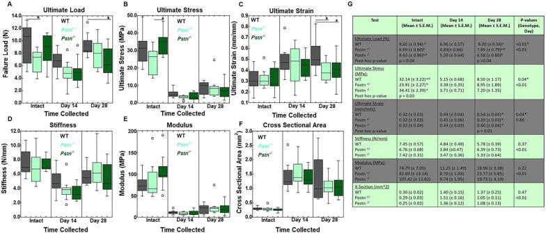
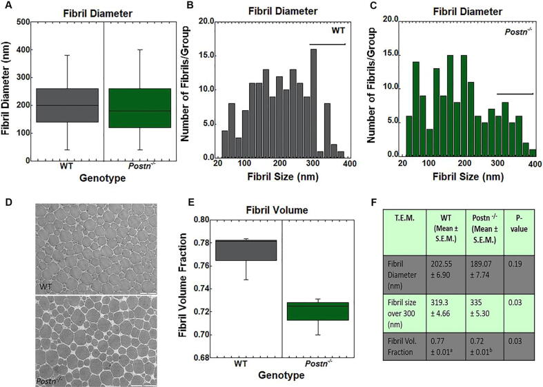
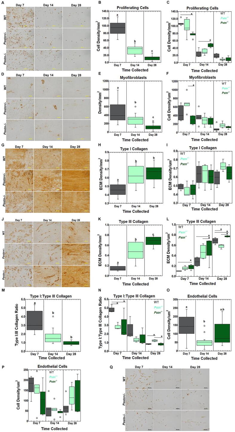
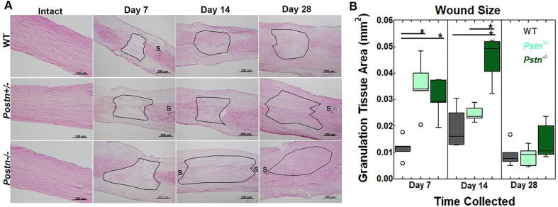
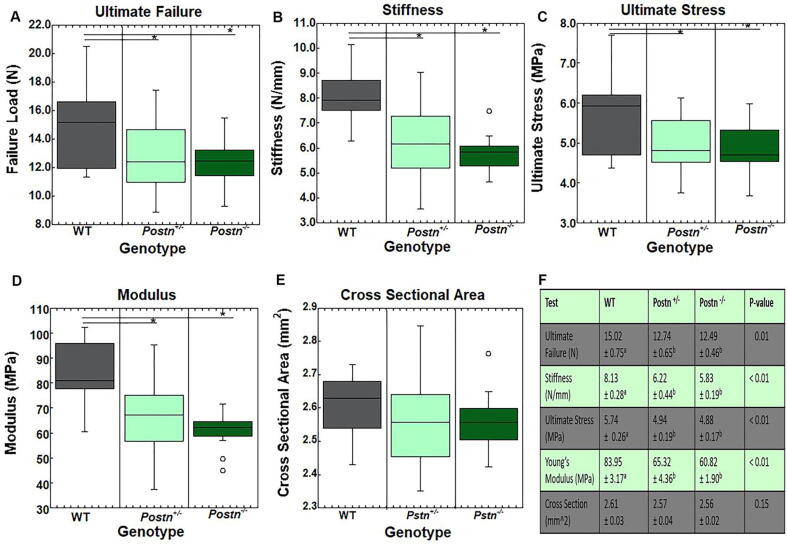
Periostin, originally named osteoblast-specific factor 2 (OSF-2) has been identified primarily in collagen rich, biomechanically active tissues where its role has been implicated in mechanisms to maintain the extracellular matrix (ECM), including collagen fibrillogenesis and crosslinking. It is well documented that periostin plays a role in wound healing and scar formation after injury, in part, by promoting cell proliferation, myofibroblast differentiation, and/or collagen fibrillogenesis. Given the significance of periostin in other scar forming models, we hypothesized that periostin will influence Achilles tendon healing by modulating ECM production. Therefore, the objective of this study was to elucidate the effects of periostin during Achilles tendon healing using periostin homozygous ( ) and heterozygous ( ) mouse models. A second experiment was included to further examine the influence of periostin on collagen composition and function using intact dorsal tail tendons. Overall, and Achilles tendons exhibited impaired healing as demonstrated by delayed wound closure, increased type III collagen production, decreased cell proliferation, and reduced tensile strength. Periostin ablation also reduced tensile strength and stiffness, and altered collagen fibril distribution in the intact dorsal tail tendons. Achilles tendon outcomes support our hypothesis that periostin influences healing, while tail tendon results indicate that periostin also affects ECM morphology and behavior in mouse tendons.
骨膜蛋白最初被命名为成骨细胞特异性因子2(OSF-2),主要在富含胶原蛋白、具有生物力学活性的组织中被发现,其作用涉及维持细胞外基质(ECM)的机制,包括胶原纤维形成和交联。有充分的文献记载,骨膜蛋白在损伤后的伤口愈合和瘢痕形成中发挥作用,部分原因是通过促进细胞增殖、肌成纤维细胞分化和/或胶原纤维形成。鉴于骨膜蛋白在其他瘢痕形成模型中的重要性,我们假设骨膜蛋白将通过调节ECM产生来影响跟腱愈合。因此,本研究的目的是使用骨膜蛋白纯合( )和杂合( )小鼠模型阐明骨膜蛋白在跟腱愈合过程中的作用。还进行了第二项实验,以使用完整的背尾肌腱进一步研究骨膜蛋白对胶原蛋白组成和功能的影响。总体而言, 和 跟腱表现出愈合受损,表现为伤口闭合延迟、III型胶原蛋白产生增加、细胞增殖减少和拉伸强度降低。骨膜蛋白缺失还降低了拉伸强度和刚度,并改变了完整背尾肌腱中胶原纤维的分布。跟腱的结果支持了我们的假设,即骨膜蛋白影响愈合,而尾腱的结果表明骨膜蛋白也影响小鼠肌腱中ECM的形态和行为。