Molecular Microscopy and Spectroscopy, Istituto Italiano di Tecnologia, Genoa, Italy.
Laboratory of Experimental Biophysics, EPFL, Lausanne, Switzerland.
Nat Commun. 2022 Dec 13;13(1):7723. doi: 10.1038/s41467-022-35333-y.
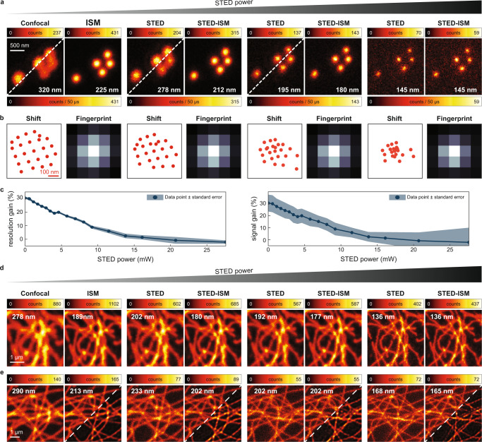
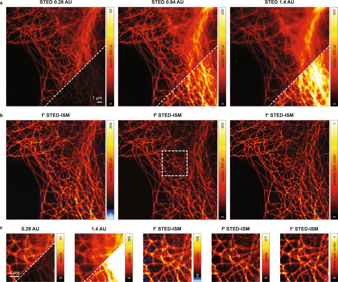
To date, the feasibility of super-resolution microscopy for imaging live and thick samples is still limited. Stimulated emission depletion (STED) microscopy requires high-intensity illumination to achieve sub-diffraction resolution, potentially introducing photodamage to live specimens. Moreover, the out-of-focus background may degrade the signal stemming from the focal plane. Here, we propose a new method to mitigate these limitations without drawbacks. First, we enhance a STED microscope with a detector array, enabling image scanning microscopy (ISM). Therefore, we implement STED-ISM, a method that exploits the working principle of ISM to reduce the depletion intensity and achieve a target resolution. Later, we develop Focus-ISM, a strategy to improve the optical sectioning and remove the background of any ISM-based imaging technique, with or without a STED beam. The proposed approach requires minimal architectural changes to a conventional microscope but provides substantial advantages for live and thick sample imaging.
迄今为止,超分辨率显微镜对活厚样本成像的可行性仍然有限。受激发射耗散(STED)显微镜需要高强度照明以实现亚衍射分辨率,这可能对活标本造成光损伤。此外,离焦背景可能会降低来自焦平面的信号。在这里,我们提出了一种新的方法来克服这些限制,而没有缺点。首先,我们使用探测器阵列增强了 STED 显微镜,实现了图像扫描显微镜(ISM)。因此,我们实现了 STED-ISM,这是一种利用 ISM 的工作原理来降低耗散强度并实现目标分辨率的方法。后来,我们开发了 Focus-ISM,这是一种用于提高光学切片和去除任何基于 ISM 的成像技术的背景的策略,无论是否有 STED 光束。该方法仅需要对传统显微镜进行最小的结构更改,但为活厚样本成像提供了显著的优势。