Institute of Science and Technology Austria, Am Campus 1, 3400 Klosterneuburg, Austria.
Institute of Science and Technology Austria, Am Campus 1, 3400 Klosterneuburg, Austria.
Methods. 2020 Mar 1;174:27-41. doi: 10.1016/j.ymeth.2019.07.019. Epub 2019 Jul 22.
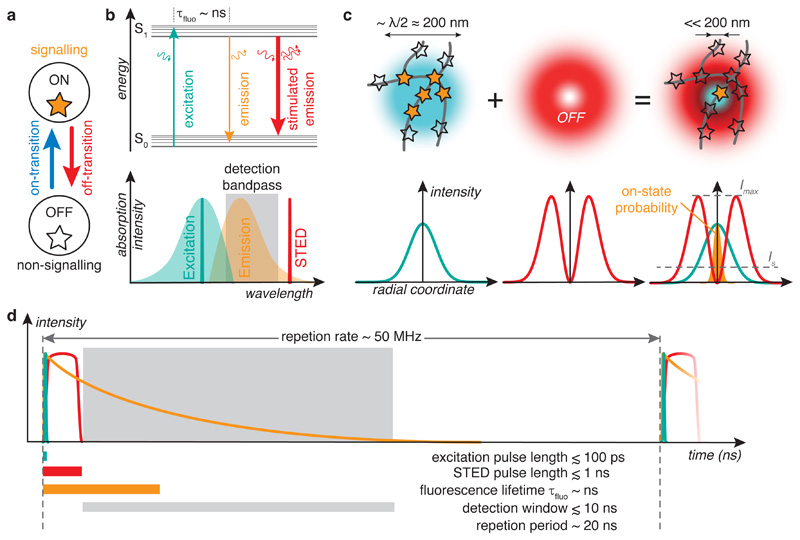
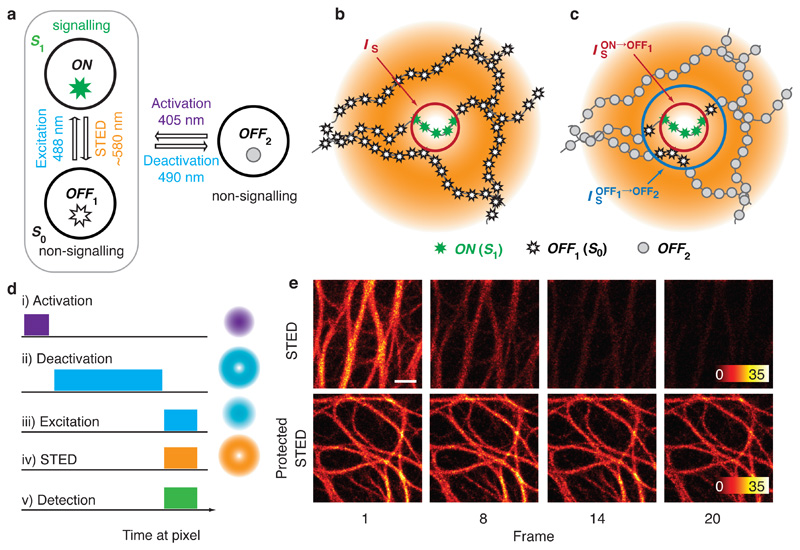
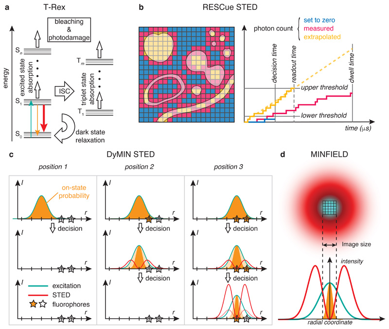
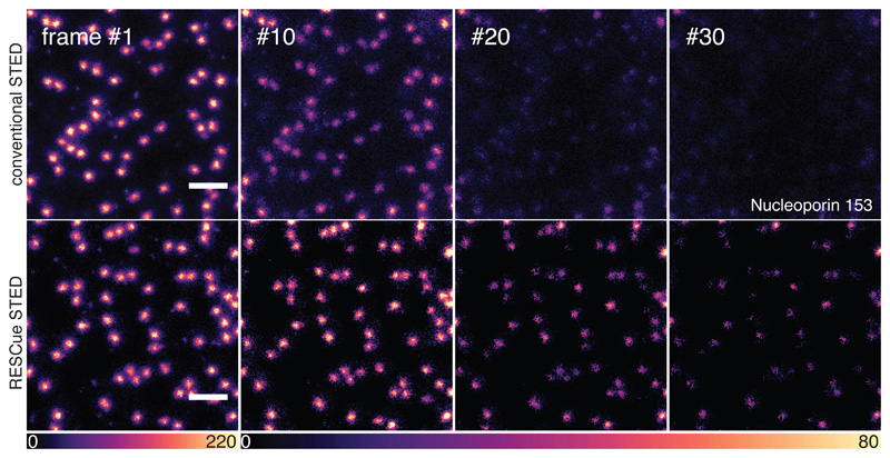
Super-resolution fluorescence microscopy has become an important catalyst for discovery in the life sciences. In STimulated Emission Depletion (STED) microscopy, a pattern of light drives fluorophores from a signal-emitting on-state to a non-signalling off-state. Only emitters residing in a sub-diffraction volume around an intensity minimum are allowed to fluoresce, rendering them distinguishable from the nearby, but dark fluorophores. STED routinely achieves resolution in the few tens of nanometers range in biological samples and is suitable for live imaging. Here, we review the working principle of STED and provide general guidelines for successful STED imaging. The strive for ever higher resolution comes at the cost of increased light burden. We discuss techniques to reduce light exposure and mitigate its detrimental effects on the specimen. These include specialized illumination strategies as well as protecting fluorophores from photobleaching mediated by high-intensity STED light. This opens up the prospect of volumetric imaging in living cells and tissues with diffraction-unlimited resolution in all three spatial dimensions.
超分辨率荧光显微镜已成为生命科学发现的重要催化剂。在受激发射损耗(STED)显微镜中,光的图案将荧光团从信号发射的开态驱动到无信号的关态。只有位于强度最小的亚衍射体积内的发射器才能发出荧光,从而使它们与附近但黑暗的荧光团区分开来。STED 通常可在生物样品中实现几十纳米范围内的分辨率,适用于活细胞成像。在这里,我们回顾了 STED 的工作原理,并提供了成功进行 STED 成像的一般指南。对更高分辨率的追求是以增加光负荷为代价的。我们讨论了降低光暴露并减轻其对标本不利影响的技术。这些技术包括专门的照明策略,以及通过高强度 STED 光保护荧光团免受光漂白。这为在所有三个空间维度上具有衍射极限分辨率的活细胞和组织中的体积成像开辟了前景。