Department of Ophthalmology, Hyogo Prefectural Amagasaki General Medical Center, Higashinaniwa-Cho 2-17-77, Amagasaki, Hyogo, 660-8550, Japan.
Department of Ophthalmology, Mitsubishi Kyoto Hospital, Kyoto, Japan.
Sci Rep. 2022 Dec 22;12(1):22139. doi: 10.1038/s41598-022-26289-6.
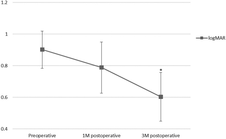
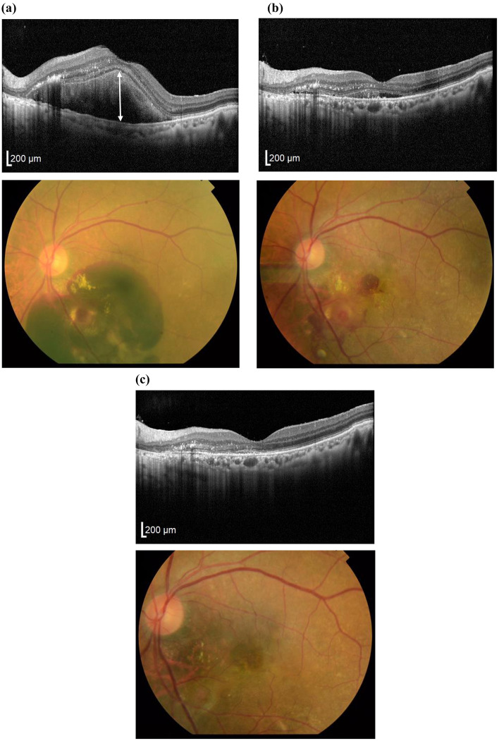
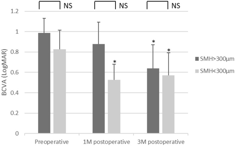
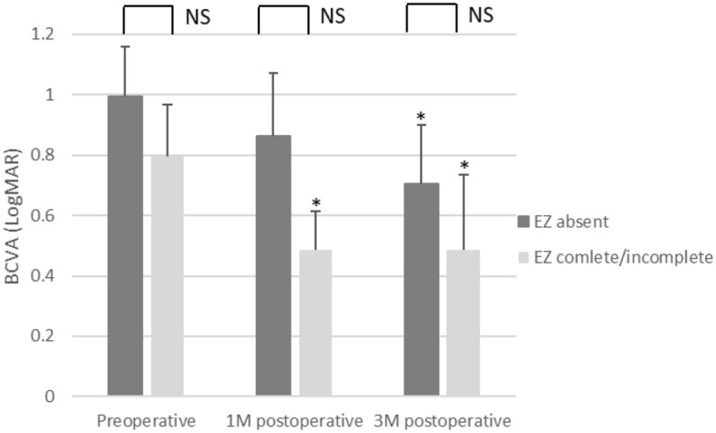
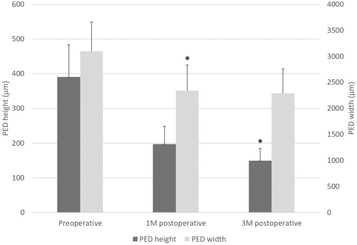
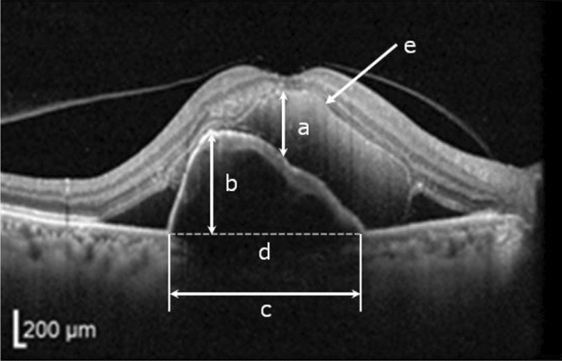
Submacular hemorrhage (SMH) can lead to devastating visual loss in patients with age-related macular degeneration. We retrospectively evaluated the surgical outcomes of vitrectomy with subretinal injection of tissue plasminogen activator, bevacizumab, and air in 13 cases. Visual prognosis, anatomical results obtained with optical coherence tomography (OCT), and their correlations were investigated. We analyzed OCT parameters including SMH height, pigment epithelial detachment (PED) height and width, and status of ellipsoid zone (EZ) line. Complete displacement of SMH was achieved in 12 eyes. At 3 months post-surgery, best-corrected visual acuity (BCVA) and SMH height exhibited significant improvements (P < 0.01). In eyes with preoperative SMH height < 300 µm and a detectable EZ line, BCVA was significantly improved at as early as 1 month, whereas the remaining eyes exhibited visual improvements only at 3 months. Postoperative BCVA positively correlated with preoperative BCVA (r = 0.86, P < 0.005), and negatively correlated with SMH size (r = 0.69, P < 0.01) and PED height (r = 0.58, P < 0.05) and width (r = 0.67, P < 0.05). Multivariate analyses confirmed preoperative BCVA as the predominant factor associated with postoperative BCVA (β = 1.093, P < 0.05). In conclusion, significant improvements in BCVA and anatomical findings can be achieved with our reported surgical technique. Preoperative OCT findings may influence the duration required for visual improvements.
黄斑下出血(SMH)可导致年龄相关性黄斑变性患者视力严重受损。我们回顾性评估了 13 例玻璃体内视网膜下注射组织纤溶酶原激活物、贝伐单抗和空气治疗的手术结果。研究了视力预后、光学相干断层扫描(OCT)获得的解剖结果及其相关性。我们分析了包括 SMH 高度、色素上皮脱离(PED)高度和宽度以及椭圆体带(EZ)线状态在内的 OCT 参数。12 只眼的 SMH 完全移位。术后 3 个月,最佳矫正视力(BCVA)和 SMH 高度均显著改善(P<0.01)。在术前 SMH 高度<300μm 和可检测到 EZ 线的眼中,BCVA 在 1 个月时显著提高,而其余眼仅在 3 个月时表现出视力提高。术后 BCVA 与术前 BCVA 呈正相关(r=0.86,P<0.005),与 SMH 大小(r=0.69,P<0.01)和 PED 高度(r=0.58,P<0.05)和宽度(r=0.67,P<0.05)呈负相关。多变量分析证实术前 BCVA 是与术后 BCVA 相关的主要因素(β=1.093,P<0.05)。总之,我们报道的手术技术可显著改善 BCVA 和解剖学结果。术前 OCT 结果可能会影响视力改善所需的时间。