• 文献检索
  • 文档翻译
  • 深度研究
  • 学术资讯
  • Suppr Zotero 插件Zotero 插件
  • 邀请有礼
  • 套餐&价格
  • 历史记录
应用&插件
Suppr Zotero 插件Zotero 插件浏览器插件Mac 客户端Windows 客户端微信小程序
定价
高级版会员购买积分包购买API积分包
服务
文献检索文档翻译深度研究API 文档MCP 服务
关于我们
关于 Suppr公司介绍联系我们用户协议隐私条款
关注我们

Suppr 超能文献

核心技术专利:CN118964589B侵权必究
粤ICP备2023148730 号-1Suppr @ 2026

文献检索

告别复杂PubMed语法,用中文像聊天一样搜索,搜遍4000万医学文献。AI智能推荐,让科研检索更轻松。

立即免费搜索

文件翻译

保留排版,准确专业,支持PDF/Word/PPT等文件格式,支持 12+语言互译。

免费翻译文档

深度研究

AI帮你快速写综述,25分钟生成高质量综述,智能提取关键信息,辅助科研写作。

立即免费体验

基于深度学习的自动肺分析对间质性肺疾病量化的影响:与肺功能测试结果及预后的相关性

Effects of Automatic Deep-Learning-Based Lung Analysis on Quantification of Interstitial Lung Disease: Correlation with Pulmonary Function Test Results and Prognosis.

作者信息

Aoki Ryo, Iwasawa Tae, Saka Tomoki, Yamashiro Tsuneo, Utsunomiya Daisuke, Misumi Toshihiro, Baba Tomohisa, Ogura Takashi

机构信息

Diagnostic Radiology, Yokohama City University Graduate School of Medicine, 3-9 Fukuura, Kanazawa-ku, Yokohama 236-0004, Kanagawa, Japan.

Department of Radiology, Kanagawa Cardiovascular and Respiratory Center, 6-16-1 Tomioka-higashi, Kanazawa-ku, Yokohama 236-0051, Kanagawa, Japan.

出版信息

Diagnostics (Basel). 2022 Dec 4;12(12):3038. doi: 10.3390/diagnostics12123038.

DOI:10.3390/diagnostics12123038
PMID:36553045
原文链接:https://pmc.ncbi.nlm.nih.gov/articles/PMC9777463/
Abstract

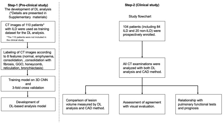
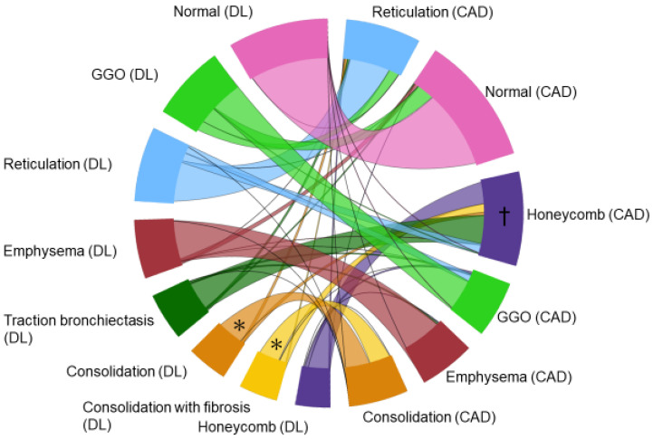
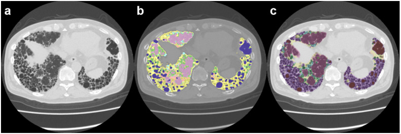
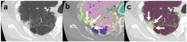
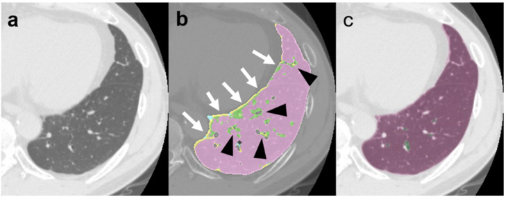
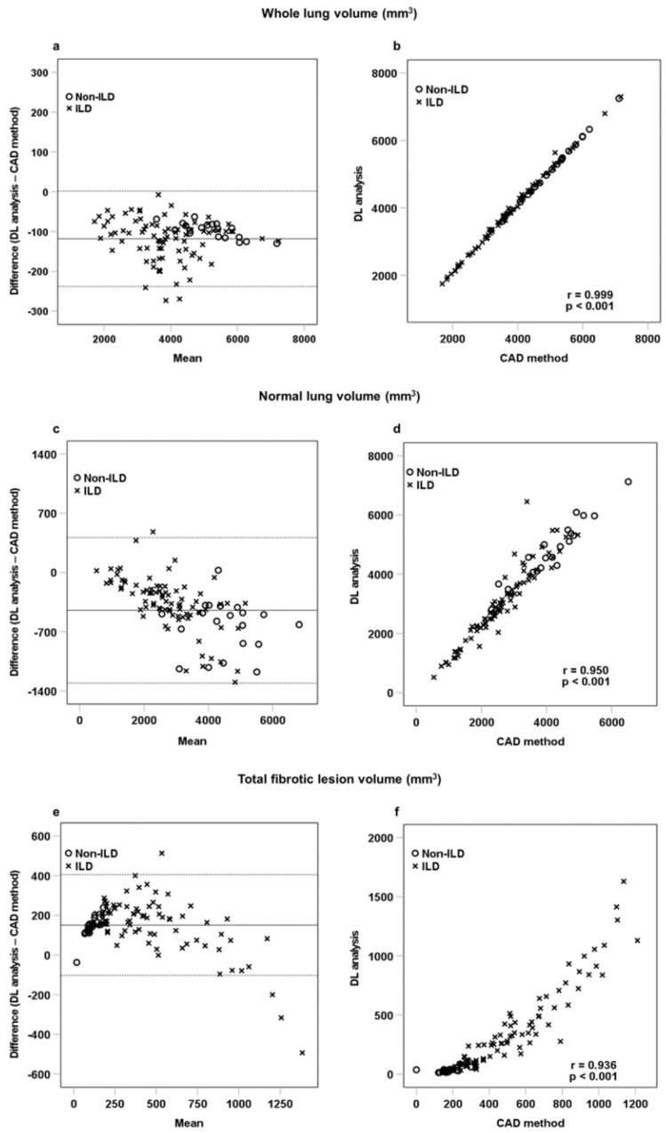
We investigated the feasibility of a new deep-learning (DL)-based lung analysis method for the evaluation of interstitial lung disease (ILD) by comparing it with evaluation using the traditional computer-aided diagnosis (CAD) system and patients’ clinical outcomes. We prospectively included 104 patients (84 with and 20 without ILD). An expert radiologist defined regions of interest in the typical areas of normal, ground-glass opacity, consolidation, consolidation with fibrosis (traction bronchiectasis), honeycombing, reticulation, traction bronchiectasis, and emphysema, and compared them with the CAD and DL-based analysis results. Next, we measured the extent of ILD lesions with the CAD and DL-based analysis and compared them. Finally, we compared the lesion extent on computed tomography (CT) images, as measured with the DL-based analysis, with pulmonary function tests results and patients’ overall survival. Pearson’s correlation analysis revealed a significant correlation between DL-based analysis and CAD results. Forced vital capacity was significantly correlated with DL-based analysis (r = 0.789, p < 0.001 for normal lung volume and r = −0.316, p = 0.001 for consolidation with fibrosis volume). Consolidation with fibrosis measured using DL-based analysis was independently associated with poor survival. The lesion extent measured using DL-based analysis showed a negative correlation with the pulmonary function test results and prognosis.

摘要

我们通过将一种基于深度学习(DL)的新型肺部分析方法与传统计算机辅助诊断(CAD)系统的评估以及患者的临床结果进行比较,来研究其用于评估间质性肺疾病(ILD)的可行性。我们前瞻性纳入了104例患者(84例患有ILD,20例未患ILD)。一名专家放射科医生在正常、磨玻璃影、实变、伴有纤维化的实变(牵拉性支气管扩张)、蜂窝状改变、网状改变、牵拉性支气管扩张和肺气肿的典型区域定义感兴趣区,并将其与CAD和基于DL的分析结果进行比较。接下来,我们用基于CAD和DL的分析测量ILD病变范围并进行比较。最后,我们将基于DL分析测量的计算机断层扫描(CT)图像上的病变范围与肺功能测试结果和患者的总生存期进行比较。Pearson相关性分析显示基于DL的分析与CAD结果之间存在显著相关性。用力肺活量与基于DL的分析显著相关(正常肺容积时r = 0.789,p < 0.001;伴有纤维化的实变容积时r = -0.316,p = 0.001)。基于DL分析测量的伴有纤维化的实变与较差的生存率独立相关。基于DL分析测量的病变范围与肺功能测试结果和预后呈负相关。

https://cdn.ncbi.nlm.nih.gov/pmc/blobs/1480/9777463/eb7d406538be/diagnostics-12-03038-g006.jpg
https://cdn.ncbi.nlm.nih.gov/pmc/blobs/1480/9777463/086778c3f23b/diagnostics-12-03038-g001.jpg
https://cdn.ncbi.nlm.nih.gov/pmc/blobs/1480/9777463/6f2a9975a93f/diagnostics-12-03038-g002.jpg
https://cdn.ncbi.nlm.nih.gov/pmc/blobs/1480/9777463/42f1f2f80388/diagnostics-12-03038-g003.jpg
https://cdn.ncbi.nlm.nih.gov/pmc/blobs/1480/9777463/46fbe3f3ffbf/diagnostics-12-03038-g004.jpg
https://cdn.ncbi.nlm.nih.gov/pmc/blobs/1480/9777463/f4be8887879e/diagnostics-12-03038-g005.jpg
https://cdn.ncbi.nlm.nih.gov/pmc/blobs/1480/9777463/eb7d406538be/diagnostics-12-03038-g006.jpg

相似文献

1
Effects of Automatic Deep-Learning-Based Lung Analysis on Quantification of Interstitial Lung Disease: Correlation with Pulmonary Function Test Results and Prognosis.基于深度学习的自动肺分析对间质性肺疾病量化的影响:与肺功能测试结果及预后的相关性
Diagnostics (Basel). 2022 Dec 4;12(12):3038. doi: 10.3390/diagnostics12123038.
2
Clinical, imaging, and blood biomarkers to assess 1-year progression risk in fibrotic interstitial lung diseases-Development and validation of the honeycombing, traction bronchiectasis, and monocyte (HTM)-score.评估纤维化间质性肺疾病1年进展风险的临床、影像学和血液生物标志物——蜂窝状改变、牵拉性支气管扩张和单核细胞(HTM)评分的开发与验证
Front Med (Lausanne). 2022 Nov 16;9:1043720. doi: 10.3389/fmed.2022.1043720. eCollection 2022.
3
Several high-resolution computed tomography findings associate with survival and clinical features in rheumatoid arthritis-associated interstitial lung disease.几种高分辨率计算机断层扫描发现与类风湿关节炎相关的间质性肺疾病的生存和临床特征相关。
Respir Med. 2018 Jan;134:24-30. doi: 10.1016/j.rmed.2017.11.013. Epub 2017 Nov 24.
4
Interstitial lung disease associated with collagen vascular disorders: disease quantification using a computer-aided diagnosis tool.与胶原血管疾病相关的间质性肺疾病:使用计算机辅助诊断工具进行疾病量化
Eur Radiol. 2009 Feb;19(2):324-32. doi: 10.1007/s00330-008-1152-1. Epub 2008 Aug 26.
5
Tomographic Fibrosis Score in the Patients with Systemic Sclerosis-Associated Interstitial Lung Disease.系统性硬化症相关间质性肺疾病患者的断层扫描纤维化评分
Eur J Rheumatol. 2023 Jul;10(3):107-113. doi: 10.5152/eurjrheum.2023.23024.
6
Evaluation of computer-based computer tomography stratification against outcome models in connective tissue disease-related interstitial lung disease: a patient outcome study.基于计算机断层扫描分层对结缔组织病相关间质性肺病结局模型的评估:一项患者结局研究。
BMC Med. 2016 Nov 23;14(1):190. doi: 10.1186/s12916-016-0739-7.
7
Pulmonary fibrosis and emphysema: Is the emphysema type associated with the pattern of fibrosis?肺纤维化与肺气肿:肺气肿类型是否与纤维化模式相关?
World J Radiol. 2015 Sep 28;7(9):294-305. doi: 10.4329/wjr.v7.i9.294.
8
Automated 3D ιnterstitial lung disease εxtent quantification: performance evaluation and correlation to PFTs.自动化三维间质性肺疾病范围定量:性能评估及其与肺功能测试的相关性
J Digit Imaging. 2014 Jun;27(3):380-91. doi: 10.1007/s10278-013-9670-z.
9
High resolution multidetector CT-aided tissue analysis and quantification of lung fibrosis.高分辨率多探测器CT辅助的肺纤维化组织分析与定量
Acad Radiol. 2007 Jul;14(7):772-87. doi: 10.1016/j.acra.2007.03.009.
10
Automatic quantitative computed tomography measurement of longitudinal lung volume loss in interstitial lung diseases.自动定量计算机断层扫描测量间质性肺疾病的肺容量纵向损失。
Eur Radiol. 2022 Jun;32(6):4292-4303. doi: 10.1007/s00330-021-08482-9. Epub 2022 Jan 14.

引用本文的文献

1
Deep-learning-based 3D content-based image retrieval system on chest HRCT: Performance assessment for interstitial lung diseases and usual interstitial pneumonia.基于深度学习的胸部高分辨率CT三维内容图像检索系统:间质性肺疾病和普通间质性肺炎的性能评估
Eur J Radiol Open. 2025 Jul 23;15:100670. doi: 10.1016/j.ejro.2025.100670. eCollection 2025 Dec.
2
Quantitative analysis of pathological findings identified clinical heterogeneity in nonspecific interstitial pneumonia with organising pneumonia overlap.病理结果的定量分析确定了非特异性间质性肺炎合并机化性肺炎重叠中的临床异质性。
Sci Rep. 2025 Jun 3;15(1):19415. doi: 10.1038/s41598-025-04259-y.
3

本文引用的文献

1
Prevalence and Long-term Outcomes of CT Interstitial Lung Abnormalities in a Health Screening Cohort.健康筛查队列中 CT 肺间质异常的患病率及长期预后。
Radiology. 2023 Feb;306(2):e221172. doi: 10.1148/radiol.221172. Epub 2022 Oct 11.
2
Different Lung Parenchyma Quantification Using Dissimilar Segmentation Software: A Multi-Center Study for COVID-19 Patients.使用不同分割软件对肺实质进行不同量化:一项针对新冠肺炎患者的多中心研究
Diagnostics (Basel). 2022 Jun 20;12(6):1501. doi: 10.3390/diagnostics12061501.
3
Effects of High-Resolution CT Changes on Prognosis Predictability in Acute Respiratory Distress Syndrome with Diffuse Alveolar Damage.
Interstitial lung disease associated with anti-aminoacyl-tRNA synthetase syndrome: quantitative evaluation of CT after initial treatment and long-term follow-up.
抗氨酰基-tRNA 合成酶综合征相关的间质性肺疾病:初始治疗和长期随访后的 CT 定量评估。
Acta Radiol. 2024 Nov;65(11):1332-1340. doi: 10.1177/02841851241281492. Epub 2024 Oct 10.
4
Advances in CT-based lung function imaging for thoracic radiotherapy.基于CT的肺功能成像在胸部放疗中的进展。
Front Oncol. 2024 Sep 2;14:1414337. doi: 10.3389/fonc.2024.1414337. eCollection 2024.
5
Evaluation of Progressive Architectural Distortion in Idiopathic Pulmonary Fibrosis Using Deformable Registration of Sequential CT Images.使用序列CT图像的可变形配准评估特发性肺纤维化中的进行性结构扭曲
Diagnostics (Basel). 2024 Jul 31;14(15):1650. doi: 10.3390/diagnostics14151650.
6
Correlation of CT-based radiomics analysis with pathological cellular infiltration in fibrosing interstitial lung diseases.基于 CT 的放射组学分析与纤维性肺间质疾病中病理性细胞浸润的相关性。
Jpn J Radiol. 2024 Oct;42(10):1157-1167. doi: 10.1007/s11604-024-01607-2. Epub 2024 Jun 18.
7
Longitudinal assessment of interstitial lung abnormalities on CT in patients with COPD using artificial intelligence-based segmentation: a prospective observational study.基于人工智能分割技术对慢性阻塞性肺疾病患者进行CT肺间质异常的纵向评估:一项前瞻性观察研究。
BMC Pulm Med. 2024 Apr 23;24(1):200. doi: 10.1186/s12890-024-03002-z.
8
The Role of Inflammation and Fibrosis in Interstitial Lung Disease Treatment Decisions.炎症和纤维化在间质性肺疾病治疗决策中的作用
Am J Respir Crit Care Med. 2024 Aug 15;210(4):392-400. doi: 10.1164/rccm.202401-0048PP.
9
Quantitative Analysis for Lung Disease on Thin-Section CT.薄层CT上肺部疾病的定量分析
Diagnostics (Basel). 2023 Sep 18;13(18):2988. doi: 10.3390/diagnostics13182988.
10
Semi-Supervised Segmentation of Interstitial Lung Disease Patterns from CT Images via Self-Training with Selective Re-Training.通过选择性再训练的自训练方法对CT图像中的间质性肺疾病模式进行半监督分割
Bioengineering (Basel). 2023 Jul 12;10(7):830. doi: 10.3390/bioengineering10070830.
高分辨率CT改变对伴有弥漫性肺泡损伤的急性呼吸窘迫综合征预后预测的影响
J Clin Med. 2022 Apr 27;11(9):2458. doi: 10.3390/jcm11092458.
4
Idiopathic Pulmonary Fibrosis (an Update) and Progressive Pulmonary Fibrosis in Adults: An Official ATS/ERS/JRS/ALAT Clinical Practice Guideline.特发性肺纤维化(更新版)和成人进展性肺纤维化:美国胸科学会/欧洲呼吸学会/日本呼吸学会/拉丁美洲胸科学会临床实践指南。
Am J Respir Crit Care Med. 2022 May 1;205(9):e18-e47. doi: 10.1164/rccm.202202-0399ST.
5
Radiomics for the Prediction of Response to Antifibrotic Treatment in Patients with Idiopathic Pulmonary Fibrosis: A Pilot Study.基于影像组学预测特发性肺纤维化患者抗纤维化治疗反应的初步研究
Diagnostics (Basel). 2022 Apr 15;12(4):1002. doi: 10.3390/diagnostics12041002.
6
Predicting Usual Interstitial Pneumonia Histopathology From Chest CT Imaging With Deep Learning.利用深度学习从胸部CT影像预测寻常型间质性肺炎组织病理学
Chest. 2022 Oct;162(4):815-823. doi: 10.1016/j.chest.2022.03.044. Epub 2022 Apr 8.
7
Radiological pleuroparenchymal fibroelastosis-like lesion in idiopathic interstitial pneumonias.特发性间质性肺炎中的放射学胸膜肺实质纤维弹性组织增生样病变。
Respir Res. 2021 Nov 11;22(1):290. doi: 10.1186/s12931-021-01892-9.
8
Content-based Image Retrieval by Using Deep Learning for Interstitial Lung Disease Diagnosis with Chest CT.基于深度学习的基于内容的图像检索用于胸部 CT 诊断间质性肺病。
Radiology. 2022 Jan;302(1):187-197. doi: 10.1148/radiol.2021204164. Epub 2021 Oct 12.
9
Novel Artificial Intelligence-based Technology for Chest Computed Tomography Analysis of Idiopathic Pulmonary Fibrosis.基于新型人工智能技术的特发性肺纤维化胸部计算机断层扫描分析。
Ann Am Thorac Soc. 2022 Mar;19(3):399-406. doi: 10.1513/AnnalsATS.202101-044OC.
10
Imaging of COVID-19: An update of current evidences.COVID-19 的影像学表现:现有证据的更新。
Diagn Interv Imaging. 2021 Sep;102(9):493-500. doi: 10.1016/j.diii.2021.05.006. Epub 2021 May 25.